Low Power Light Microscope Micrograph Thin Skin Showing Form Left — Stock Photo, Image

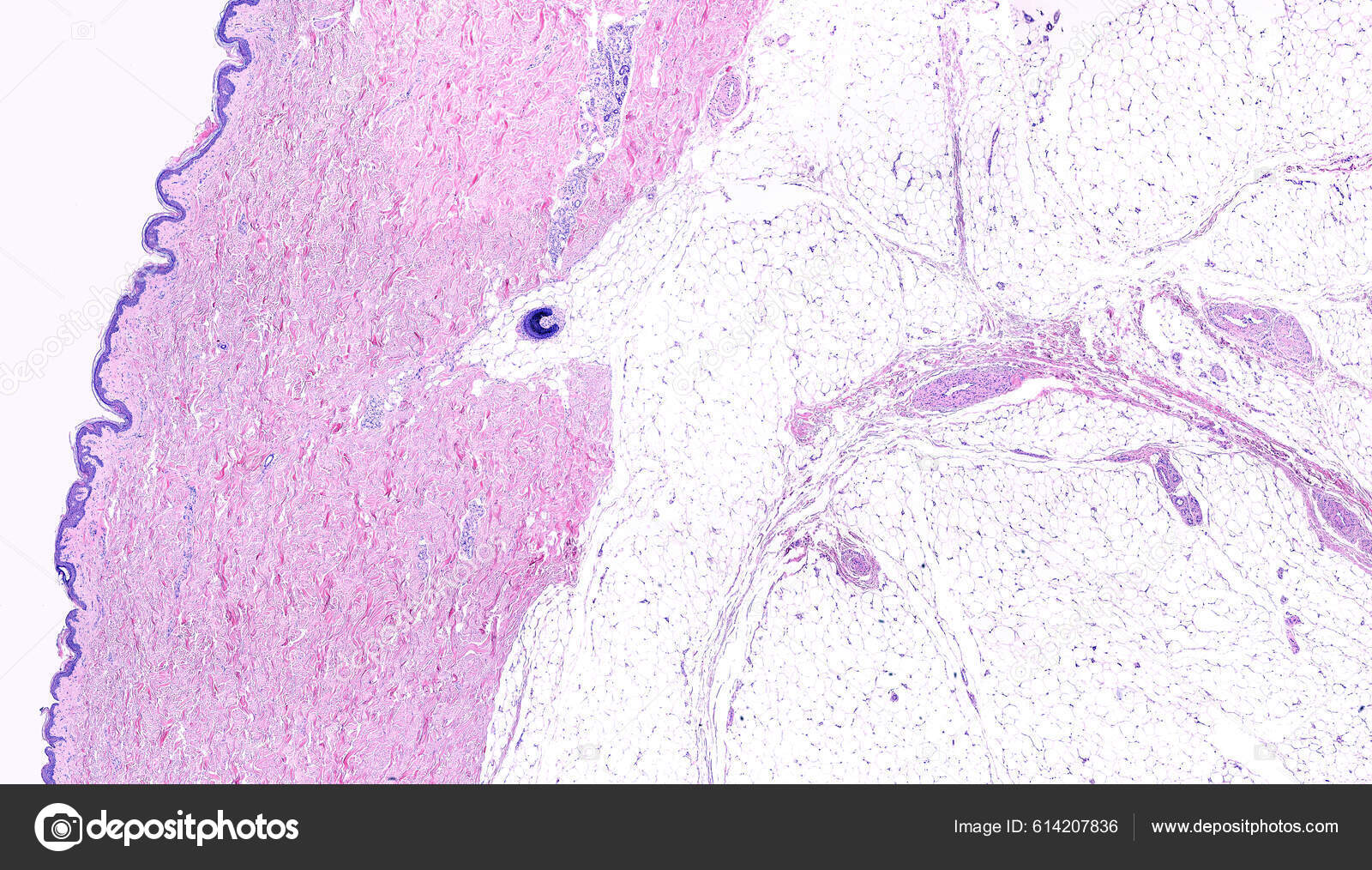
Low power light microscope micrograph of thin skin showing form left to right, the epidermis, a very thick dermis showing sweat glands and a hair follicle in the deep region, and the adipose tissue of the hypodermis. Hematoxylin-eosin. — Illustration

Low power light microscope micrograph of thin skin showing form left to right, the epidermis, a very thick dermis showing sweat glands and a hair follicle in the deep region, and the adipose tissue of the hypodermis. Hematoxylin-eosin.

 — Illustration by jlcalvo@ucm.es

Same Series:

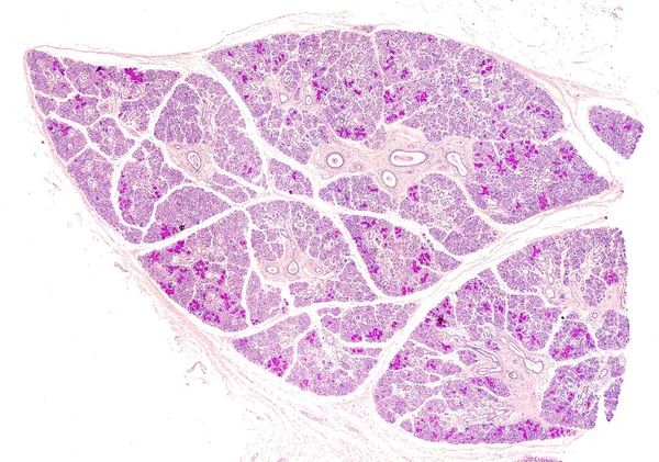
Low power light microscope micrograph of a human submandibular or submaxillary gland. This is a mixed salivary gland with predominance of serous acini. The PAS stain highlights the mucous component of the mixed tubule acini.
Low Power Light Microscope Micrograph Human Submandibular Submaxillary Gland Mixed — Stock Photo, Image
Ovarian mucinous cystadenoma (15-20% of ovarian tumours) is a usually large and benign tumour showing glands and cysts, with filiform papillae with fibrovascular cores, lined by benign columnar epithelium.
Ovarian Mucinous Cystadenoma Ovarian Tumours Usually Large Benign Tumour Showing — Stock Photo, Image
Low magnification of a human prostate gland in a 70-year-old man. The prostate gland appears with dilated alveoli, which contains many corpora amylacea (prostatic concretions) in their lumen. Light microscope micrograph. Hematoxylin & eosin stain.
Human prostate. Corpora amylacea — Stock Photo, Image
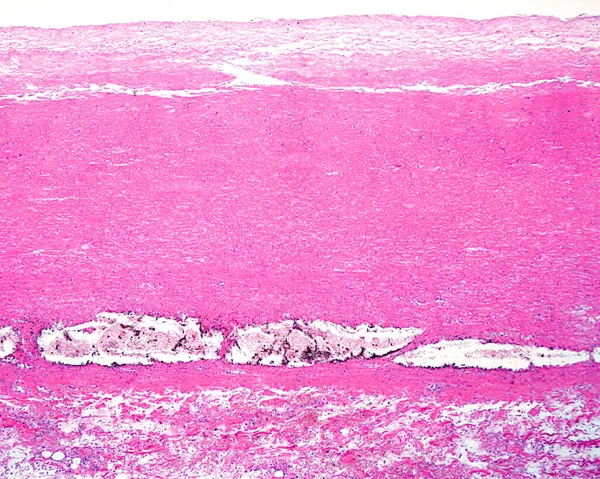
Human aorta. Dissecting aneurysm or aortic dissection. Low magnification micrograph showing a dissecting aneurysm located in the media layer of the aorta wall. Macrophages loaded with iron pigment appear in the detachment zone.
Human Aorta Dissecting Aneurysm Aortic Dissection Low Magnification Micrograph Showing — Stock Photo, Image
Low power light microscope micrograph of a cat thyroid. The thyroid parenchyma is composed of many follicles filled with colloid. On the right side, a parathyroid gland is located inside the thyroid. Light microscope micrograph. H&E stain.
Low Power Light Microscope Micrograph Cat Thyroid Thyroid Parenchyma Composed — Stock Photo, Image
Ovarian mucinous cystadenoma (15-20% of ovarian tumours) is a usually large and benign tumour showing glands and cysts, with filiform papillae with fibrovascular cores, lined by benign columnar epithelium.
Ovarian Mucinous Cystadenoma Ovarian Tumours Usually Large Benign Tumour Showing — Stock Photo, Image
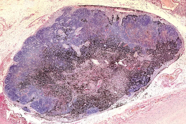
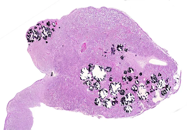
Anthracotic lymph node. Accumulation of carbon is most commonly found in intrapulmonary lymph nodes, due to coal dust, smoke or pollution. Hematoxylin & eosin stai
Anthracotic lymph node — Stock Photo, Image
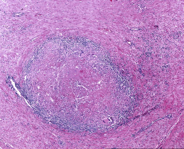
Tuberculosis granuloma with a central necrosis (caseous necrosis), lymphocytic infiltrate and multinucleated Langhans cell.
Tuberculosis granuloma. Langhans cell — Stock Photo, Image
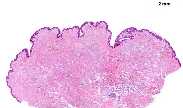
Low power light microscope micrograph of a human breast nipple. It is covered by a pigmented thin skin. On the connective tissue there are many fascicles of smooth muscle fibers (nipple erection). On the bottom there are two sweat glands, apocrine an
Low Power Light Microscope Micrograph Human Breast Nipple Covered Pigmented — Stock Photo, Image
Low power light microscope micrograph of a cat thyroid. The thyroid parenchyma is composed of many follicles filled with colloid. On the right side, a parathyroid gland is located inside the thyroid. Light microscope micrograph. H&E stain.
Low Power Light Microscope Micrograph Cat Thyroid Thyroid Parenchyma Composed — Stock Photo, Image
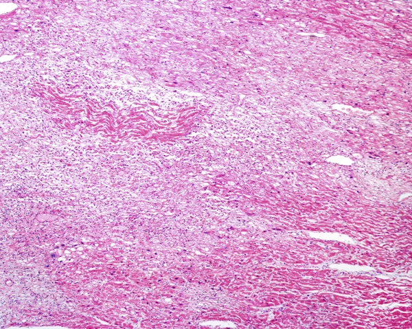
Light micrograph of a human heart showing several pale areas of a recent infarct. In these areas, there is an ischemic, coagulative necrosis and inflammatory infiltrates of macrophages, suggesting an evolution of at least one week.
Light Micrograph Human Heart Showing Several Pale Areas Recent Infarct — Stock Photo, Image
Sagittal section of a human pineal gland. Numerous calcareous concretions can be seen, most of them broken by artefact. Light microscope micrograph stained with hematoxylin-eosin.
Human pineal gland. Calcareous concretions — Stock Photo, Image
Low magnification micrograph showing a metastasis of a malignant melanoma invading a lymph node. The normal lymphoid tissue has almost completely disappeared and has been replaced by malignant melanoma pigmented and non-pigmented cells.
Low Magnification Micrograph Showing Metastasis Malignant Melanoma Invading Lymph Node — Stock Photo, Image
Low power light microscope micrograph of a human breast nipple. It is covered by a pigmented thin skin. On the connective tissue there are many fascicles of smooth muscle fibers (nipple erection). On the bottom there are two sweat glands, apocrine an
Low Power Light Microscope Micrograph Human Breast Nipple Covered Pigmented — Stock Photo, Image
Corpus albicans. Human ovary. The corpus albicans is the regressed form of the corpus luteum. After the corpus luteum degenerates, it is replaced by white scar of fibrous tissue made by type I collagen.
Ovary. Corpus albicans — Stock Photo, Image
Low power light microscope micrograph of a human breast nipple. It is covered by a pigmented thin skin. On the connective tissue there are many fascicles of smooth muscle fibers (nipple erection). On the bottom there are two sweat glands, apocrine an
Low Power Light Microscope Micrograph Human Breast Nipple Covered Pigmented — Stock Photo, Image

Usage Information

You can use this royalty-free illustration "Low power light microscope micrograph of thin skin showing form left to right, the epidermis, a very thick dermis showing sweat glands and a hair follicle in the deep region, and the adipose tissue of the hypodermis. Hematoxylin-eosin." for personal and commercial purposes according to the Standard or Extended License. The Standard License covers most use cases, including advertising, UI designs, and product packaging, and allows up to 500,000 print copies. The Extended License permits all use cases under the Standard License with unlimited print rights and allows you to use the downloaded stock illustrations for merchandise, product resale, or free distribution.

You can buy this illustration and download it in high resolution up to 5378x3072. Upload Date: Oct 8, 2022