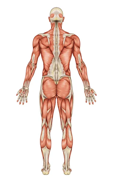
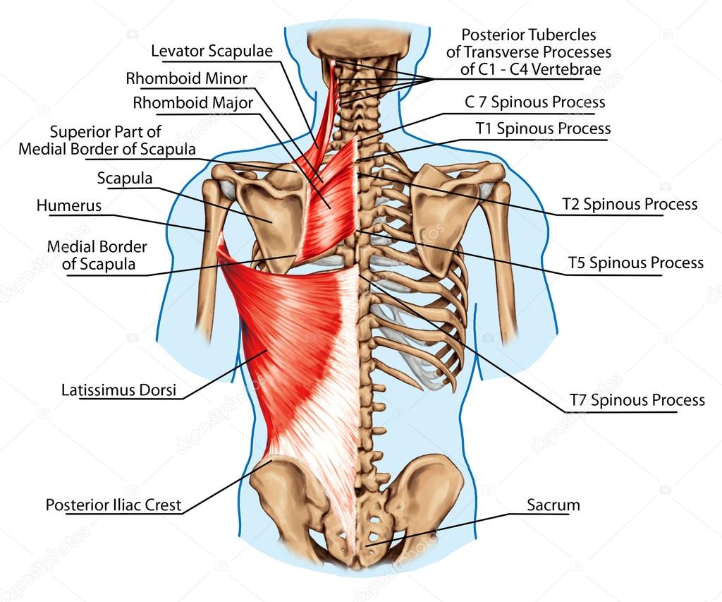
Rhomboid minor and rhomboid major, levator scapulae and latissimus dorsi muscles - didactic board of anatomy of human bony and muscular system, posterior view — Stock Photo, Image

Rhomboid minor and rhomboid major, levator scapulae and latissimus dorsi muscles - didactic board of anatomy of human bony and muscular system, posterior view — Illustration

Rhomboid minor and rhomboid major, levator scapulae and latissimus dorsi muscles - didactic board of anatomy of human bony and muscular system, posterior view

 — Illustration by stihii

Same Series:

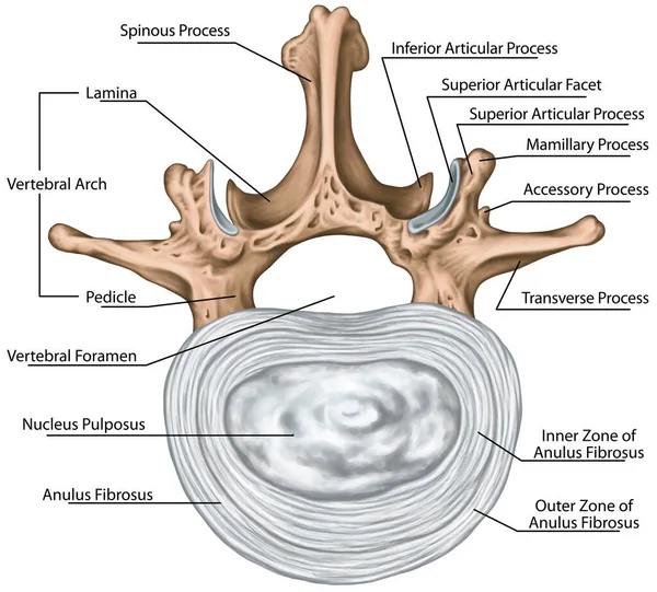
Structure of an intervertebral disk, second lumbar vertebra, lumbar spine, outer and inner zone of the anulus fibrosus, vertebral bones, vertebra, trunk wall, anatomy of human skeletal system, superior view
Structure Intervertebral Disk Second Lumbar Vertebra Lumbar Spine Outer Inner — Stock Photo, Image
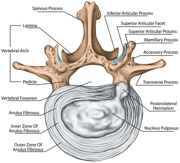
Lumbar disk herniation, herniated disc, lumbar vertebra, lumbar spine, intervertebral disk, anulus fibrosus, nucleus pulposus, vertebral bones, vertebra, anatomy of human skeletal system, superior view
Lumbar Disk Herniation Herniated Disc Lumbar Vertebra Lumbar Spine Intervertebral — Stock Photo, Image
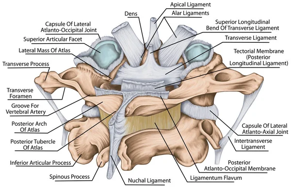
The ligaments of the median Atlantoaxial joint. Atlas and axis ligaments. Cervical spine, vertebral morphology, first and second cervical vertebra, cervical vertebrae, atlas, axis, atlantoaxial joint, posterosuperior view
Ligaments Median Atlantoaxial Joint Atlas Axis Ligaments Cervical Spine Vertebral — Stock Photo, Image
Anatomy of male muscular system posterior view full body
Anatomy of male muscular system posterior view full body — Stock Photo, Image
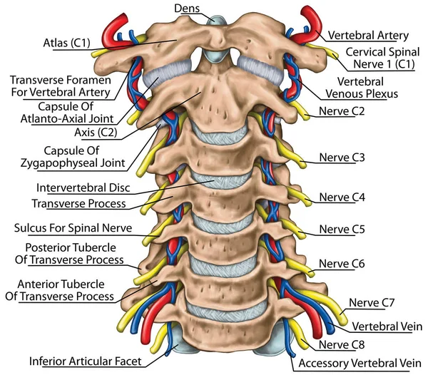
Cervical spine with both vertebral arteries in transverse foramen and the emerging spinal nerves. Topographic relationship of the spinal nerve and vertebral artery. Anterior view.
Cervical Spine Both Vertebral Arteries Transverse Foramen Emerging Spinal Nerves — Stock Photo, Image
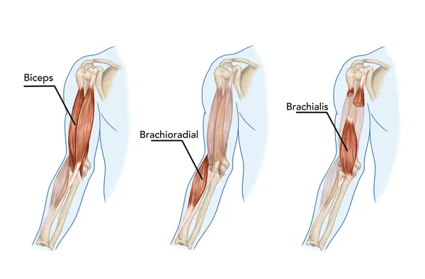
Biceps, Brachii, Brachioradial, Brachialis muscles – didactic
Biceps, Brachii, Brachioradial, Brachialis muscles – didactic — Stock Photo, Image
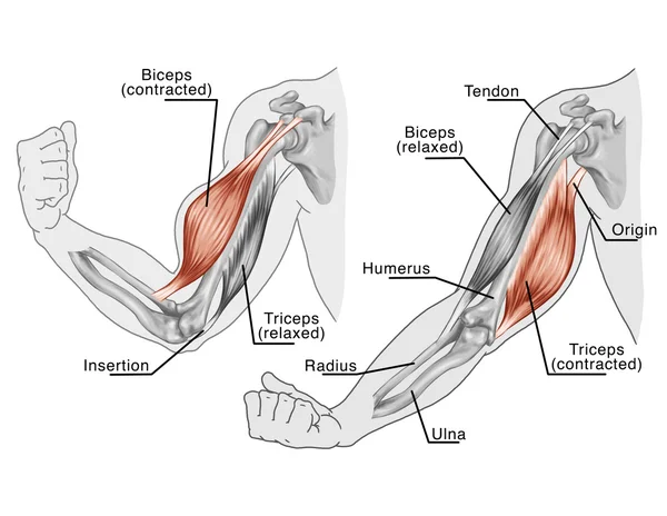
Biceps, Triceps - movement of the arm and hand muscles – board - Black White
Movement of the arm and hand muscles — Stock Photo, Image
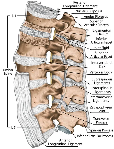
Ligaments and lumbar spine structure, the ligaments surrounding the lumbar spine, posterior longitudinal ligament, intertransverse ligaments, human bony system, human skeletal system, posterior view
Ligaments Lumbar Spine Structure Ligaments Surrounding Lumbar Spine Posterior Longitudinal — Stock Photo, Image
Ligaments, lumbar spine structure, anterior longitudinal, intertransverse, interspinous, supraspinous ligaments, vertebral bones, anatomy of human bony system, human skeletal system, left lateral view
Ligaments Lumbar Spine Structure Anterior Longitudinal Intertransverse Interspinous Supraspinous Ligaments — Stock Photo, Image
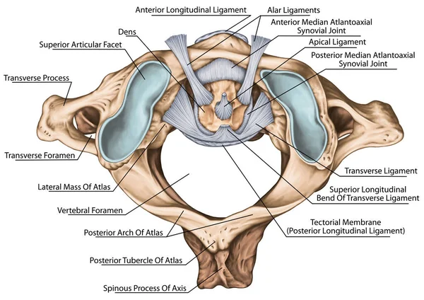
The ligaments of the median Atlantoaxial joint. Atlas and axis ligaments. Cervical spine, vertebral morphology, first and second cervical vertebra, cervical vertebrae, atlas, axis, atlantoaxial joint, superior view
Ligaments Median Atlantoaxial Joint Atlas Axis Ligaments Cervical Spine Vertebral — Stock Photo, Image
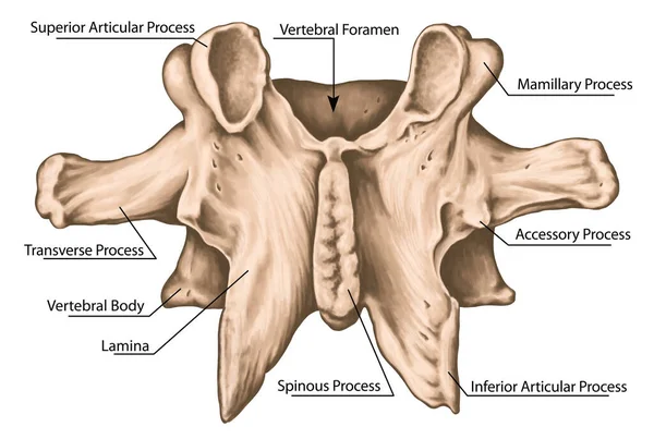
Second lumbar vertebra, lumbar vertebrae, vertebral bone, vertebra, vertebral body, transverse, spinous, mamillary process, inferior vertebral notch, posterior view
Second Lumbar Vertebra Lumbar Vertebrae Vertebral Bone Vertebra Vertebral Body — Stock Photo, Image
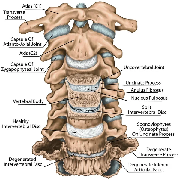
Degenerative changes in the cervical spine, uncinate process, uncovertebral arthrosis, anatomy of human bone system, uncovertebral joint, split intervertebral disc, coronal section, anterior view
Degenerative Changes Cervical Spine Uncinate Process Uncovertebral Arthrosis Anatomy Human — Stock Photo, Image
Structure of an intervertebral disk, outer and inner zone of the anulus fibrosus, hyaline cartilage endplate, vertebral bones, vertebra, trunk wall, anatomy of human skeletal system, motion segment
Structure Intervertebral Disk Outer Inner Zone Anulus Fibrosus Hyaline Cartilage — Stock Photo, Image
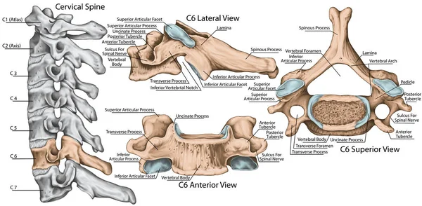
Didactic board, cervical spine, common vertebral morphology, sixth cervical vertebra, cervical vertebrae, anterior, lateral and superior view
Didactic Board Cervical Spine Common Vertebral Morphology Sixth Cervical Vertebra — Stock Photo, Image
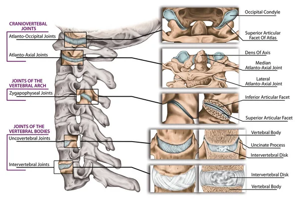
Joints of the vertebral column, cervical spine structure, vertebral bones, cervical bones, anatomy of human bone system, craniovertebral joints, joints of the vertebral bodies and arch, lateral view
Joints Vertebral Column Cervical Spine Structure Vertebral Bones Cervical Bones — Stock Photo, Image
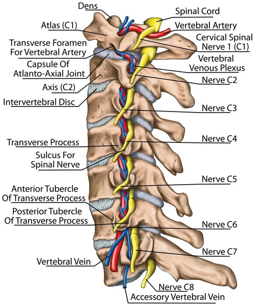
Cervical spine with both vertebral arteries in transverse foramen and the emerging spinal nerves. Topographic relationship of the spinal nerve and vertebral artery. Lateral view.
Cervical Spine Both Vertebral Arteries Transverse Foramen Emerging Spinal Nerves — Stock Photo, Image

Similar Stock Videos:
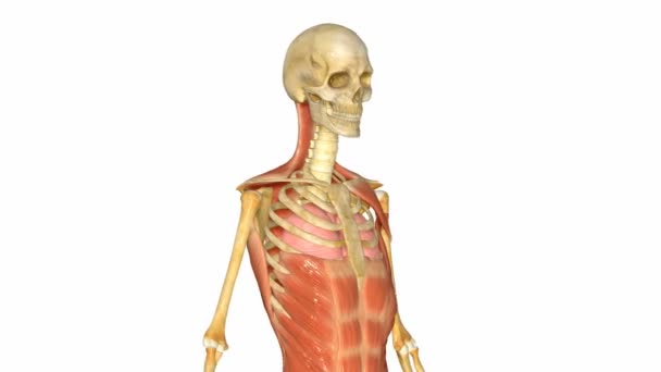
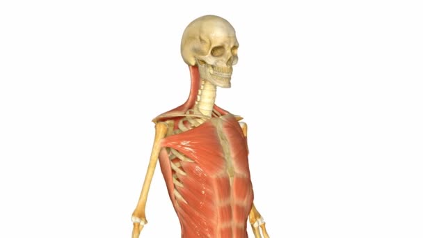
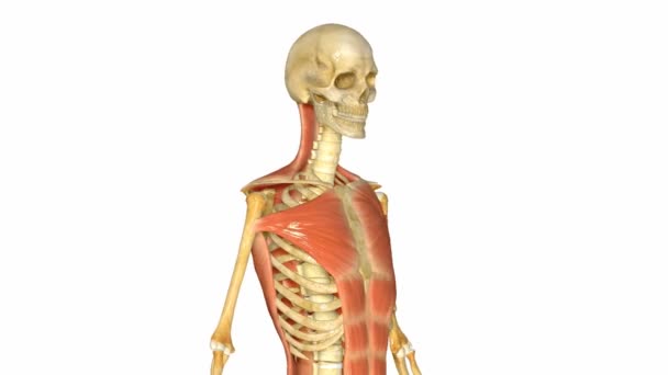
Muscle is a soft tissue found in most animals. Muscle cells contain protein filaments of actin and myosin that slide past one another, producing a contraction that changes both the length and the shape of the cell.
Muscles — Stock Video
Muscle is a soft tissue found in most animals. Muscle cells contain protein filaments of actin and myosin that slide past one another, producing a contraction that changes both the length and the shape of the cell.
Muscles — Stock Video
Muscle is a soft tissue found in most animals. Muscle cells contain protein filaments of actin and myosin that slide past one another, producing a contraction that changes both the length and the shape of the cell.
Muscles — Stock Video
Muscle is a soft tissue found in most animals. Muscle cells contain protein filaments of actin and myosin that slide past one another, producing a contraction that changes both the length and the shape of the cell.
Muscles — Stock Video
Muscle is a soft tissue found in most animals. Muscle cells contain protein filaments of actin and myosin that slide past one another, producing a contraction that changes both the length and the shape of the cell.
Muscles — Stock Video
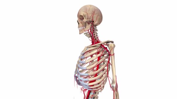
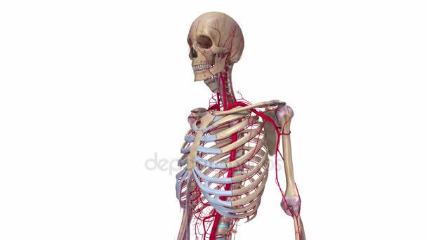
Skeleton with Arteries system, isolated on white background
Skeleton with Arteries system — Stock Video
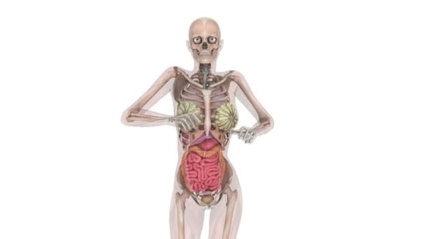
Walking woman
Walking woman — Stock Video
Skeleton Ligaments and Arteries system, isolated on white background
Skeleton Ligaments and Arteries — Stock Video
Running woman
Running woman — Stock Video
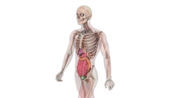
Walking man
Walking man — Stock Video
Running man
Running man — Stock Video
Woman Belly dance in red- look and smile
Woman Belly dance in red- look and smile — Stock Video
Male scoliosis in time lapse view
Male scoliosis in time lapse — Stock Video
Walking man
Walking man — Stock Video
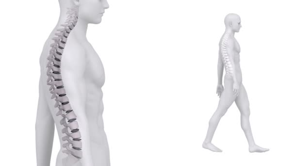
Walking man backbone zoom
Walking man backbone zoom — Stock Video
Futuristic humanoid female robot is idle . Realistic motion and reflections. 4K green screen footage
Futuristic humanoid female robot is idle . Realistic motion and reflections. 4K green screen footage. — Stock Video
Woman in traditiona indian outfit isolated on white upset
Woman in traditiona indian outfit — Stock Video
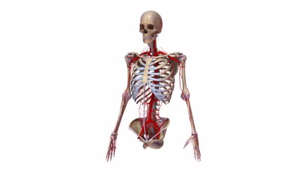
Upper body Ligaments and Arteries system, isolated on white background
Upper body Ligaments and Arteries — Stock Video

Usage Information

You can use this royalty-free illustration "Rhomboid minor and rhomboid major, levator scapulae and latissimus dorsi muscles - didactic board of anatomy of human bony and muscular system, posterior view" for personal and commercial purposes according to the Standard or Extended License. The Standard License covers most use cases, including advertising, UI designs, and product packaging, and allows up to 500,000 print copies. The Extended License permits all use cases under the Standard License with unlimited print rights and allows you to use the downloaded stock illustrations for merchandise, product resale, or free distribution.

You can buy this illustration and download it in high resolution up to 6000x5000. Upload Date: Apr 17, 2013