Cerebellar Granule Cells Light Micrograph Cerebellar Granular Layer Stained Golgi — Stock Photo, Image

Cerebellar granule cells, light micrograph. Cerebellar granular layer stained with Golgi silver chromate showing the abundance of granule cells. They have a very small rounded soma with three to five dendrites, ending in an enlargement called a dendr — Photo

Cerebellar granule cells, light micrograph. Cerebellar granular layer stained with Golgi silver chromate showing the abundance of granule cells. They have a very small rounded soma with three to five dendrites, ending in an enlargement called a dendr

 — Photo by jlcalvo@ucm.es

Same Series:

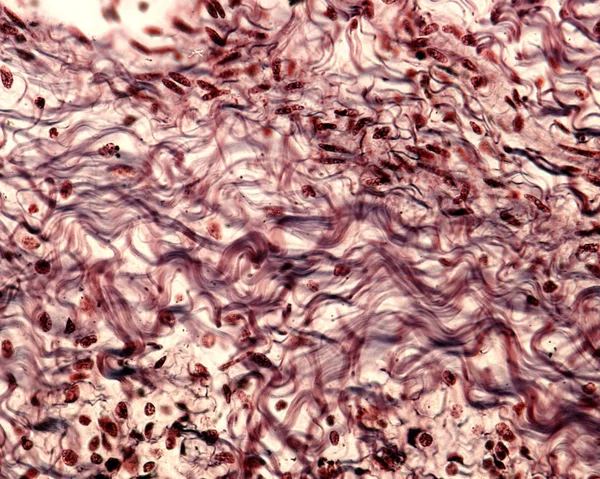
Bundles of collagen fibers in a connective tissue stained with a silver method. The collagen fibers show a typical wavy appearance.
Collagen fibers — Stock Photo, Image
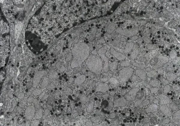
Transmission electron microscope (TEM) micrograph of a gonadotropic cell of a castrated animal showing dark FSL-LH granules and large swellings of the RER, due to the castration.
Gonadotropic cell, TEM — Stock Photo, Image
Transmission electron micrograph (TEM) showing two desmosomes (maculae adherentes) with prominent dense plaques where keratin intermediate filaments were attached.
Transmission Electron Micrograph Tem Showing Two Desmosomes Maculae Adherentes Prominent — Stock Photo, Image
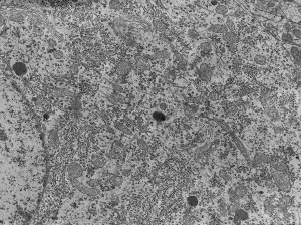
Transmission electron microscope (TEM) micrograph of a neuron cell body showing mitochondria, lysosomes, RER, Golgi system, and the nucleus (right).
Neuron. Cell organelles, TEM — Stock Photo, Image
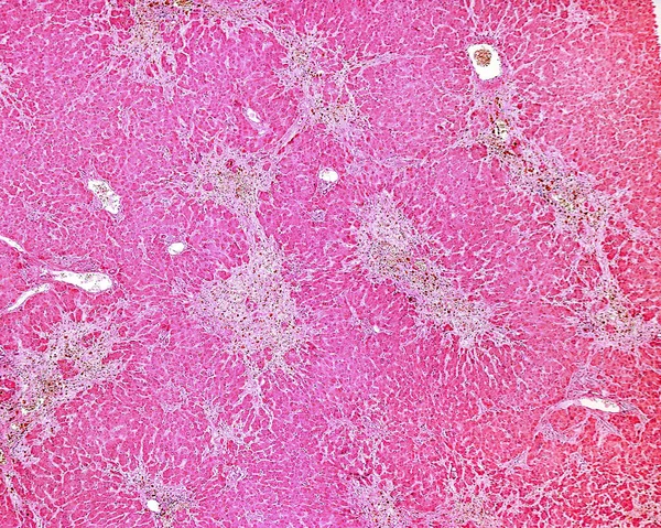
Human liver. Drug-induced liver injury by carbon tetrachloride. Low magnification micrograph showing several clear areas of degeneration and necrosis of hepatocytes with little or no inflammation.
Human Liver Drug Induced Liver Injury Carbon Tetrachloride Low Magnification — Stock Photo, Image
Light microscope micrograph of a senile plaque, stain with a silver method, frequently seen in the Alzheimer disease. In the bottom of the image, there are three normal pyramidal neurons.
Senile plaques. Alzheimer disease — Stock Photo, Image
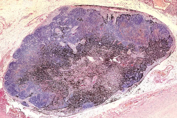
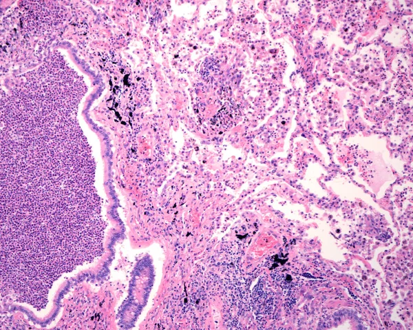
Anthracotic lymph node. Accumulation of carbon is most commonly found in intrapulmonary lymph nodes, due to coal dust, smoke or pollution. Hematoxylin & eosin stai
Anthracotic lymph node — Stock Photo, Image
Light microscopy micrograph showing white (bottom) and brown (top) fat. The larger holes correspond to white adipocytes, while brown adipocytes shows a spongy aspect because they stored fat as small lipid droplets.
White and brown fat — Stock Photo, Image
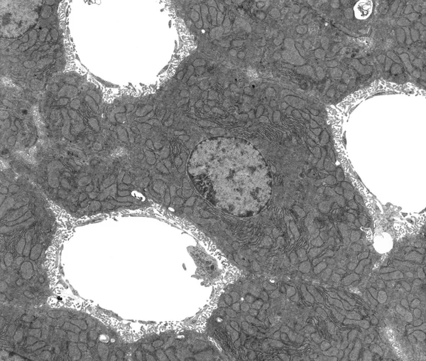
TEM micrograph showing hepatocyte plates located among three sinusoids. The hepatocyte cytoplasm shows many mitochondria and elongated cisterns of rough endoplasmic reticulum (RER). The liver sinusoids with discontinuous endothelium are surrounded by
Tem Micrograph Showing Hepatocyte Plates Located Three Sinusoids Hepatocyte Cytoplasm — Stock Photo, Image
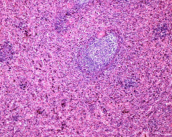
Human spleen. Lymphoid follicle, a component of white pulp. On top, a periarteriolar lymphoid sheath, another component of white pulp. The rest of micrograph is the red pulp, composed by splenic sinusoids and cord of Billroth showing macrophages with
Human Spleen Lymphoid Follicle Component White Pulp Top Periarteriolar Lymphoid — Stock Photo, Image
Light microscope micrograph of an influenza associated encephalopathy (IAE). The meninx and the nervous parenchyma show severe inflammatory infiltrates.
Light Microscope Micrograph Influenza Associated Encephalopathy Iae Meninx Nervous Parenchyma — Stock Photo, Image
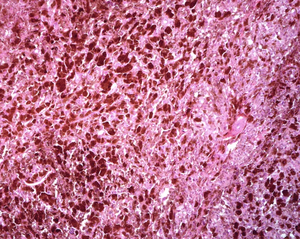
Human lymph node. Metastasis of a malignant melanoma. The normal lymphoid tissue of lymph node has completely disappeared and has been replaced by malignant melanoma pigmented and non-pigmented cells.
Human Lymph Node Metastasis Malignant Melanoma Normal Lymphoid Tissue Lymph — Stock Photo, Image
Neuron stained with cresyl violet showing a Barr body (sex chromatin) located near to a big nucleolus. Light microscope micrograph.
Barr body. Neuron — Stock Photo, Image
Human lung affected by an acute bronchopneumonia, commonly a hospital-acquired bacterial pneumonia. The lumen of alveoli is occupied by liquid of oedema which contains acute inflammatory infiltrates (with predominance of neutrophil granulocytes). On
Human Lung Affected Acute Bronchopneumonia Commonly Hospital Acquired Bacterial Pneumonia — Stock Photo, Image
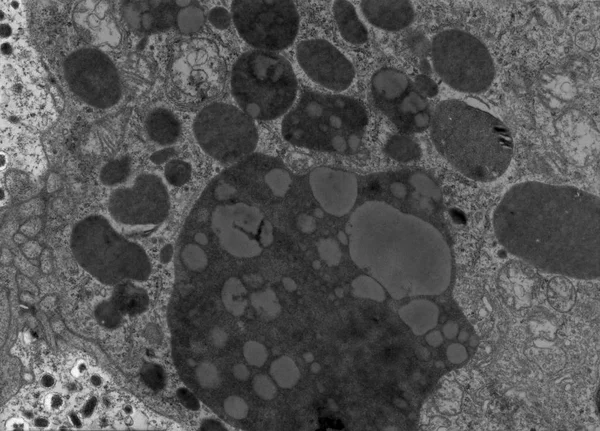
Transmission electron microscope (TEM) micrograph showing many lysosomes (dark granules). Some of the, with rounded grey zones inside, are lipofuscin granules. On the left side, there is a noradrenaline cell.
Lysosomes and lipofuscin. False colour TEM — Stock Photo, Image
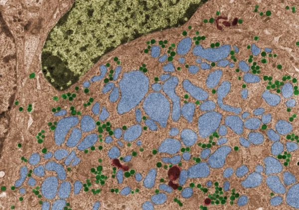
False colour transmission electron microscope (TEM) micrograph of a gonadotropic cell of a castrated animal showing FSL-LH granules (green), lysosomes (red) and large swellings of the RER (blue), due to the castration.
Gonadotropic cell. False colour TEM — Stock Photo, Image

Usage Information

You can use this royalty-free photo "Cerebellar granule cells, light micrograph. Cerebellar granular layer stained with Golgi silver chromate showing the abundance of granule cells. They have a very small rounded soma with three to five dendrites, ending in an enlargement called a dendr" for personal and commercial purposes according to the Standard or Extended License. The Standard License covers most use cases, including advertising, UI designs, and product packaging, and allows up to 500,000 print copies. The Extended License permits all use cases under the Standard License with unlimited print rights and allows you to use the downloaded stock images for merchandise, product resale, or free distribution.

You can buy this stock photo and download it in high resolution up to 4696x3136. Upload Date: Sep 28, 2023