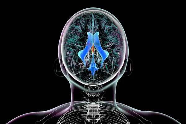
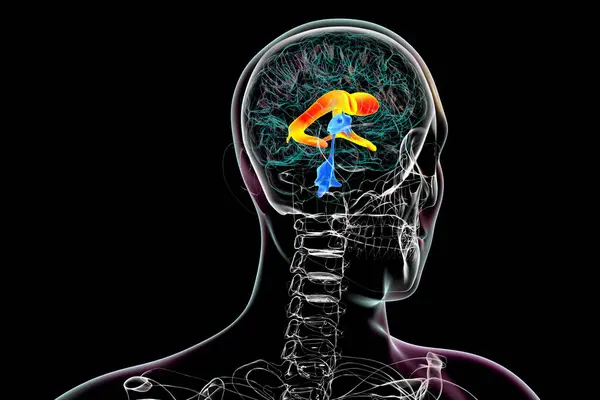
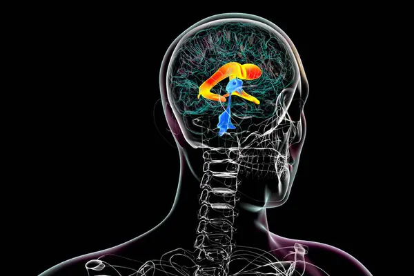
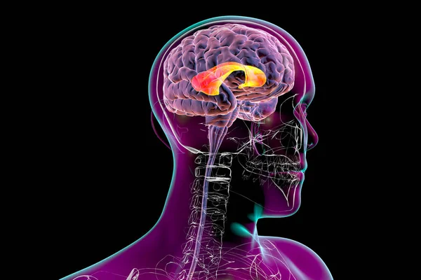
Cerebral Aqueduct Marked Orange Color Narrow Channel Midbrain Connecting Third — Stock Photo, Image

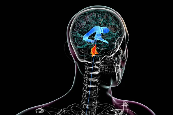
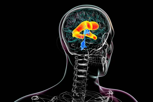
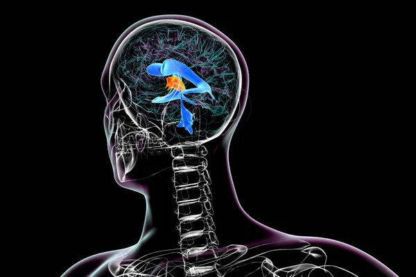
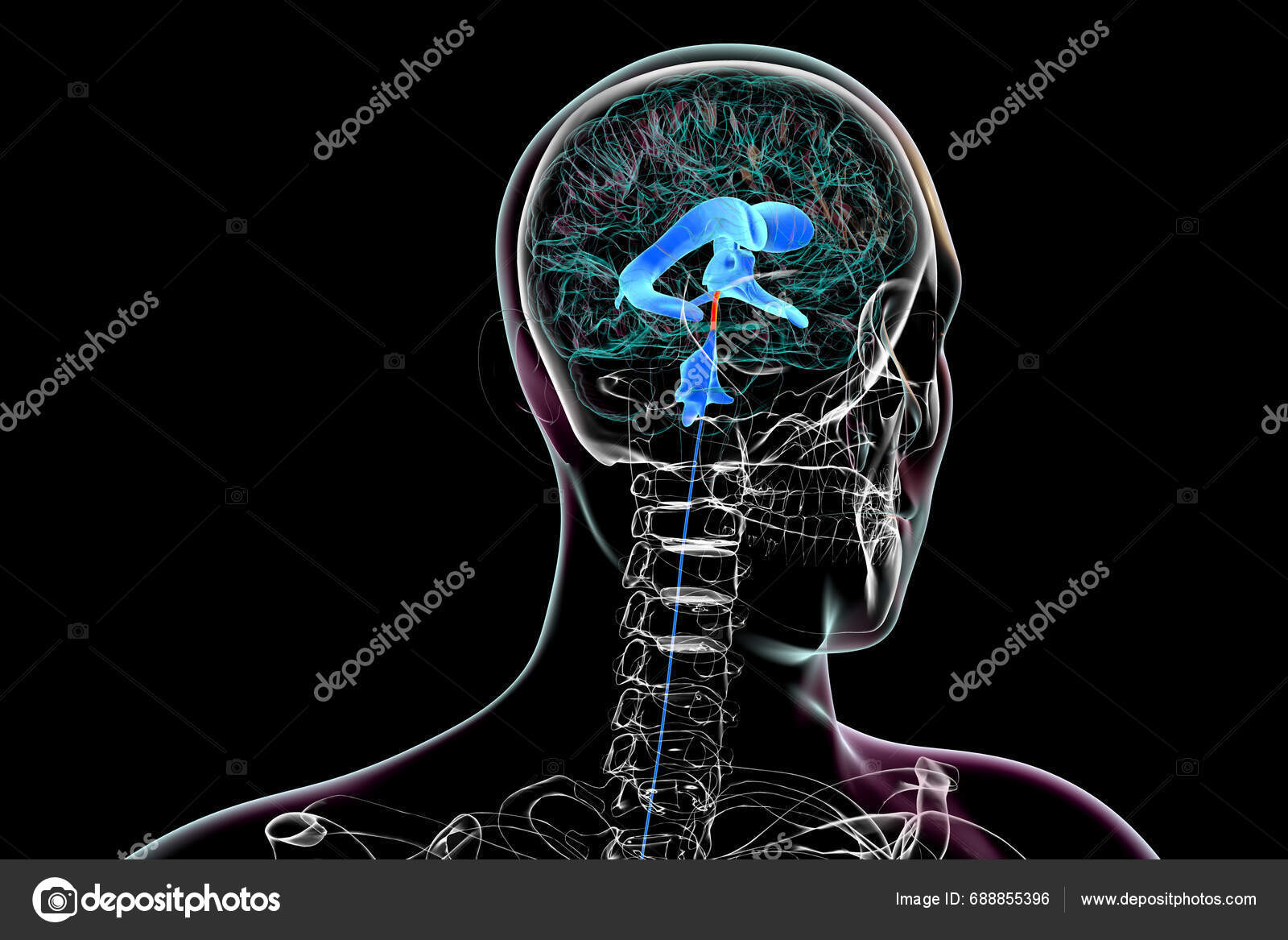
The cerebral aqueduct (marked in orange color), a narrow channel in the midbrain connecting the third and fourth ventricles, facilitating cerebrospinal fluid flow, 3D illustration. — Photo

The cerebral aqueduct (marked in orange color), a narrow channel in the midbrain connecting the third and fourth ventricles, facilitating cerebrospinal fluid flow, 3D illustration.

 — Photo by katerynakon

Same Series:

Anatomy of the fourth brain ventricle (highlighted in orange color), a fluid-filled cavity situated at the posterior of the brainstem, involved in cerebrospinal fluid circulation, 3D illustration.
Anatomy Fourth Brain Ventricle Highlighted Orange Color Fluid Filled Cavity — Stock Photo, Image
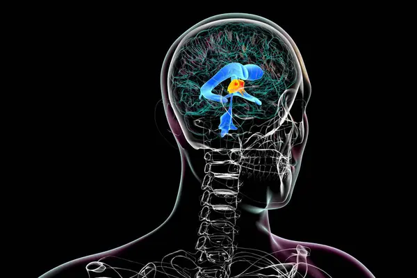
The third brain ventricle, a vital component of the brain's ventricular system, 3D illustration.
Third Brain Ventricle Vital Component Brain Ventricular System Illustration — Stock Photo, Image
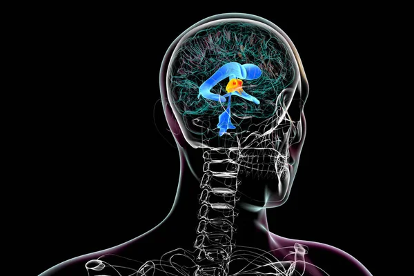
The third brain ventricle, a vital component of the brain's ventricular system, 3D illustration.
Third Brain Ventricle Vital Component Brain Ventricular System Illustration — Stock Photo, Image
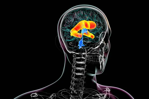
A 3D scientific illustration depicting enlarged lateral ventricles of the brain (hydrocephalus).
Scientific Illustration Depicting Enlarged Lateral Ventricles Brain Hydrocephalus — Stock Photo, Image
A 3D scientific illustration depicting enlarged lateral ventricles of the brain (hydrocephalus).
Scientific Illustration Depicting Enlarged Lateral Ventricles Brain Hydrocephalus — Stock Photo, Image
The third brain ventricle, a vital component of the brain's ventricular system, 3D illustration.
Third Brain Ventricle Vital Component Brain Ventricular System Illustration — Stock Photo, Image
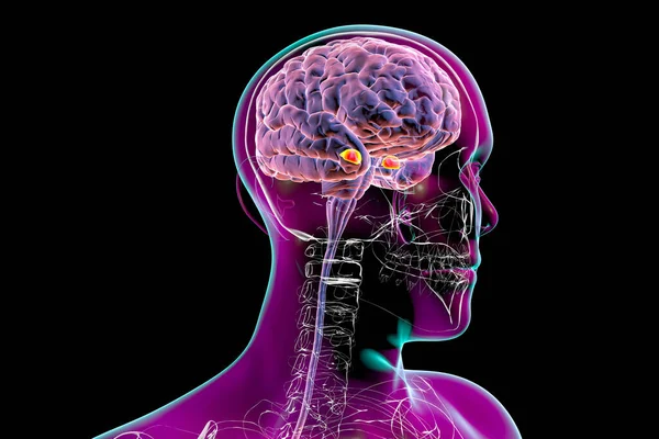
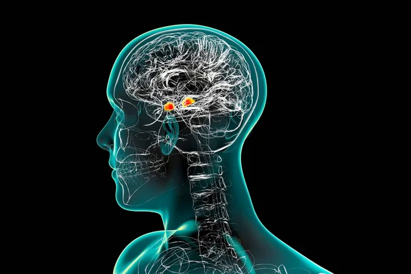
Amygdala, also known as corpus amygdaloideum, in the brain, 3D illustration. Two almond-shaped clusters of nuclei within the temporal lobes, part of the limbic system, play role in memory and emotions
Amygdala Also Known Corpus Amygdaloideum Brain Illustration Two Almond Shaped — Stock Photo, Image
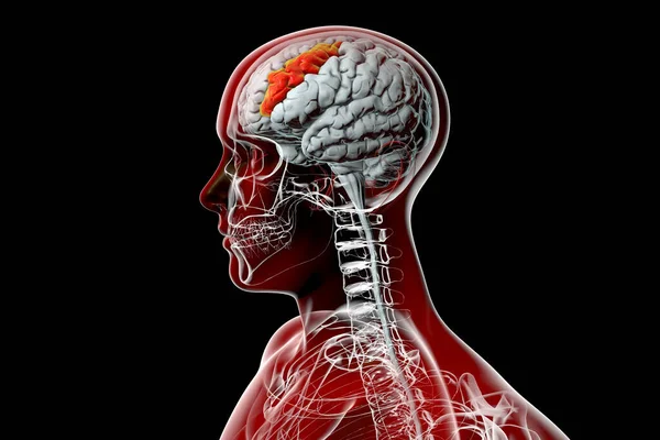
Human brain in the body with highlighted middle frontal gyrus, 3D illustration. It is the part of prefrontal cortex of the frontal lobe. It is involved in the language learning and attention
Human Brain Body Highlighted Middle Frontal Gyrus Illustration Part Prefrontal — Stock Photo, Image
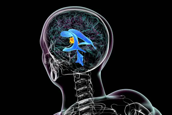
The third brain ventricle, a vital component of the brain's ventricular system, 3D illustration, top view.
Third Brain Ventricle Vital Component Brain Ventricular System Illustration Top — Stock Photo, Image
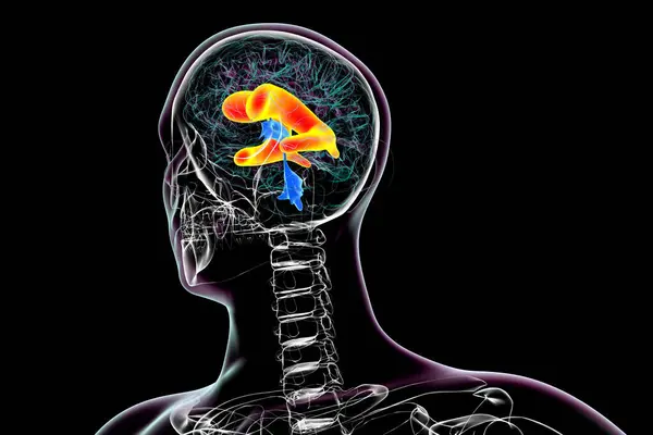
The lateral brain ventricles, the largest paired cavities in the brain, responsible for producing and circulating cerebrospinal fluid, 3D illustration.
Lateral Brain Ventricles Largest Paired Cavities Brain Responsible Producing Circulating — Stock Photo, Image
The lateral brain ventricles, the largest paired cavities in the brain, responsible for producing and circulating cerebrospinal fluid, 3D illustration.
Lateral Brain Ventricles Largest Paired Cavities Brain Responsible Producing Circulating — Stock Photo, Image
Corpus callosum, also known as callosal commissure, highlighted in human brain, 3D illustration. It is a wide, thick nerve tract connecting the left and right cerebral hemispheres
Corpus Callosum Also Known Callosal Commissure Highlighted Human Brain Illustration — Stock Photo, Image
Amygdala, also known as corpus amygdaloideum, in the brain, 3D illustration. Two almond-shaped clusters of nuclei within the temporal lobes, part of the limbic system, play role in memory and emotions
Amygdala Also Known Corpus Amygdaloideum Brain Illustration Two Almond Shaped — Stock Photo, Image
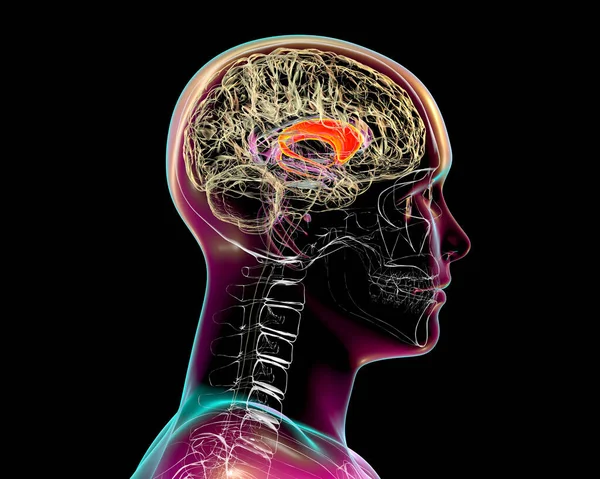
Caudate nuclei highlighted in human brain, 3D illustration. The caudate nucleus is a component of the basal ganglia, it is associated with motor processes and plays role in Parkinson's disease
Caudate Nuclei Highlighted Human Brain Illustration Caudate Nucleus Component Basal — Stock Photo, Image
A 3D scientific illustration depicting enlarged lateral ventricles of the brain (hydrocephalus).
Scientific Illustration Depicting Enlarged Lateral Ventricles Brain Hydrocephalus — Stock Photo, Image
The third brain ventricle, a vital component of the brain's ventricular system, 3D illustration.
Third Brain Ventricle Vital Component Brain Ventricular System Illustration — Stock Photo, Image

Usage Information

You can use this royalty-free photo "The cerebral aqueduct (marked in orange color), a narrow channel in the midbrain connecting the third and fourth ventricles, facilitating cerebrospinal fluid flow, 3D illustration." for personal and commercial purposes according to the Standard or Extended License. The Standard License covers most use cases, including advertising, UI designs, and product packaging, and allows up to 500,000 print copies. The Extended License permits all use cases under the Standard License with unlimited print rights and allows you to use the downloaded stock images for merchandise, product resale, or free distribution.

You can buy this stock photo and download it in high resolution up to 6000x4000. Upload Date: Nov 23, 2023