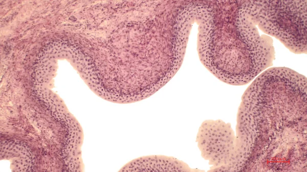
Cross Section Cerebellum Light Micrograph — Stock Photo, Image

Cross section of a cerebellum. Light micrograph. — Photo

Cross section of a cerebellum. Light micrograph.

 — Photo by BioFoto

Same Series:

Punch Biopsy of the Skin. Layers of human skin. Hematoxlyn and eosin stain.
Punch Biopsy Skin Layers Human Skin Hematoxlyn Eosin Stain — Stock Photo, Image
The cerebellar cortex is divided into three layers. At the bottom lies the thick granular layer, densely packed with granule cells. In the middle lies the Purkinje layer. Silver-nitrate impregnated.
Cerebellar Cortex Divided Three Layers Bottom Lies Thick Granular Layer — Stock Photo, Image
Cross section of a cerebellum. Light micrograph.
Cross Section Cerebellum Light Micrograph — Stock Photo, Image
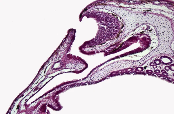
Cross section of the olfactory organ (nasal cavity, vomeronasal organ) of the marsh frog (Pelophylax ridibundus). Hematoxylin and Eosin Staining (H&E stain).
Cross Section Olfactory Organ Nasal Cavity Vomeronasal Organ Marsh Frog — Stock Photo, Image
Cross section of a cerebellum. Light micrograph.
Cross Section Cerebellum Light Micrograph — Stock Photo, Image
Pseudounipolar neurons. Pseudounipolar neurons of rounded soma of a dorsal root ganglion.ell body is surrounded by glia cells called satellite cells. Hematoxlyn and eosin stain. Magnification: x1000.
Pseudounipolar Neurons Pseudounipolar Neurons Rounded Soma Dorsal Root Ganglion Ell — Stock Photo, Image
Trachea, light micrograph. Hyaline cartilage from trachea showing chondrocytes, collagen fibers and matrix.
Trachea Light Micrograph Hyaline Cartilage Trachea Showing Chondrocytes Collagen Fibers — Stock Photo, Image
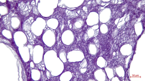
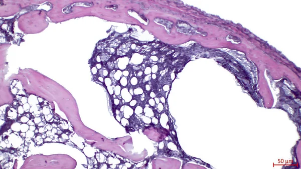
Light microscopy: white adipose tissue of the tubular bone. Large bright holes - the remains of lipocytes.
Light Microscopy White Adipose Tissue Tubular Bone Large Bright Holes — Stock Photo, Image
Cross section of a cerebellum. Light micrograph.
Cross Section Cerebellum Light Micrograph — Stock Photo, Image
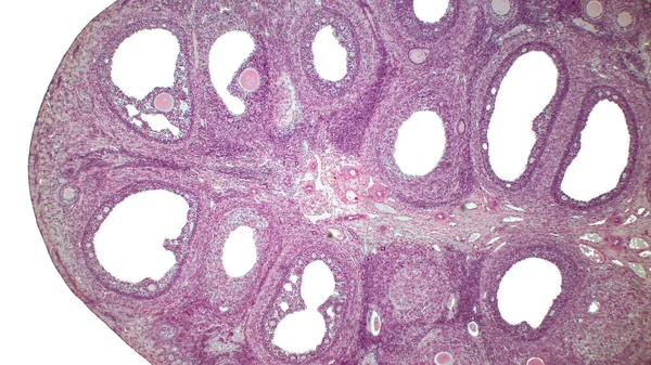
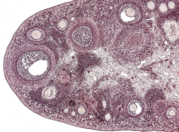
Light micrograph of the Cat's ovarian. Micrograph of ovary showing primordial, primary and secondary follicles isolated on white background. Hematoxylin and eosin stain.
Light Micrograph Cat Ovarian Micrograph Ovary Showing Primordial Primary Secondary — Stock Photo, Image
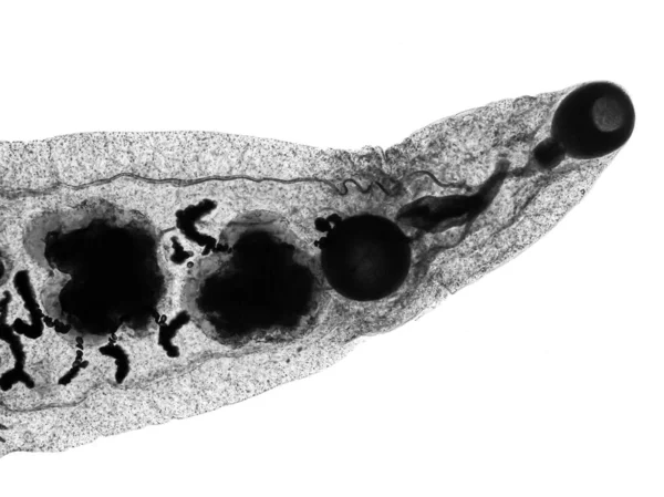
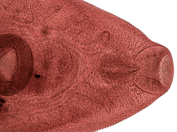
Parasitic flatworms in humans. Liver fluke (Fasciola hepatica) adult stage in light microscope.
Parasitic Flatworms Humans Liver Fluke Fasciola Hepatica Adult Stage Light — Stock Photo, Image
Cross section of a cerebellum. Light micrograph.
Cross Section Cerebellum Light Micrograph — Stock Photo, Image
An interesting photo taken with a microscope. Unmyelinated fibers in peripheral nerves. Longitudinal section. Hematoxylin and Eosin Stainit.
Interesting Photo Taken Microscope Unmyelinated Fibers Peripheral Nerves Longitudinal Section — Stock Photo, Image
Light micrograph of a cross-section of the bone. Epiphysis of a developing long bone.
Light Micrograph Cross Section Bone Epiphysis Developing Long Bone — Stock Photo, Image
Cross section of a cerebellum. Light micrograph.
Cross Section Cerebellum Light Micrograph — Stock Photo, Image
Light microscopy of the internal lining of the urinary bladder. The tissue is a wide transitional epithelium (urothelium) so called because it accommodates to the changes in size of the bladder.
Light Microscopy Internal Lining Urinary Bladder Tissue Wide Transitional Epithelium — Stock Photo, Image

Usage Information

You can use this royalty-free photo "Cross section of a cerebellum. Light micrograph." for personal and commercial purposes according to the Standard or Extended License. The Standard License covers most use cases, including advertising, UI designs, and product packaging, and allows up to 500,000 print copies. The Extended License permits all use cases under the Standard License with unlimited print rights and allows you to use the downloaded stock images for merchandise, product resale, or free distribution.

You can buy this stock photo and download it in high resolution up to 3000x2248. Upload Date: Apr 12, 2022