Epididymis. Pseudostratified epithelium — Stock Photo, Image

Epididymis. Pseudostratified epithelium — Photo

Two sections of the epididymal duct surrounded by concentric layers of fibromuscular tissue. The pseudostratified epithelium consists of tall columnar cells showing stereocilia, and basal cells. The lumen is filled with sperm cells.

 — Photo by jlcalvo@ucm.es

Same Series:

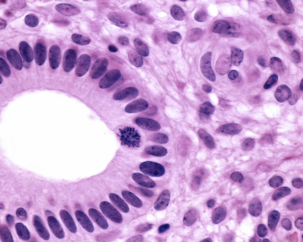
Cross section of a human uterine gland. The epithelium is columnar simple and show a mitotic cell near the lumen.
Uterine gland — Stock Photo, Image
Light microscope micrograph showing a bone joint. The epiphysis are constituted by hyaline cartilage; the epiphyseal secondary ossification center has not yet been formed. However, in the bone on the right can be seen a primary ossification center in
Bone joint — Stock Photo, Image
Wall of the small intestine stained with PAS technique. Goblet cells appear as fuchsia points in the epithelium lining the villi and the crypts of Lieberkuhn. Light microscopy image.
Goblet cells. Small intestine epithelium — Stock Photo, Image
Pseudostratified columnar epithelium of the epididymis. The epithelial cells show stereocilia in the apical surface.
Human epididymis — Stock Photo, Image
Skeletal striated muscle fibers in cross section showing the presence of several nuclei (multinucleated cells) located in the cell periphery. Light microscope photomicrograph.
Striated muscle fibers — Stock Photo, Image
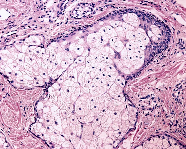
The sebaceous gland is a holocrine gland which secrete sebum. The cells accumulate oil droplets as clear vacuoles. Finally, the cells die, which is indicated by their pyknotic nucleus, visible on top half of the image.
Sebaceous gland — Stock Photo, Image
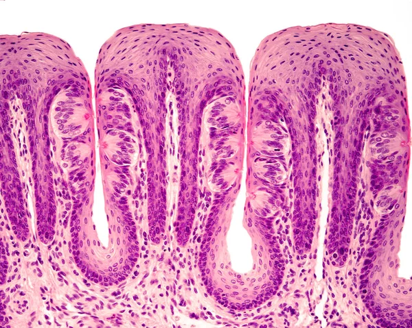
Taste buds in foliate tongue papillae. Many of them show the taste or gustatory pore. Hematoxylin & eosin stain.
Taste buds — Stock Photo, Image
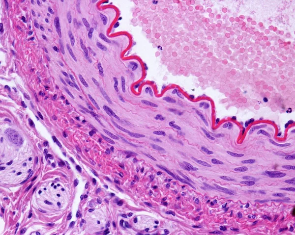
Light micrograph of a cross-sectioned muscular artery, showing a thick and wavy internal elastic lamina, a middle layer with smooth muscle fibers, and an outer connective tissue adventitia. Hematoxylin & eosin stain
Muscular artery — Stock Photo, Image
Stratified columnar epithelium. It is a rare type of epithelium composed of columnar cells arranged in several layers (usually only two). Large excretory duct of a salivary gland.
Stratified columnar epithelium — Stock Photo, Image
Light micrograph of goblet cells stained with the PAS technique. They are located in the epithelium lining the villi of the small intestine. The striated border of enterocytes is also stained with the PAS technique.
Goblet cells. Small intestine epithelium — Stock Photo, Image
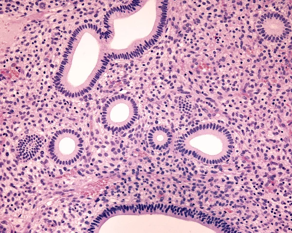
Human endometrium. Proliferative phase. Several tubular endometrial glands are cross sectioned showing their simple columnar epithelium. Among them, the uterine stroma shows many cells and blood vessels.
Endometrium. Proliferative phase — Stock Photo, Image
High magnification light micrograph showing several taste buds with their taste or gustatory pores. H&E stain.
Taste buds — Stock Photo, Image
Respiratory prismatic ciliated pseudostratified epithelium. The apical border of the epithelium has a layer of cilia supported in their basal bodies. Light microscope photomicrograph.
Respiratory epithelium — Stock Photo, Image
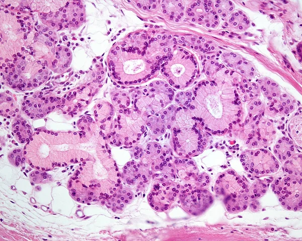
Mixed gland showing both mucous cells (with pale cytoplasm) and serous cells in the same gland. Light microscope micrograph. Hematoxylin & eosin stain.
Mixed exocrine gland — Stock Photo, Image
A septum of connective tissue, with numerous adipocytes (fat cells) which contain a large lipid droplet. The sinuous structures correspond to nerves. Light micrograph. Hematoxylin & eosin stain
Adipose tissue — Stock Photo, Image
Low power light microscope micrograph showing a cross section of a rat small intestine. The most prominent feature of the mucosal layer is the abundance of villi, finger-like projections that extend into the lumen.
Low Power Light Microscope Micrograph Showing Cross Section Rat Small — Stock Photo, Image

Usage Information

You can use this royalty-free photo "Epididymis. Pseudostratified epithelium" for personal and commercial purposes according to the Standard or Extended License. The Standard License covers most use cases, including advertising, UI designs, and product packaging, and allows up to 500,000 print copies. The Extended License permits all use cases under the Standard License with unlimited print rights and allows you to use the downloaded stock images for merchandise, product resale, or free distribution.

You can buy this stock photo and download it in high resolution up to 3840x3072. Upload Date: Jan 13, 2018