Normal Electrocardiogram Ecg Illustration Displaying Electrical Activity Heart Healthy Individual — Stock Photo, Image

A normal electrocardiogram ECG, 3D illustration displaying the electrical activity of the heart in a healthy individual. — Photo

A normal electrocardiogram ECG, 3D illustration displaying the electrical activity of the heart in a healthy individual.

 — Photo by katerynakon

Same Series:

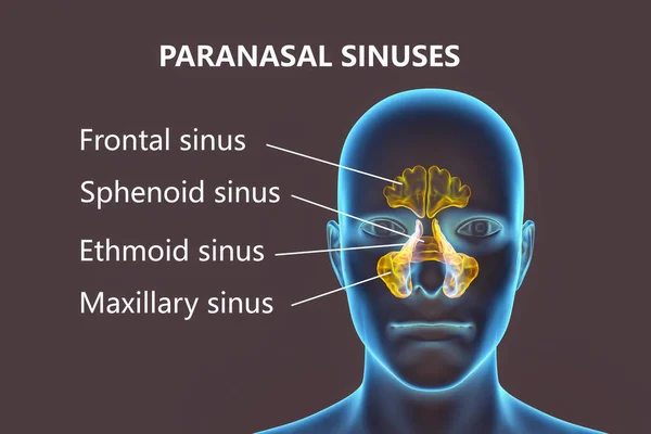
Anatomy of paranasal sinuses. 3D illustration showing female with highlighted paranasal sinuses, frontal, maxillary, ethmoid, and sphenoid. Labelled image
Anatomy Paranasal Sinuses Illustration Showing Female Highlighted Paranasal Sinuses Frontal — Stock Photo, Image
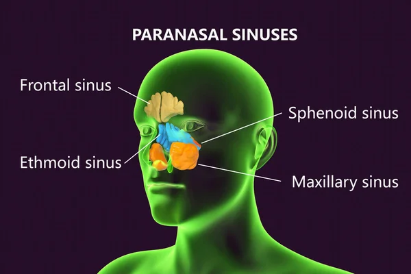
Anatomy of paranasal sinuses. 3D illustration showing male with highlighted paranasal sinuses, frontal, maxillary, ethmoid, and sphenoid. Labelled image
Anatomy Paranasal Sinuses Illustration Showing Male Highlighted Paranasal Sinuses Frontal — Stock Photo, Image
Anatomy of paranasal sinuses. 3D illustration showing male with highlighted paranasal sinuses, frontal, maxillary, ethmoid, and sphenoid. Labelled image
Anatomy Paranasal Sinuses Illustration Showing Male Highlighted Paranasal Sinuses Frontal — Stock Photo, Image
Anatomy of paranasal sinuses. 3D illustration showing male with highlighted paranasal sinuses, frontal, maxillary, ethmoid, and sphenoid. Labelled image
Anatomy Paranasal Sinuses Illustration Showing Male Highlighted Paranasal Sinuses Frontal — Stock Photo, Image
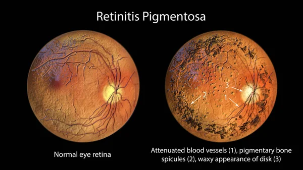
Retinitis pigmentosa, a genetic eye disease. 3D illustration shows normal eye retina and attenuated blood vessels, pigmentary bone spicules and waxy appearance of the optic disk in the affected retina
Retinitis Pigmentosa Genetic Eye Disease Illustration Shows Normal Eye Retina — Stock Photo, Image
A 3D photorealistic illustration of the upper half part of an African man with transparent skin, revealing an ascending aortic aneurysm
Photorealistic Illustration Upper Half Part African Man Transparent Skin Revealing — Stock Photo, Image
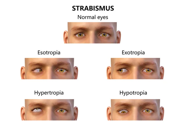
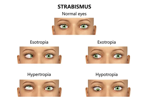
Strabismus, 3D illustration showing different types of eye deviations
Strabismus Illustration Showing Different Types Eye Deviations — Stock Photo, Image
Strabismus, 3D illustration showing different types of eye deviations
Strabismus Illustration Showing Different Types Eye Deviations — Stock Photo, Image
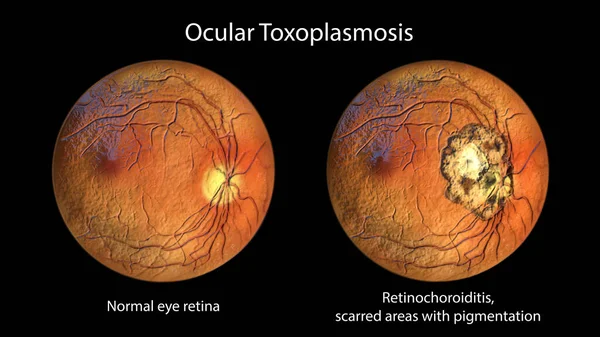
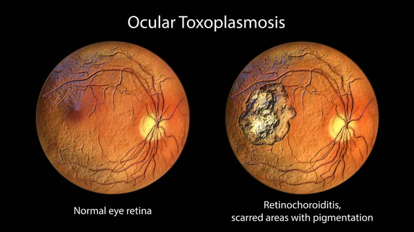
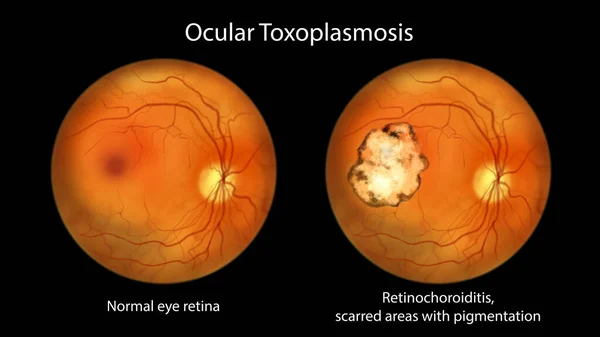
Retinal scar in toxoplasmosis, a disease caused by the single-celled protozoan Toxoplasma gondii, and the same healthy eye retina for comparison, ophthalmoscope view, 3D illustration
Retinal Scar Toxoplasmosis Disease Caused Single Celled Protozoan Toxoplasma Gondii — Stock Photo, Image
Spinal muscular atrophy, SMA, a genetic neuromuscular disorder with progressive muscle wasting because of loss of motor neurons due to mutation in the SMN1 gene, conceptual 3D illustration
Spinal Muscular Atrophy Sma Genetic Neuromuscular Disorder Progressive Muscle Wasting — Stock Photo, Image
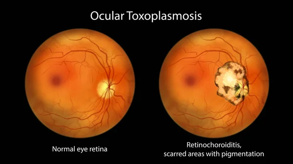
Retinal scar in toxoplasmosis, a disease caused by the single-celled protozoan Toxoplasma gondii, and the same healthy eye retina for comparison, ophthalmoscope view, scientific illustration
Retinal Scar Toxoplasmosis Disease Caused Single Celled Protozoan Toxoplasma Gondii — Stock Photo, Image
Retinal scar in toxoplasmosis, a disease caused by the single-celled protozoan Toxoplasma gondii, and the same healthy eye retina for comparison, ophthalmoscope view, 3D illustration
Retinal Scar Toxoplasmosis Disease Caused Single Celled Protozoan Toxoplasma Gondii — Stock Photo, Image
Retinal scar in toxoplasmosis, a disease caused by the single-celled protozoan Toxoplasma gondii, and the same healthy eye retina for comparison, ophthalmoscope view, scientific illustration
Retinal Scar Toxoplasmosis Disease Caused Single Celled Protozoan Toxoplasma Gondii — Stock Photo, Image
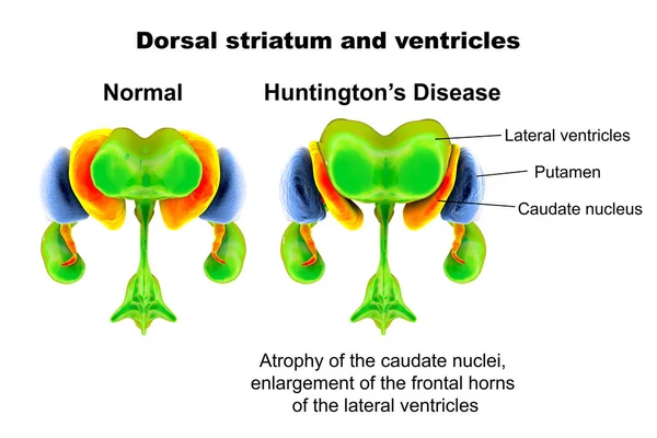
Dorsal striatum and lateral ventricles normal and in Huntington's disease (HD), 3D illustration showing enlargement of anterior horns of lateral ventricles and atrophy of the caudate nuclei in HD
Dorsal Striatum Lateral Ventricles Normal Huntington Disease Illustration Showing Enlargement — Stock Photo, Image
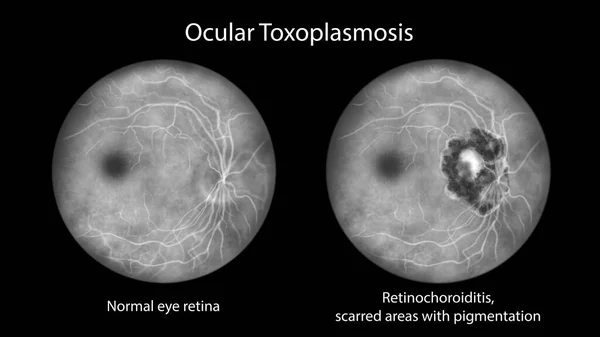
Retinal scar in toxoplasmosis, a disease caused by the single-celled protozoan Toxoplasma gondii, and the same healthy eye retina for comparison, fluorescein angiography, scientific illustration
Retinal Scar Toxoplasmosis Disease Caused Single Celled Protozoan Toxoplasma Gondii — Stock Photo, Image
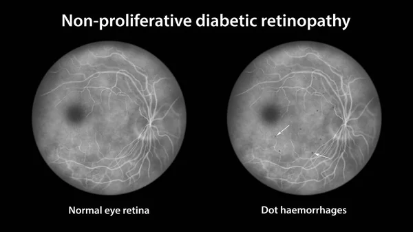
Non-proliferative diabetic retinopathy, ophthalmoscope view, illustration showing normal eye retina and retina with multiple dot haemorrhages (dark grey dots)
Non Proliferative Diabetic Retinopathy Ophthalmoscope View Illustration Showing Normal Eye — Stock Photo, Image

Usage Information

You can use this royalty-free photo "A normal electrocardiogram ECG, 3D illustration displaying the electrical activity of the heart in a healthy individual." for personal and commercial purposes according to the Standard or Extended License. The Standard License covers most use cases, including advertising, UI designs, and product packaging, and allows up to 500,000 print copies. The Extended License permits all use cases under the Standard License with unlimited print rights and allows you to use the downloaded stock images for merchandise, product resale, or free distribution.

You can buy this stock photo and download it in high resolution up to 9000x6000. Upload Date: Jun 18, 2023