Pathology Study Human Stem Cell Microscope — Stock Photo, Image

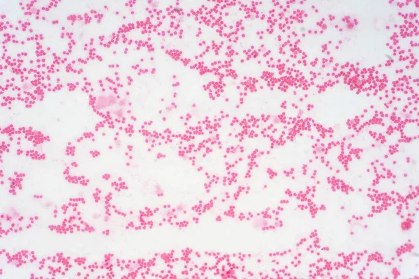
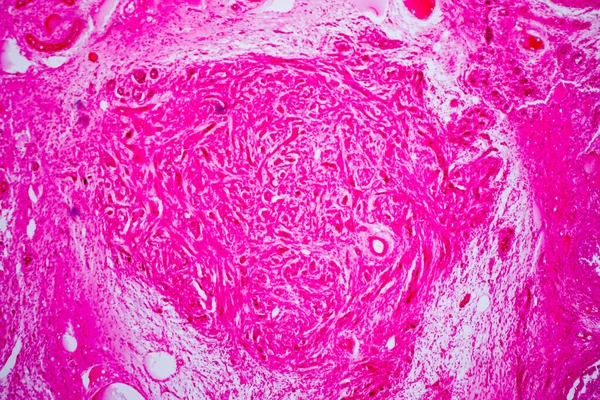
Pathology study of human stem cell under the microscope. — Photo

Pathology study of human stem cell under the microscope.

 — Photo by tonaquatic19

Same Series:

Pathology study of human stem cell under the microscope.
Pathology Study Human Stem Cell Microscope — Stock Photo, Image
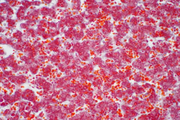
Human liver tissue under the microscope view. Histological for human physiology.
Human Liver Tissue Microscope View Histological Human Physiology — Stock Photo, Image
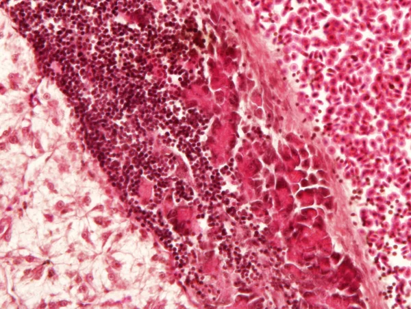
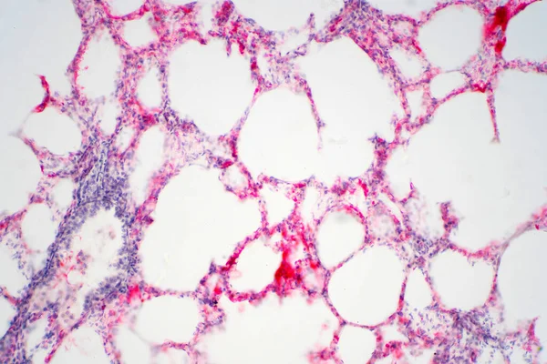
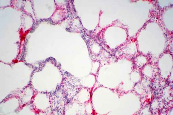
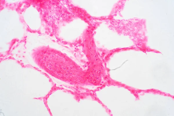
Human lung tissue under microscope view. Lungs are the primary organs of the respiratory system in humans and many other animals
Human Lung Tissue Microscope View Lungs Primary Organs Respiratory System — Stock Photo, Image
Human lung tissue under microscope view. Lungs are the primary organs of the respiratory system in humans and many other animals
Human Lung Tissue Microscope View Lungs Primary Organs Respiratory System — Stock Photo, Image
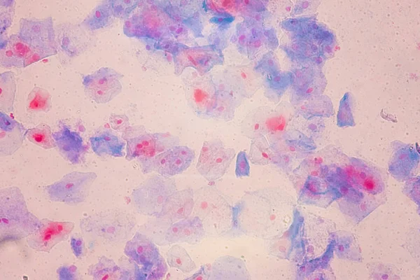
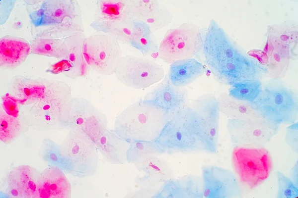
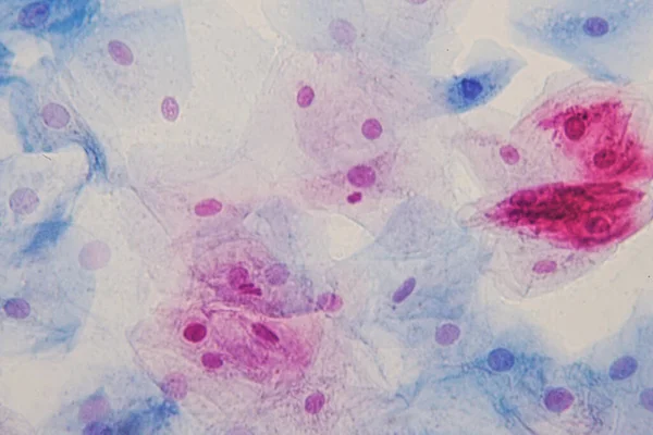
Squamous epithelial cells under microscope view for education histology. Human tissue.
Squamous Epithelial Cells Microscope View Education Histology Human Tissue — Stock Photo, Image
Liver animal tissue under microscope view, Histology of liver.
Liver Animal Tissue Microscope View Histology Liver — Stock Photo, Image
Cross section human ovary under microscope view.The ovary is an ovum-producing reproductive organ, found in double pairs in the female as part of the vertebrate female reproductive system.
Cross Section Human Ovary Microscope View Ovary Ovum Producing Reproductive — Stock Photo, Image
Human lung tissue under microscope view. Lungs are the primary organs of the respiratory system in humans and many other animals
Human Lung Tissue Microscope View Lungs Primary Organs Respiratory System — Stock Photo, Image
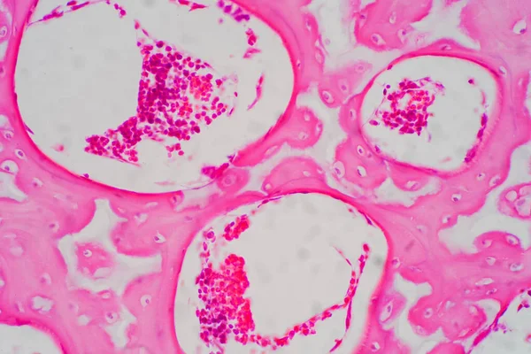
Human Hyaline cartilage bone under microscope view for education pathology. Human tissue.
Human Hyaline Cartilage Bone Microscope View Education Pathology Human Tissue — Stock Photo, Image
Squamous epithelial cells under microscope view for education histology. Histological for human physiology.
Squamous Epithelial Cells Microscope View Education Histology Histological Human Physiology — Stock Photo, Image
Squamous epithelial cells under microscope view for education histology. Human tissue.
Squamous Epithelial Cells Microscope View Education Histology Human Tissue — Stock Photo, Image
Human lung tissue under microscope view. Lungs are the primary organs of the respiratory system in humans and many other animals
Human Lung Tissue Microscope View Lungs Primary Organs Respiratory System — Stock Photo, Image
Human lung tissue under microscope view. Lungs are the primary organs of the respiratory system in humans and many other animals
Human Lung Tissue Microscope View Lungs Primary Organs Respiratory System — Stock Photo, Image
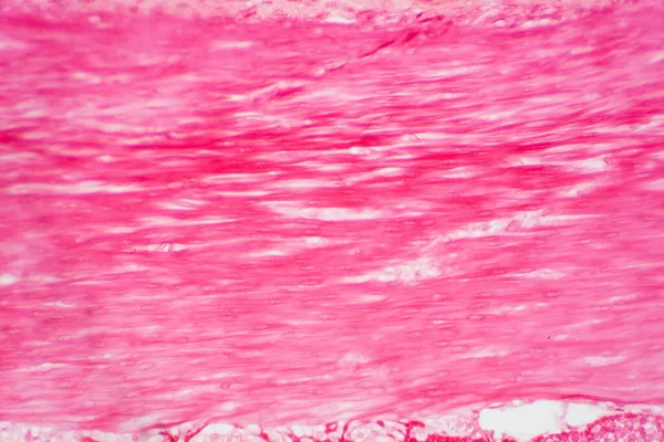
Histology of human smooth muscle under microscope view tissue anatomy for physiology education.
Histology Human Smooth Muscle Microscope View Tissue Anatomy Physiology Education — Stock Photo, Image
Human lung tissue under microscope view. Lungs are the primary organs of the respiratory system in humans and many other animals
Human Lung Tissue Microscope View Lungs Primary Organs Respiratory System — Stock Photo, Image
Human fat body tissue under microscope view for physiology education.
Human Fat Body Tissue Microscope View Physiology Education — Stock Photo, Image

Usage Information

You can use this royalty-free photo "Pathology study of human stem cell under the microscope." for personal and commercial purposes according to the Standard or Extended License. The Standard License covers most use cases, including advertising, UI designs, and product packaging, and allows up to 500,000 print copies. The Extended License permits all use cases under the Standard License with unlimited print rights and allows you to use the downloaded stock images for merchandise, product resale, or free distribution.

You can buy this stock photo and download it in high resolution up to 6000x4000. Upload Date: Aug 25, 2020