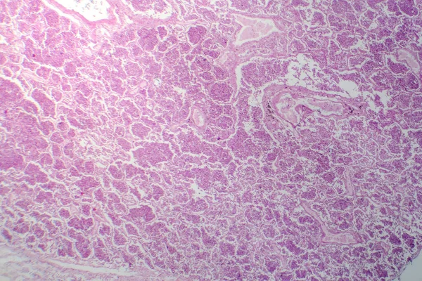
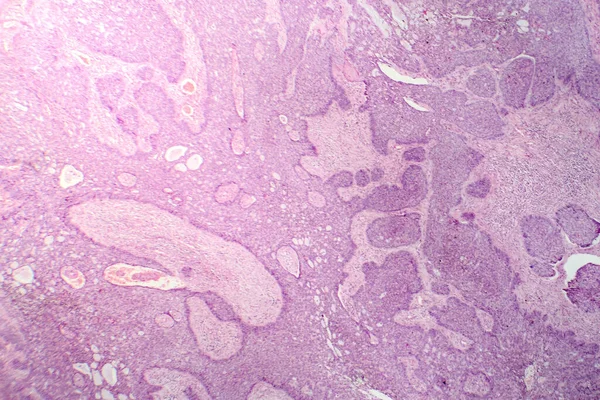
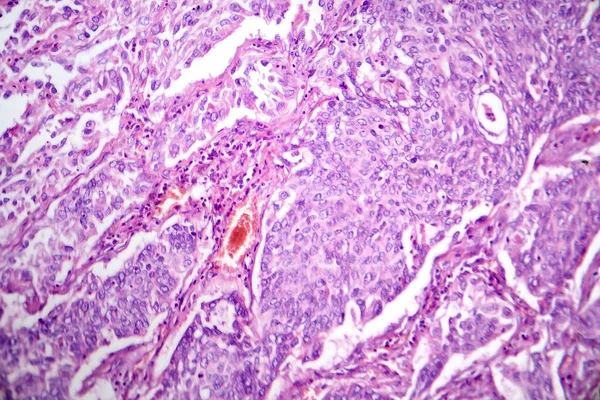
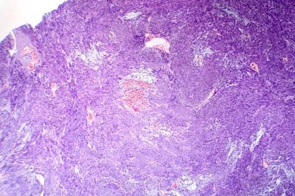
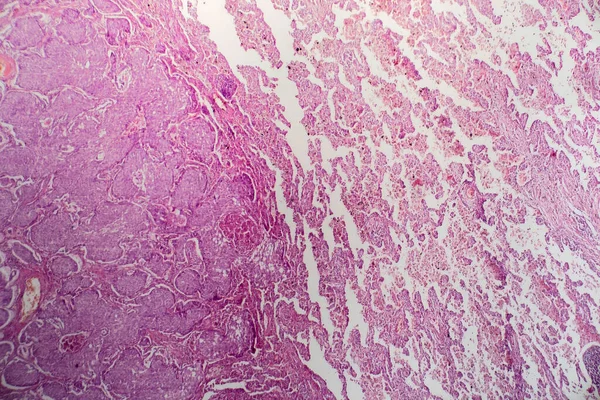
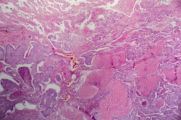
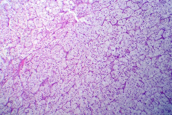
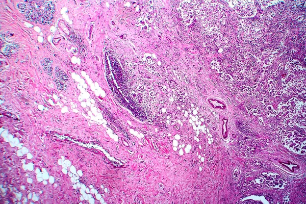
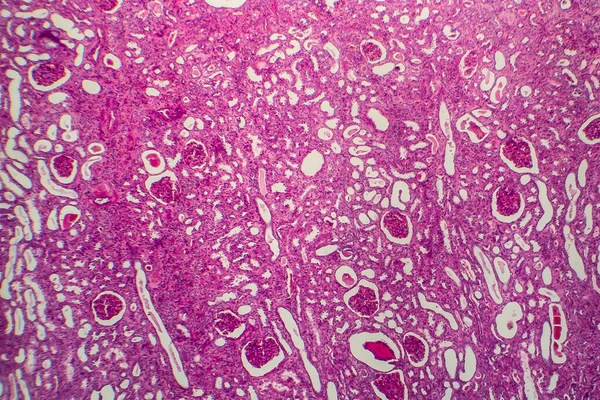
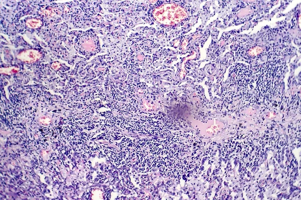
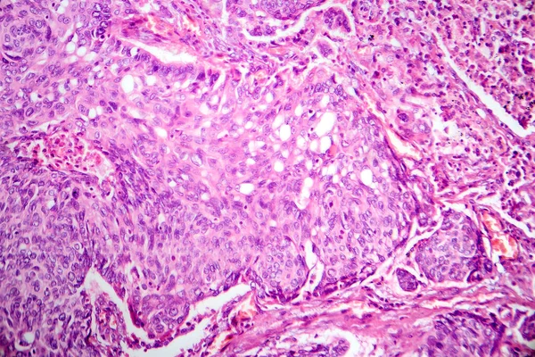
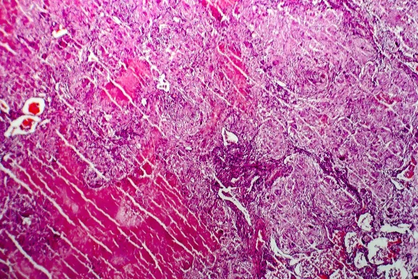
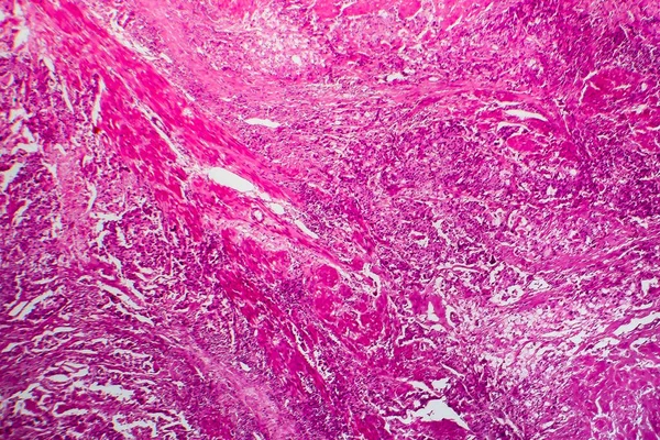
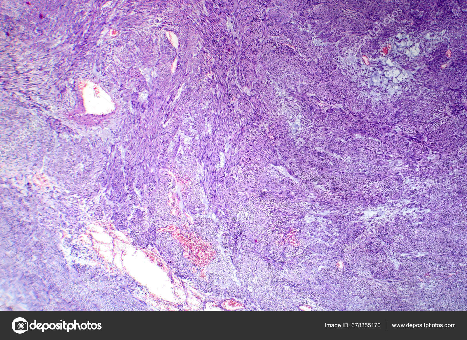
Photomicrograph Leiomyoma Illustrating Benign Smooth Muscle Tumor Cells Uterine Tissue — Stock Photo, Image

Photomicrograph of leiomyoma, illustrating benign smooth muscle tumor cells within the uterine tissue. — Photo

Photomicrograph of leiomyoma, illustrating benign smooth muscle tumor cells within the uterine tissue.

 — Photo by katerynakon

Same Series:

Photomicrograph of lobar pneumonia in grey hepatic phase, revealing lung tissue transition with exudate-filled alveoli.
Photomicrograph Lobar Pneumonia Grey Hepatic Phase Revealing Lung Tissue Transition — Stock Photo, Image
Photomicrograph of leiomyoma, illustrating benign smooth muscle tumor cells within the uterine tissue.
Photomicrograph Leiomyoma Illustrating Benign Smooth Muscle Tumor Cells Uterine Tissue — Stock Photo, Image
Photomicrograph of basal cell carcinoma, displaying malignant basal cells typical of the most common skin cancer.
Photomicrograph Basal Cell Carcinoma Displaying Malignant Basal Cells Typical Most — Stock Photo, Image
Photomicrograph of lung adenocarcinoma, illustrating malignant glandular cells characteristic of the most common type of lung cancer.
Photomicrograph Lung Adenocarcinoma Illustrating Malignant Glandular Cells Characteristic Most Common — Stock Photo, Image
Photomicrograph of leiomyoma, illustrating benign smooth muscle tumor cells within the uterine tissue.
Photomicrograph Leiomyoma Illustrating Benign Smooth Muscle Tumor Cells Uterine Tissue — Stock Photo, Image
Photomicrograph of lung adenocarcinoma, illustrating malignant glandular cells characteristic of the most common type of lung cancer.
Photomicrograph Lung Adenocarcinoma Illustrating Malignant Glandular Cells Characteristic Most Common — Stock Photo, Image
Photomicrograph of lung adenocarcinoma, illustrating malignant glandular cells characteristic of the most common type of lung cancer.
Photomicrograph Lung Adenocarcinoma Illustrating Malignant Glandular Cells Characteristic Most Common — Stock Photo, Image
Adrenocortical adenoma, light micrograph, photo under microscope. Benign tumor that emerges from the cells of the adrenal cortex
Adrenocortical Adenoma Light Micrograph Photo Microscope Benign Tumor Emerges Cells — Stock Photo, Image
Breast ductal carcinoma, light micrograph, photo under microscope
Breast Ductal Carcinoma Light Micrograph Photo Microscope — Stock Photo, Image
Miliary tuberculosis, light micrograph, photo under microscope
Miliary tuberculosis, light micrograph — Stock Photo, Image
Chronic pyelonephritis, light micrograph, photo under microscope
Chronic Pyelonephritis Light Micrograph Photo Microscope — Stock Photo, Image
Histopathology of tuberculosis, light micrograph of tuberculosis lesion, photo under microscope
Histopathology of tuberculosis lesion — Stock Photo, Image
Photomicrograph of lung adenocarcinoma, illustrating malignant glandular cells characteristic of the most common type of lung cancer.
Photomicrograph Lung Adenocarcinoma Illustrating Malignant Glandular Cells Characteristic Most Common — Stock Photo, Image
Hepatic congestion, light micrograph, photo under microscope
Hepatic congestion, light micrograph — Stock Photo, Image
Miliary tuberculosis, light micrograph, photo under microscope
Miliary tuberculosis, light micrograph — Stock Photo, Image
Bladder cancer, light micrograph, photo under microscope
Bladder Cancer Light Micrograph Photo Microscope — Stock Photo, Image

Usage Information

You can use this royalty-free photo "Photomicrograph of leiomyoma, illustrating benign smooth muscle tumor cells within the uterine tissue." for personal and commercial purposes according to the Standard or Extended License. The Standard License covers most use cases, including advertising, UI designs, and product packaging, and allows up to 500,000 print copies. The Extended License permits all use cases under the Standard License with unlimited print rights and allows you to use the downloaded stock images for merchandise, product resale, or free distribution.

You can buy this stock photo and download it in high resolution up to 7124x4749. Upload Date: Sep 29, 2023