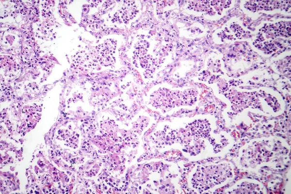
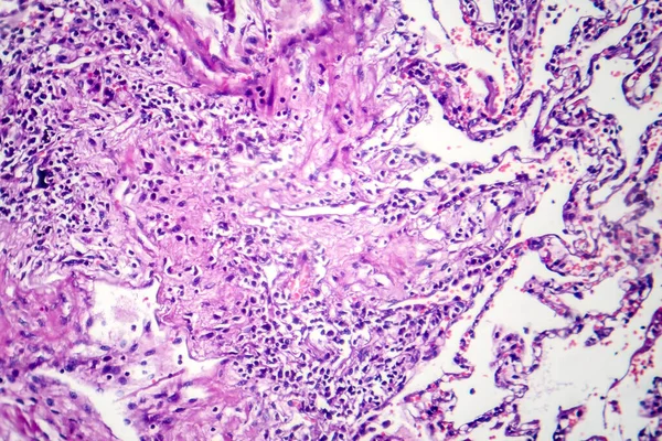
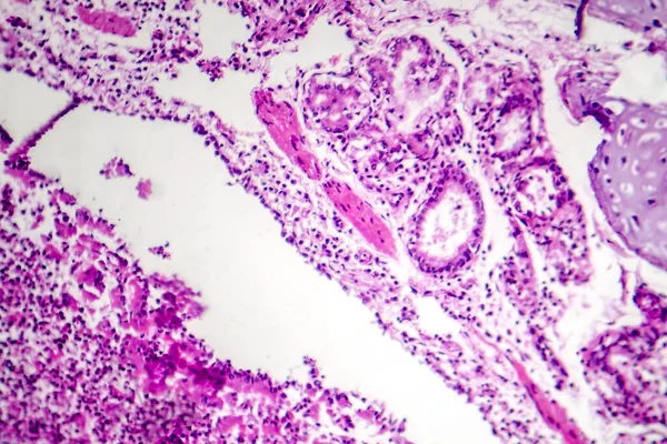
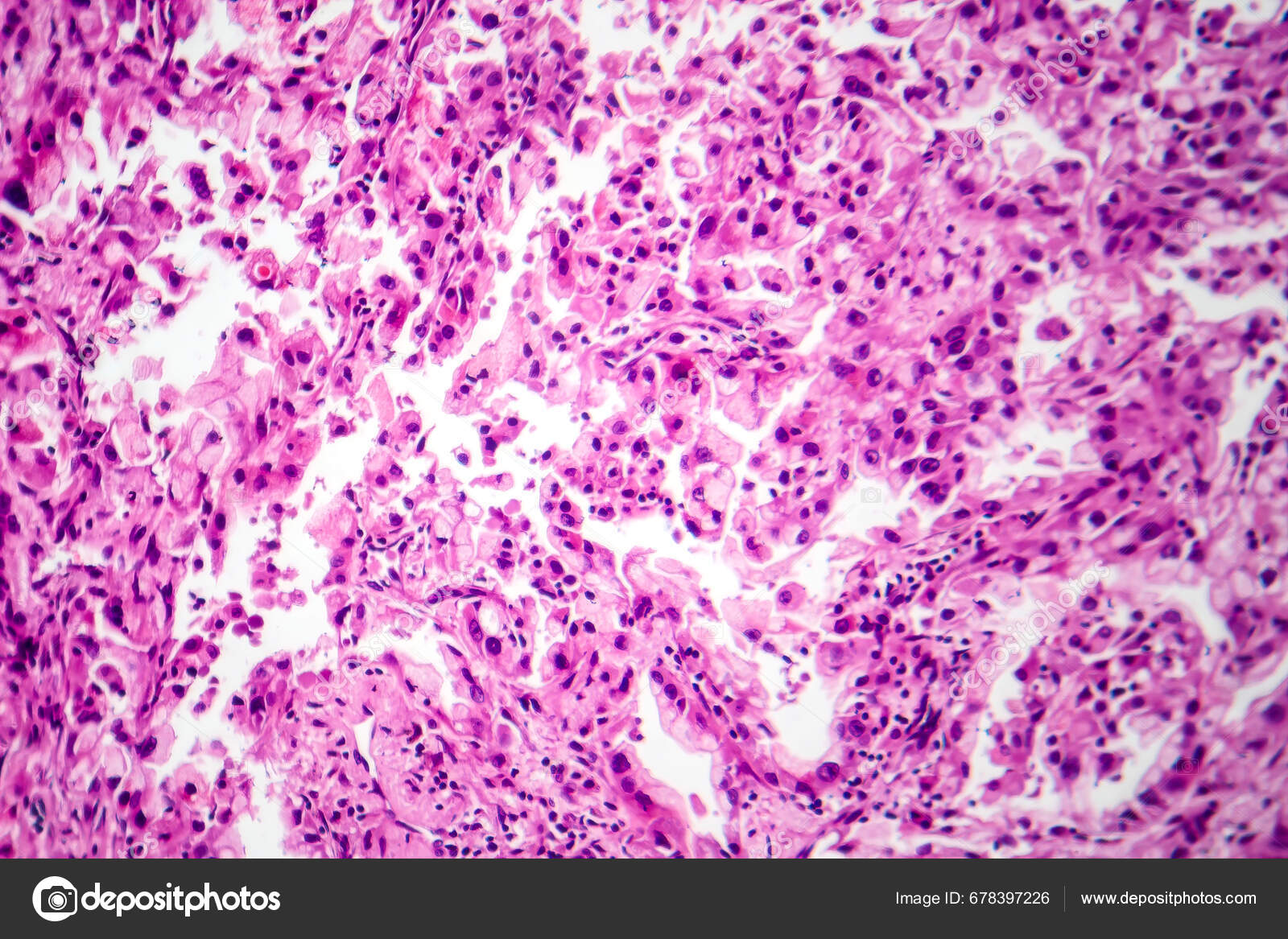
Photomicrograph Lung Cancer Tissue Revealing Malignant Cells Abnormal Growth Characteristic — Stock Photo, Image

Photomicrograph of lung cancer tissue, revealing malignant cells and the abnormal growth characteristic of lung malignancy. — Photo

Photomicrograph of lung cancer tissue, revealing malignant cells and the abnormal growth characteristic of lung malignancy.

 — Photo by katerynakon

Same Series:

Photomicrograph of lobar pneumonia in red hepatic phase, displaying inflamed lung tissue with red hepatization characteristic of the disease.
Photomicrograph Lobar Pneumonia Red Hepatic Phase Displaying Inflamed Lung Tissue — Stock Photo, Image
Histopathology of postnecrotic liver cirrhosis, light micrograph, photo under microscope
Histopathology of postnecrotic liver cirrhosis — Stock Photo, Image
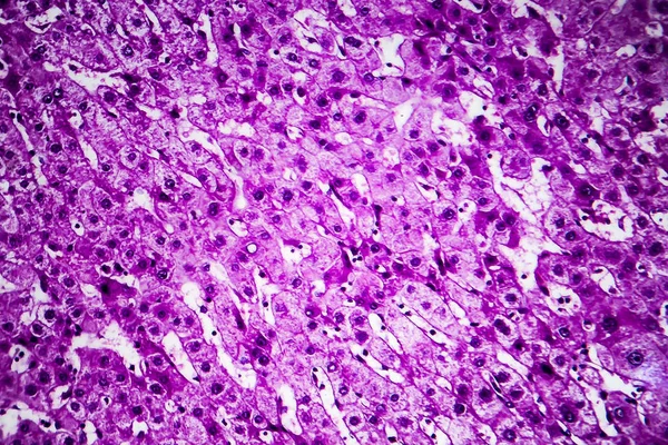
Histopathology of portal liver cirrhosis, light micrograph, photo under microscope
Histopathology of portal cirrhosis — Stock Photo, Image
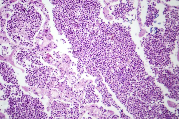
Photomicrograph of lobar pneumonia in grey hepatic phase, revealing lung tissue transition with exudate-filled alveoli.
Photomicrograph Lobar Pneumonia Grey Hepatic Phase Revealing Lung Tissue Transition — Stock Photo, Image
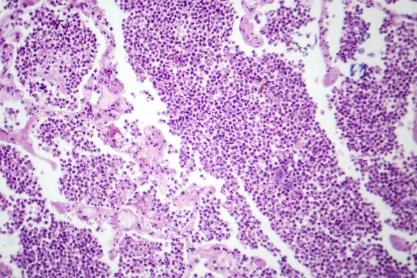
Photomicrograph of lobar pneumonia in grey hepatic phase, revealing lung tissue transition with exudate-filled alveoli.
Photomicrograph Lobar Pneumonia Grey Hepatic Phase Revealing Lung Tissue Transition — Stock Photo, Image
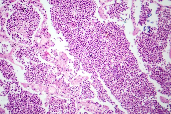
Photomicrograph of lobar pneumonia in grey hepatic phase, revealing lung tissue transition with exudate-filled alveoli.
Photomicrograph Lobar Pneumonia Grey Hepatic Phase Revealing Lung Tissue Transition — Stock Photo, Image
Histopathology of postnecrotic liver cirrhosis, light micrograph, photo under microscope
Histopathology of postnecrotic liver cirrhosis — Stock Photo, Image
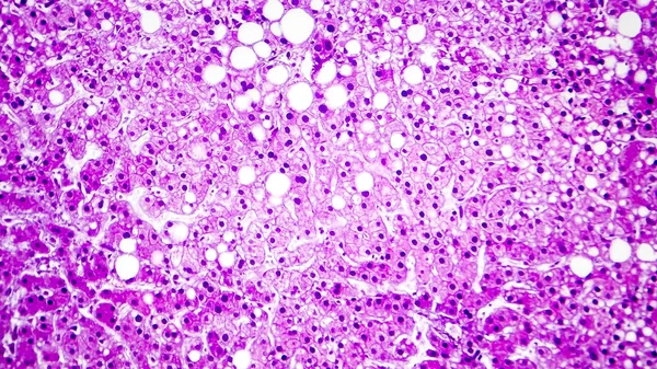
Histopathology of liver steatosis, or fatty liver. Light micrograph, photo under microscope
Histopathology of liver steatosis, or fatty liver — Stock Photo, Image
Histopathology of portal liver cirrhosis, light micrograph, photo under microscope
Histopathology of portal cirrhosis — Stock Photo, Image
Photomicrograph of smoker's lung, revealing characteristic changes and damage associated with long-term smoking habits.
Photomicrograph Smoker Lung Revealing Characteristic Changes Damage Associated Long Term — Stock Photo, Image
Histopathology of portal liver cirrhosis, light micrograph, photo under microscope
Histopathology of portal cirrhosis — Stock Photo, Image
Histopathology of portal liver cirrhosis, light micrograph, photo under microscope
Histopathology of portal cirrhosis — Stock Photo, Image
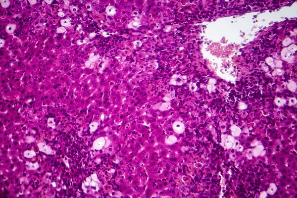
Viral hepatitis, light micrograph, photo under microscope
Viral hepatitis, light micrograph — Stock Photo, Image
Photomicrograph of bronchopneumonia, illustrating inflammation and consolidation of lung tissue due to bacterial infection.
Photomicrograph Bronchopneumonia Illustrating Inflammation Consolidation Lung Tissue Due Bacterial Infection — Stock Photo, Image
Lung adenocarcinoma, light micrograph, photo under microscope
Lung adenocarcinoma, light micrograph — Stock Photo, Image
Histopathology of portal liver cirrhosis, light micrograph, photo under microscope
Histopathology of portal cirrhosis — Stock Photo, Image

Usage Information

You can use this royalty-free photo "Photomicrograph of lung cancer tissue, revealing malignant cells and the abnormal growth characteristic of lung malignancy." for personal and commercial purposes according to the Standard or Extended License. The Standard License covers most use cases, including advertising, UI designs, and product packaging, and allows up to 500,000 print copies. The Extended License permits all use cases under the Standard License with unlimited print rights and allows you to use the downloaded stock images for merchandise, product resale, or free distribution.

You can buy this stock photo and download it in high resolution up to 7008x4673. Upload Date: Sep 29, 2023