Scientific Illustration Showcasing Third Brain Ventricle Vital Component Brain Ventricular — Stock Photo, Image

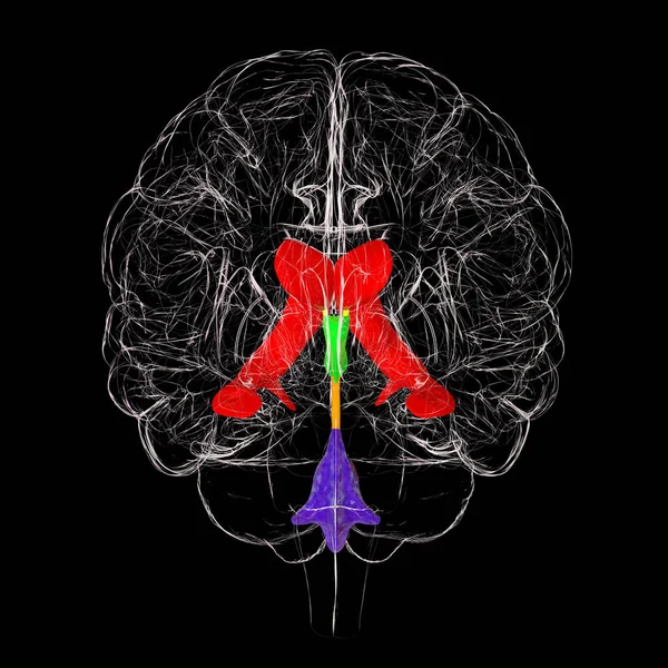
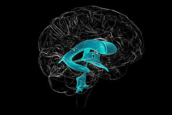
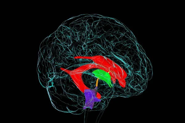
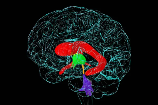
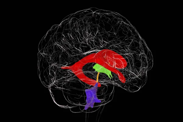
A 3D scientific illustration showcasing the third brain ventricle, a vital component of the brain's ventricular system, side view. — Photo

A 3D scientific illustration showcasing the third brain ventricle, a vital component of the brain's ventricular system, side view.

 — Photo by katerynakon

Same Series:

Dorsal striatum and lateral ventricles in the brain of a person with Huntington's disease, 3D illustration showing enlargement of anterior horns of lateral ventricles and atrophy of the caudate nuclei
Dorsal Striatum Lateral Ventricles Brain Person Huntington Disease Illustration Showing — Stock Photo, Image
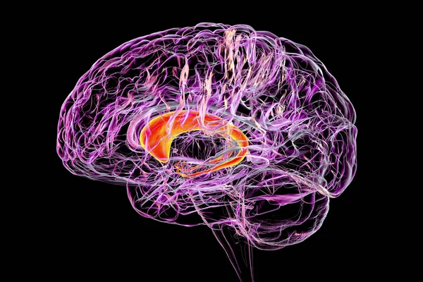
Caudate nuclei highlighted in human brain, 3D illustration. The caudate nucleus is a component of the basal ganglia, it is associated with motor processes and plays role in Parkinson's disease
Caudate Nuclei Highlighted Human Brain Illustration Caudate Nucleus Component Basal — Stock Photo, Image
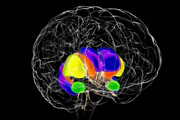
Brain dorsal striatum anatomy, 3D illustration. The dorsal striatum consists of the caudate nucleus (orange) and the putamen (yellow). Lateral ventricle is colored blue, amygdala is green
Brain Dorsal Striatum Anatomy Illustration Dorsal Striatum Consists Caudate Nucleus — Stock Photo, Image
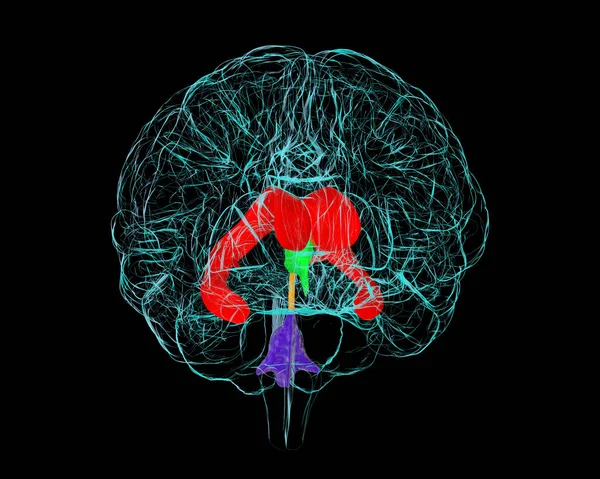
Ventricular system of brain, front view, 3D illustration. The ventricles are cavities in the brain that are filled with cerebrospinal fluid, CSF.
Ventricular System Brain Front View Illustration Ventricles Cavities Brain Filled — Stock Photo, Image
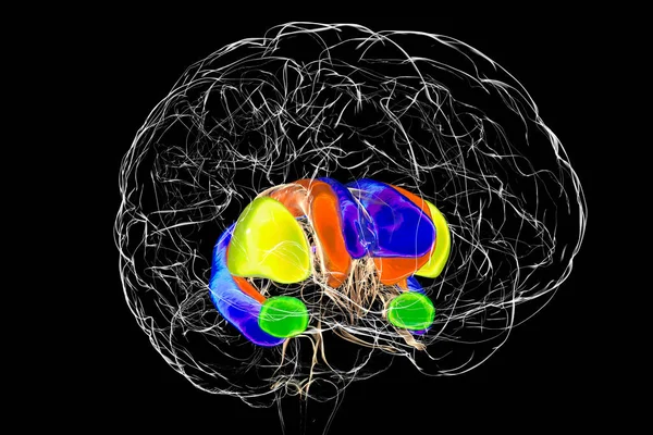
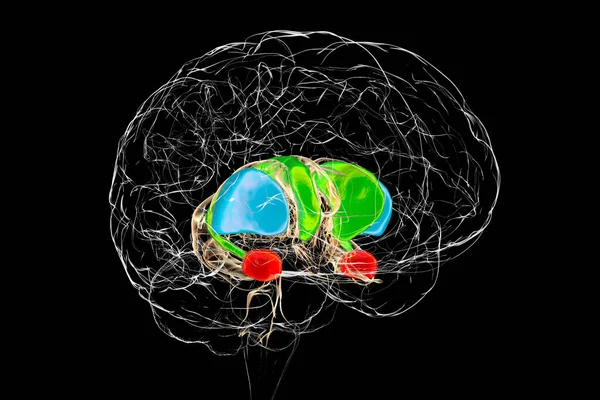
Brain dorsal striatum anatomy, 3D illustration. The dorsal striatum consists of the caudate nucleus (green) and the putamen (blue). Amygdala is colored in red
Brain Dorsal Striatum Anatomy Illustration Dorsal Striatum Consists Caudate Nucleus — Stock Photo, Image
Ventricular system of brain, 3D illustration. The ventricles are cavities in the brain that are filled with cerebrospinal fluid, CSF.
Ventricular System Brain Illustration Ventricles Cavities Brain Filled Cerebrospinal Fluid — Stock Photo, Image
The interventricular foramen, the foramen of Monro, a passage that connects the brain lateral ventricles, allowing cerebrospinal fluid to flow between them and the third ventricle, 3D illustration.
Interventricular Foramen Foramen Monro Passage Connects Brain Lateral Ventricles Allowing — Stock Photo, Image
Ventricular system of brain, 3D illustration. The ventricles are cavities in the brain that are filled with cerebrospinal fluid, CSF.
Ventricular System Brain Illustration Ventricles Cavities Brain Filled Cerebrospinal Fluid — Stock Photo, Image
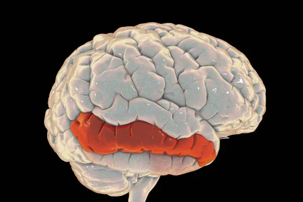
Human brain with highlighted middle temporal gyrus, 3D illustration. It is located in the temporal lobe, is involved in recognition of known faces, accessing word meaning while reading
Human Brain Highlighted Middle Temporal Gyrus Illustration Located Temporal Lobe — Stock Photo, Image
Ventricular system of brain, 3D illustration. The ventricles are cavities in the brain that are filled with cerebrospinal fluid, CSF.
Ventricular System Brain Illustration Ventricles Cavities Brain Filled Cerebrospinal Fluid — Stock Photo, Image
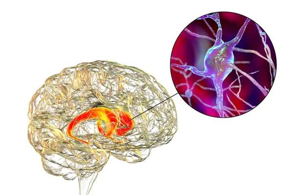
Caudate nuclei in human brain and its neurons, 3D illustration. The caudate nucleus is a component of the basal ganglia, it plays role in choreas, neurodegenerative and other brain diseases
Caudate Nuclei Human Brain Its Neurons Illustration Caudate Nucleus Component — Stock Photo, Image
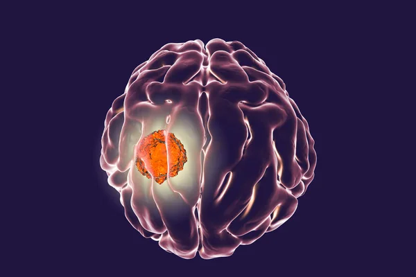
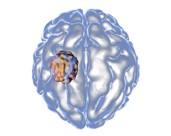
Brain cancer, 3D illustration showing presence of tumor inside brain
Brain tumor, illustration — Stock Photo, Image
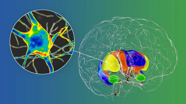
Dorsal striatum highlighted in brain and its neurons, 3D illustration. The dorsal striatum consists of the caudate nucleus (orange) and the putamen (yellow)
Dorsal Striatum Highlighted Brain Its Neurons Illustration Dorsal Striatum Consists — Stock Photo, Image
Aspergilloma of the brain, 3D illustration. Also known as mycetoma, or fungus ball, an intracranial lesion produced by fungi Aspergillus in immunocompromised patients
Aspergilloma of the brain — Stock Photo, Image
Ventricular system of brain, 3D illustration. The ventricles are cavities in the brain that are filled with cerebrospinal fluid, CSF.
Ventricular System Brain Illustration Ventricles Cavities Brain Filled Cerebrospinal Fluid — Stock Photo, Image
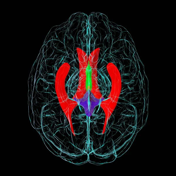
Ventricular system of brain, bottom view, 3D illustration. The ventricles are cavities in the brain that are filled with cerebrospinal fluid, CSF.
Ventricular System Brain Bottom View Illustration Ventricles Cavities Brain Filled — Stock Photo, Image

Usage Information

You can use this royalty-free photo "A 3D scientific illustration showcasing the third brain ventricle, a vital component of the brain's ventricular system, side view." for personal and commercial purposes according to the Standard or Extended License. The Standard License covers most use cases, including advertising, UI designs, and product packaging, and allows up to 500,000 print copies. The Extended License permits all use cases under the Standard License with unlimited print rights and allows you to use the downloaded stock images for merchandise, product resale, or free distribution.

You can buy this stock photo and download it in high resolution up to 6000x6000. Upload Date: Jul 25, 2023