Structure Main Female Hormone Estradiol Molecule Combined Surface Ball Stick — Stock Photo, Image

Structure of the main female hormone estradiol molecule, combined surface-ball-and-stick model, white background, 3D illustration — Photo

Structure of the main female hormone estradiol molecule, combined surface-ball-and-stick model, white background, 3D illustration

 — Photo by unnaugan

Same Series:

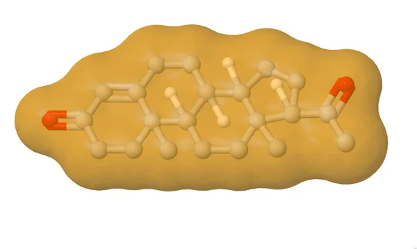
Structure of the human sex hormone progesteron molecule, combined surface-ball-and-stick model, white background, 3D illustration
Structure Human Sex Hormone Progesteron Molecule Combined Surface Ball Stick — Stock Photo, Image
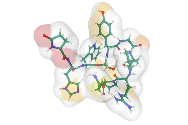
Structure of the human gonadotropin-releasing hormone, 3D ball-and-stick model with the accessible surface area shown, white background
Structure Human Gonadotropin Releasing Hormone Ball Stick Model Accessible Surface — Stock Photo, Image
A 3D-model of the recombinant human insulin chains A and B quaternary structure, white background, surface model
Model Recombinant Human Insulin Chains Quaternary Structure White Background Surface — Stock Photo, Image
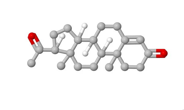
Structure of steroid hormone progesterone, ball-and-stick model, white background, 3D illustration
Structure Steroid Hormone Progesterone Ball Stick Model White Background Illustration — Stock Photo, Image
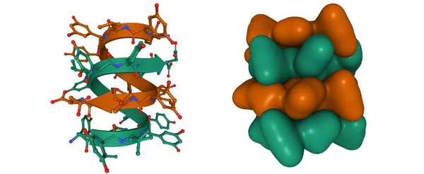
Antibiotic feglymycin P64 crystal form, 3D cartoon and Gaussian surface models, based on PDB 1w7r, white background
Antibiotic Feglymycin P64 Crystal Form Cartoon Gaussian Surface Models Based — Stock Photo, Image
Bovine insulin, 3D-model of the heterodimer quaternary structure, chains A and B, surface filled model, white background
Bovine Insulin Model Heterodimer Quaternary Structure Chains Surface Filled Model — Stock Photo, Image
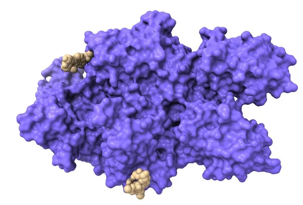
The tertiary structure of Human Coagulation Factor VIII with attached ligands (gray color). Mutation in the respective gene causes hemophilia a.
Tertiary Structure Human Coagulation Factor Viii Attached Ligands Gray Color — Stock Photo, Image
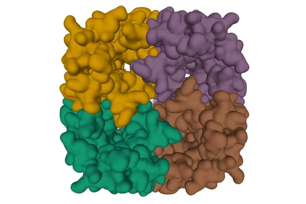
Crystal structure of human Aquaporin 7, 3D surface model isolated, white background
Crystal Structure Human Aquaporin Surface Model Isolated White Background — Stock Photo, Image
A 3D surface model of the human CD69 transmembrane C-Type lectin protein, white background
Surface Model Human Cd69 Transmembrane Type Lectin Protein White Background — Stock Photo, Image
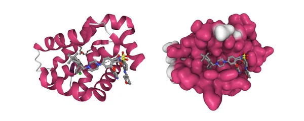
Crystal structure of BCL-2 with venetoclax. 3D cartoon and Gaussian surface models, secondary structure color scheme, PDB 6o0k, white background
Crystal Structure Bcl Venetoclax Cartoon Gaussian Surface Models Secondary Structure — Stock Photo, Image
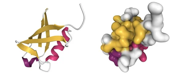
Structure of human ubiquitin, 3D cartoon and Gaussian surface model with the differently colored secondary structure elements, white background
Structure Human Ubiquitin Cartoon Gaussian Surface Model Differently Colored Secondary — Stock Photo, Image
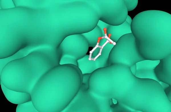
Crystal structure of a complex formed between group II phospholipase A2 (green) and aspirin, 3D model, black background
Crystal Structure Complex Formed Group Phospholipase Green Aspirin Model Black — Stock Photo, Image
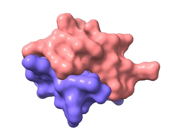
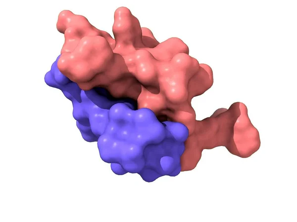
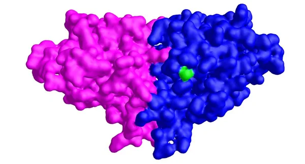
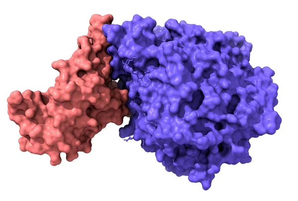
Structure of novel coronavirus spike receptor-binding domain (pink) complexed with its receptor ACE2 (blue), white background
Structure Novel Coronavirus Spike Receptor Binding Domain Pink Complexed Its — Stock Photo, Image
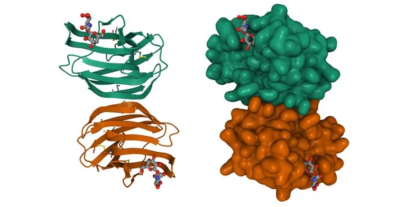
Crystal structure of human galectin-1 in complex with type 1 N-acetyllactosamine. 3D cartoon and Gaussian surface model, chain id color scheme, PDB 4xbl, white background
Crystal Structure Human Galectin Complex Type Acetyllactosamine Cartoon Gaussian Surface — Stock Photo, Image
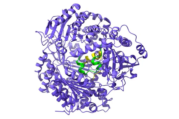
Structure of human insulin-degrading enzyme (blue) in complex with insulin (green-yellow). 3D cartoon model, PDB 2wby, white background.
Structure Human Insulin Degrading Enzyme Blue Complex Insulin Green Yellow — Stock Photo, Image
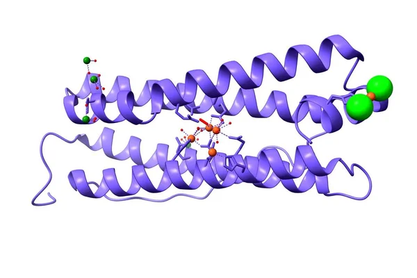
Human H ferritin heavy chain with ions of iron (orange), chlorine (green), and magnesium (dark green). 3D cartoon model, PDB 4y08, white background
Human Ferritin Heavy Chain Ions Iron Orange Chlorine Green Magnesium — Stock Photo, Image

Usage Information

You can use this royalty-free photo "Structure of the main female hormone estradiol molecule, combined surface-ball-and-stick model, white background, 3D illustration" for personal and commercial purposes according to the Standard or Extended License. The Standard License covers most use cases, including advertising, UI designs, and product packaging, and allows up to 500,000 print copies. The Extended License permits all use cases under the Standard License with unlimited print rights and allows you to use the downloaded stock images for merchandise, product resale, or free distribution.

You can buy this stock photo and download it in high resolution up to 5000x3380. Upload Date: Jun 19, 2020