Animal cell Stock Photos

100,000 Animal cell pictures are available under a royalty-free license

  • Best Match
  • Fresh
  • Popular
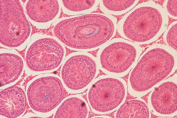
1-100 of 100,000
Cell structure
Cell structure — Stock Photo, Image
Blue cell under microscope. Life and biology, medicine scientific, molecular research dna. 3d illustration
Blue cell under microscope. Life and biology, medicine scientific, molecular research dna. 3d illustration — Stock Photo, Image
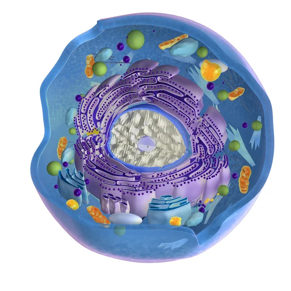
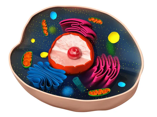
Animal cell on white
Animal cell on white — Stock Photo, Image
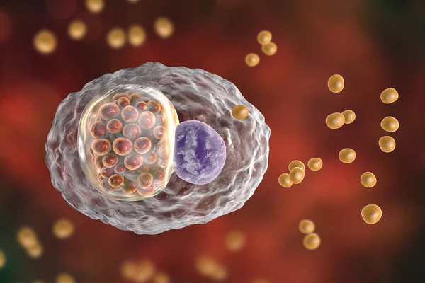
Chlamydophila psittaci, intracellular bacteria that cause psittacosis
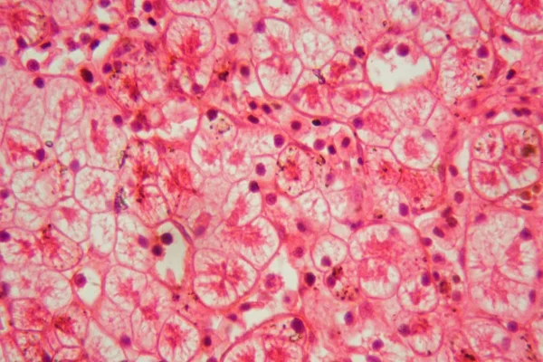
Chlamydophila psittaci, intracellular bacteria that cause psittacosis — Stock Photo, Image
Human (animal) cell under microscope. 3d illustration
Human (animal) cell under microscope. 3d illustration — Stock Photo, Image
Human cells or animal. Cell colony. Concept of science and medicine, the regeneration of cells, the renewal of cells in the living organism. 3D illustration
Human cells or animal. Cell colony. Concept of science and medicine, the regeneration of cells, the renewal of cells in the living organism. 3D illustration — Stock Photo, Image
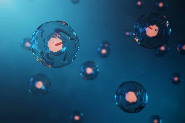
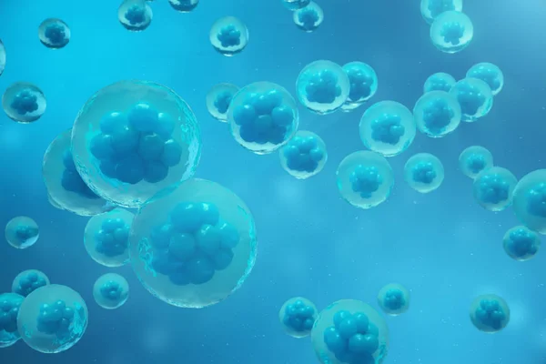
3D Rendering human or animal cells on blue background. Concept Early stage embryo Medicine scientific concept, Stem cell research and treatment.
3D Rendering human or animal cells on blue background. Concept Early stage embryo Medicine scientific concept, Stem cell research and treatment. — Stock Photo, Image
T Cells attacking Cancer Cells 3D illustration
Cells Attacking Cancer Cells Illustration — Stock Photo, Image
Cell structure
Cell structure — Stock Photo, Image
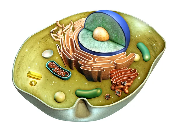
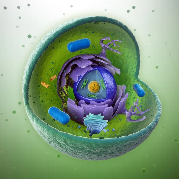
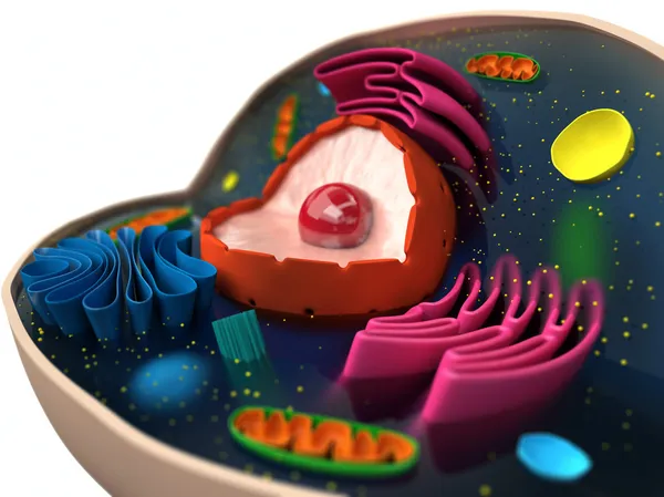
Animal cell cut-away - scientifically correct 3d illustration
Animal cell cut-away - scientifically correct 3d illustration — Stock Photo, Image
Human (animal) cell under microscope. 3d illustration
Human (animal) cell under microscope. 3d illustration — Stock Photo, Image
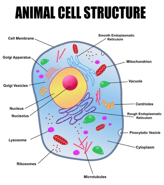
An illustration of the animal cell structure with names isolated on a white background
Illustration Animal Cell Structure Names Isolated White Background — Stock Photo, Image
Cell structure
Cell structure — Stock Photo, Image
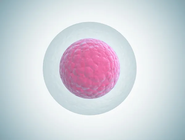
Human embryo cell illustration
Human embryo cell illustration — Stock Photo, Image
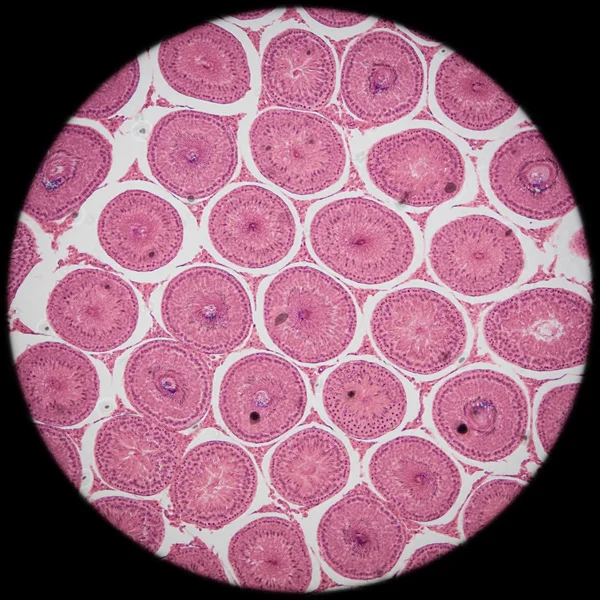
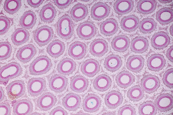
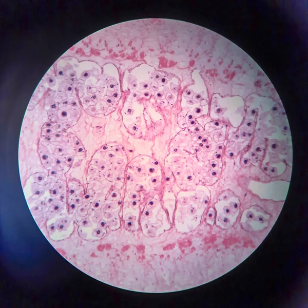
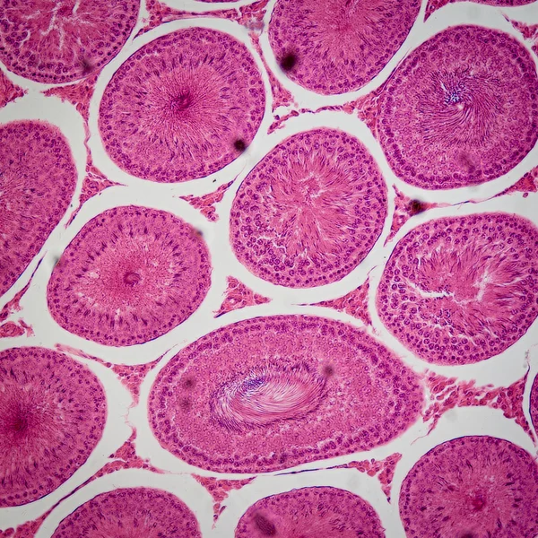
Microscopic section of Testis T.S tissue
Microscopic section of Testis T.S tissue — Stock Photo, Image
Abstract black ink stain. Chinese ink background on watercolor paper.
Abstract Black Ink Stain Chinese Ink Background Watercolor Paper — Stock Photo, Image
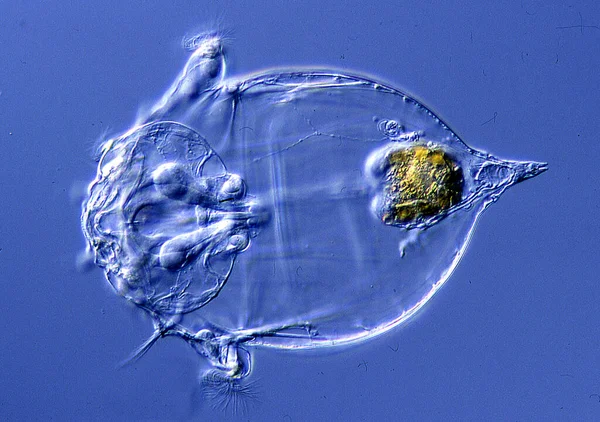
Asplanchna rotifer in drops of water under a microscope 200x
Asplanchna Rotifer Drops Water Microscope 200X — Stock Photo, Image
Microscope shot of abstract cell element on gray background
Microscope Shot Abstract Cell Element Gray Background — Stock Photo, Image
Pink abstract background in watercolor style
Pink Abstract Background Watercolor Style — Stock Photo, Image
Bazex syndrome or paraneoplastic acrokeratosis is a psoriasiform eruption that is seen more frequently in adult Caucasian males and is characterized by a predominantly acral distribution with nail dystrophy.
Bazex Syndrome Paraneoplastic Acrokeratosis Psoriasiform Eruption Seen More Frequently Adult — Stock Photo, Image
Characteristics of fungi living in wood as a group, are polyphyletic under the microscope for education.
Characteristics Fungi Living Wood Group Polyphyletic Microscope Education — Stock Photo, Image
Characteristics of fungi living in wood as a group, are polyphyletic under the microscope for education.
Characteristics Fungi Living Wood Group Polyphyletic Microscope Education — Stock Photo, Image
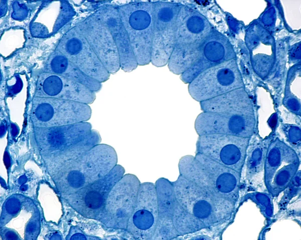
Kidney. Collecting duct
Kidney. Collecting duct — Stock Photo, Image
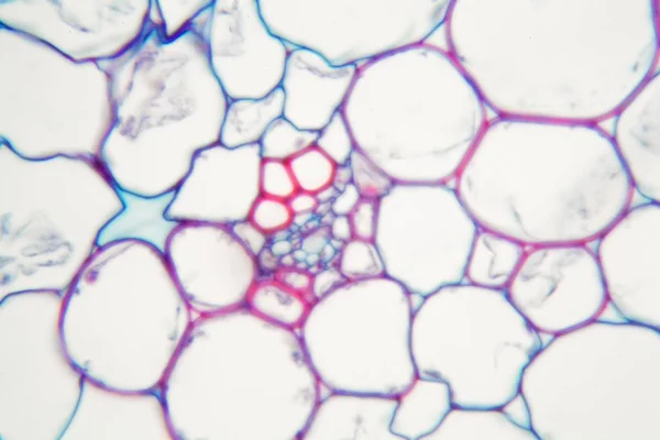
The study Plant tissue of under the microscope for classroom education.
Study Plant Tissue Microscope Classroom Education — Stock Photo, Image
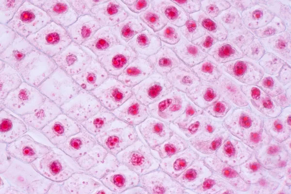
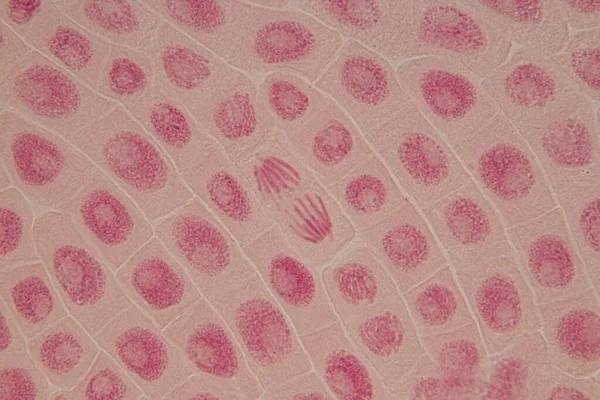
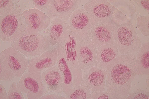
Root tip of Onion and Mitosis cell in the Root tip of Onion under a microscope.
Root Tip Onion Mitosis Cell Root Tip Onion Microscope — Stock Photo, Image
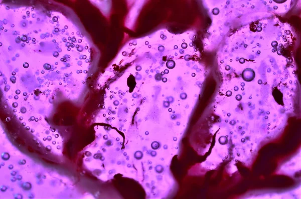
Purple liquid ink, digital wallpaper
Purple Liquid Ink Digital Wallpaper — Stock Photo, Image
Muscle cells
Muscle cells — Stock Photo, Image
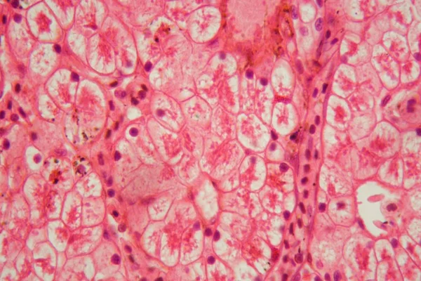
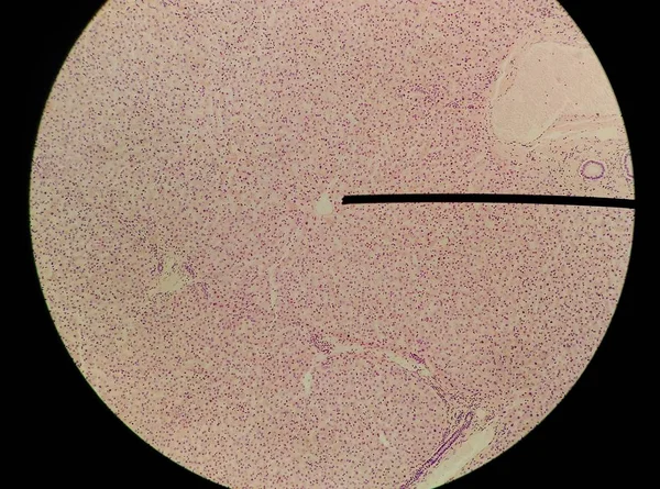
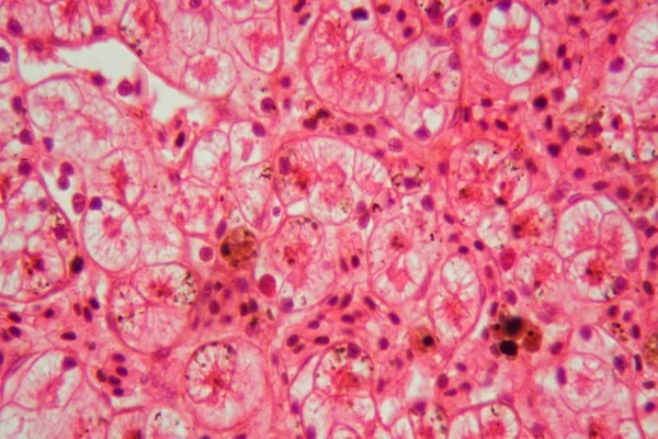
Microscope photo of a section through a frog liver.
Microscope photo of a section through a frog liver. — Stock Photo, Image
Characteristics of fungi living in wood as a group, are polyphyletic under the microscope for education.
Characteristics Fungi Living Wood Group Polyphyletic Microscope Education — Stock Photo, Image
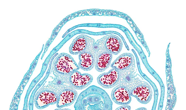
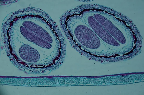
Rapeseed flower bud halve, cross section, 20X light micrograph. Brassica napus subsp. napus, also known as oilseed rape, a flowering plant, under the light microscope. Isolated on white background.
Rapeseed Flower Bud Halve Cross Section 20X Light Micrograph Brassica — Stock Photo, Image
Root tip of Onion and Mitosis cell in the Root tip of Onion under a microscope.
Root Tip Onion Mitosis Cell Root Tip Onion Microscope — Stock Photo, Image
Egg cell oocyte in ovary tissue
Egg cell oocyte in ovary tissue — Stock Photo, Image
Pork tapeworm Taenia solium, section through the body, light micrograph
Pork Tapeworm Taenia Solium Section Body Light Micrograph — Stock Photo, Image
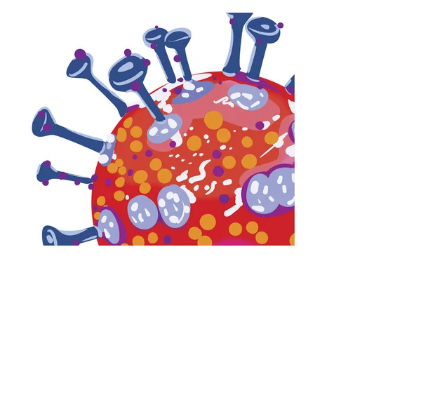
Virus Pandemic Koronavirus Covid-19 Bred
Virus Pandemic Koronavirus Covid Bred — Stock Photo, Image
Mould on spoiled food.
Mould Spoiled Food — Stock Photo, Image
Granite surface texture for background, detail of the polished granite texture
Granite Surface Texture Background Detail Polished Granite Texture — Stock Photo, Image
Lime juice in glass jar
Lime juice in glass jar — Stock Photo, Image
Microscope photo of a section through a frog liver.
Microscope photo of a section through a frog liver. — Stock Photo, Image
Water droplets bathroom shower texture background
Water Droplets Bathroom Shower Texture Background — Stock Photo, Image
Root tip of Onion and Mitosis cell in the Root tip of Onion under a microscope.
Root Tip Onion Mitosis Cell Root Tip Onion Microscope — Stock Photo, Image
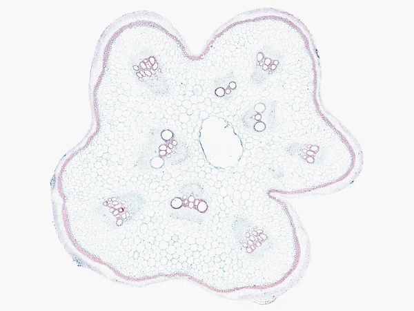
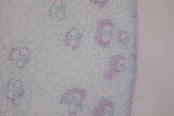
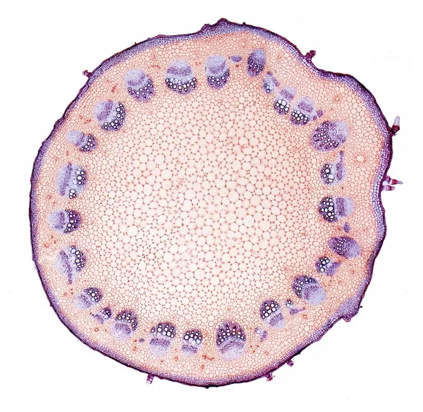
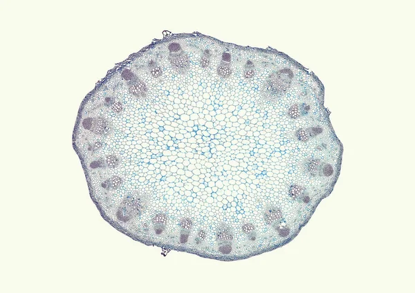
Biological specimen of pumpkin stalk under a microscope
Biological Specimen Pumpkin Stalk Microscope — Stock Photo, Image
Thyroid follicular carcinoma, light micrograph, photo under microscope
Thyroid Follicular Carcinoma Light Micrograph Photo Microscope — Stock Photo, Image
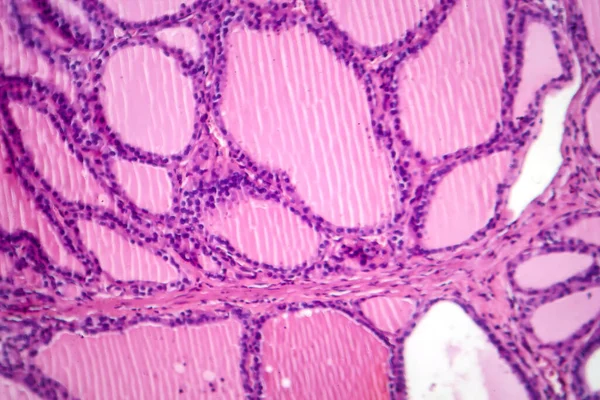
Helianthus stem. Light micrograph (LM) of stem of perennial sunflower (Helianthus sp.) in cross-section showing vascular bundles (green, yellow). Vascular tissues transport water and dissolved substances inside plant
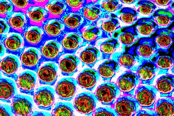
Helianthus Stem Light Micrograph Stem Perennial Sunflower Helianthus Cross Section — Stock Photo, Image
White blood cells in synovial body fluid.
White Blood Cells Synovial Body Fluid — Stock Photo, Image
Animal Pattern with Alligator Skin.
Animal Pattern with Alligator Skin. — Stock Photo, Image
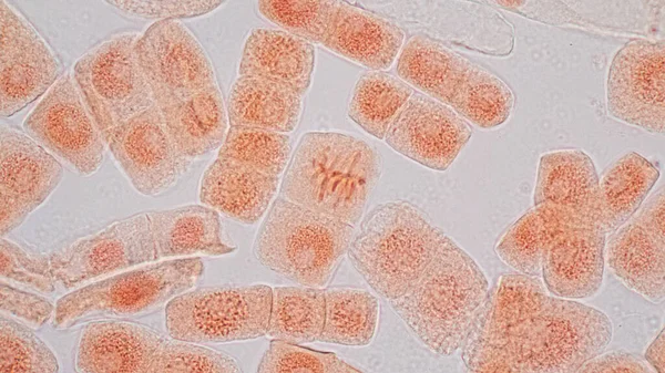
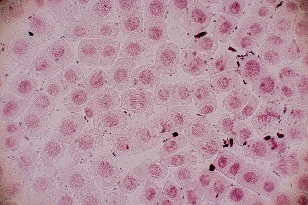
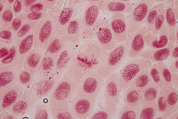
Microscopic image of onion root tip cells undergoing mitosis. Anaphases and metaphases. 1000x magnified
Microscopic Image Onion Root Tip Cells Undergoing Mitosis Anaphases Metaphases — Stock Photo, Image
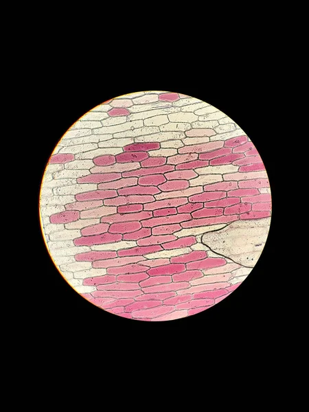
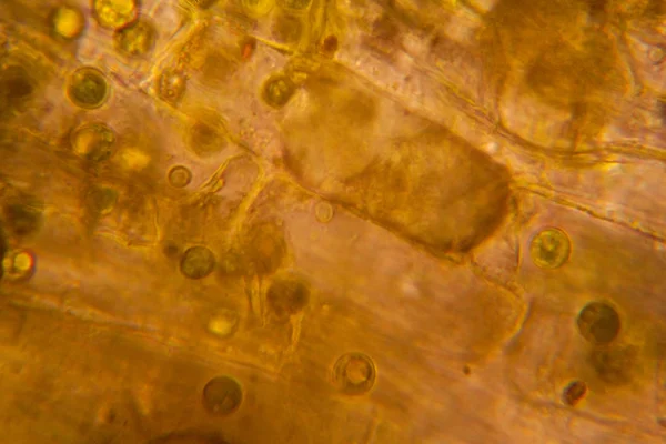
Biological preparation of onion plant cells under a microscope with the phenomenon of plasmolysis
Biological Preparation Onion Plant Cells Microscope Phenomenon Plasmolysis — Stock Photo, Image
Freeze Motion Shot of Crushing Raspberries.
Freeze Motion Shot of Crushing Raspberries. — Stock Photo, Image
Pink liquid ink, digital wallpaper
Pink Liquid Ink Digital Wallpaper — Stock Photo, Image
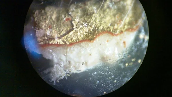
A macro shot of gold under the microscope
Macro Shot Gold Microscope — Stock Photo, Image
Wall Decor for interior home decoration, Ceramic Tile Design For Bathroom. it can be used for ceramic tile, wallpaper, linoleum, textile, web page background.
Wall Decor Interior Home Decoration Ceramic Tile Design Bathroom Can — Stock Photo, Image
Microscopic view of organisms in the fusty water with rotten vegetation
Microscopic View Organisms Fusty Water Rotten Vegetation — Stock Photo, Image
Bone cells of a bone seen from the microscope.
Bone Cells Bone Seen Microscope — Stock Photo, Image
Root tip of Onion and Mitosis cell in the Root tip of Onion under a microscope.
Root Tip Onion Mitosis Cell Root Tip Onion Microscope — Stock Photo, Image
Abstract violet background, acrylic painting decorative texture
Abstract Violet Background Acrylic Painting Decorative Texture — Stock Photo, Image
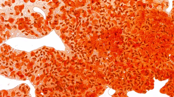
Red blood cells medical science background.
Red blood cells medical science background. — Stock Photo, Image
Symmetrical growth of bacteria
Symmetrical growth of bacteria — Stock Photo, Image
Watercolor seamless pattern with floral bouquets. Vintage botanical illustration. Elegant decoration for any kind of a design. Fashion print with colorful abstract flowers. Watercolor texture.
Watercolor Seamless Pattern Floral Bouquets Vintage Botanical Illustration Elegant Decoration — Stock Photo, Image
Root tip of Onion and Mitosis cell in the Root tip of Onion under a microscope.
Root Tip Onion Mitosis Cell Root Tip Onion Microscope — Stock Photo, Image
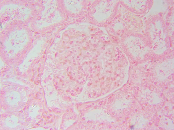
Kidney's Star: Single Glomerulus Closeup at 400x
Kidney Star Single Glomerulus Closeup 400X — Stock Photo, Image
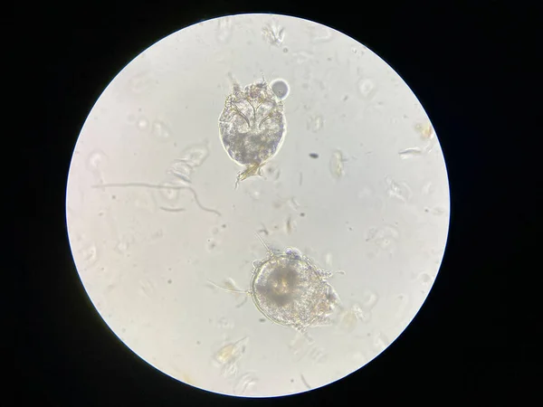
Notoedres cati under the microscope. Notoedric mange, also referred to as Feline scabies, is a highly contagious skin infestation caused by an ectoparasitic and skin burrowing mite Notoedres cati.
Notoedres Cati Microscope Notoedric Mange Also Referred Feline Scabies Highly — Stock Photo, Image
In iran the religion architecture
In iran  the religion     architecture — Stock Photo, Image
Shiny pink leaf gold foil texture background
Shiny Pink Leaf Gold Foil Texture Background — Stock Photo, Image
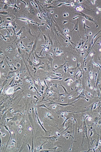
Cells of human find with microscope.
Cells of human find with microscope. — Stock Photo, Image
Capsella old embryo sec. under microscope
Capsella Old Embryo Sec Microscope — Stock Photo, Image
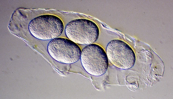
Tardigrade skin with eggs for the winter
Tardigrade Skin Eggs Winter — Stock Photo, Image
Pink abstract background in watercolor style
Pink Abstract Background Watercolor Style — Stock Photo, Image
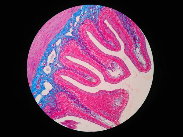
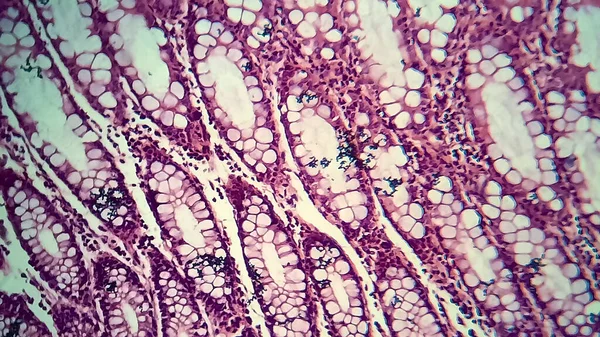
Histology of human intestine tissue under microscope view
Histology Human Intestine Tissue Microscope View — Stock Photo, Image
Root tip of Onion and Mitosis cell in the Root tip of Onion under a microscope.
Root Tip Onion Mitosis Cell Root Tip Onion Microscope — Stock Photo, Image
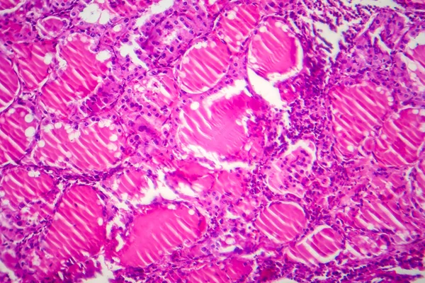
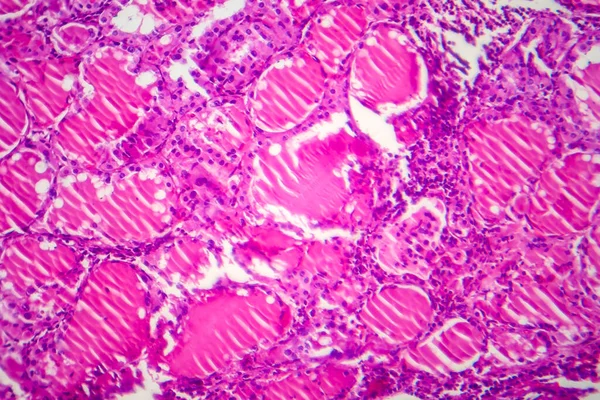
Simple goiter, light micrograph, photo under microscope. Enlargement of the thyroid gland. The most common cause of the simple goiter is iodine deficiency
Simple Goiter Light Micrograph Photo Microscope Enlargement Thyroid Gland Most — Stock Photo, Image
Simple goiter, light micrograph, photo under microscope. Enlargement of the thyroid gland. The most common cause of the simple goiter is iodine deficiency
Simple Goiter Light Micrograph Photo Microscope Enlargement Thyroid Gland Most — Stock Photo, Image
Eastern Equine Encephalitis virus
Eastern Equine Encephalitis virus — Stock Photo, Image
Imaginatory fractal abstract background Image
Imaginatory fractal abstract background Image — Stock Photo, Image
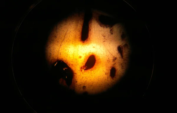
Mitosis cell in the Root tip of Onion under a microscope.
Mitosis Cell Root Tip Onion Microscope — Stock Photo, Image
Tissues with the fungus candida grow through 100x
Tissues Fungus Candida Grow 100X — Stock Photo, Image
Orange dream
Orange dream — Stock Photo, Image
Rotten leaves at the microscope
Rotten Leaves Microscope — Stock Photo, Image
Microscope photo of a section through a frog liver.
Microscope photo of a section through a frog liver. — Stock Photo, Image
Hand drawn pattern, background - illustration
Hand Drawn Pattern Background Illustration — Stock Photo, Image
Microscope shot of abstract cell element on beige background
Microscope Shot Abstract Cell Element Beige Background — Stock Photo, Image
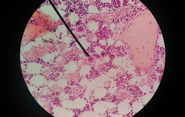
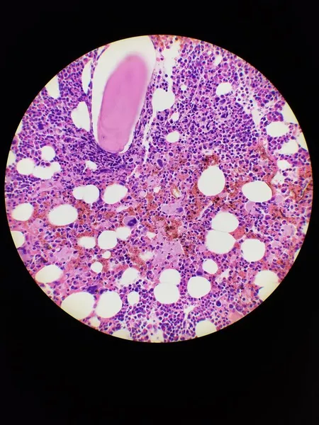
Bone marrow biopsy H and E stain showing eosinophilia
Bone marrow biopsy H and E stain showing eosinophilia — Stock Photo, Image
Very nice paint.
Very nice paint. — Stock Photo, Image
Rotten leaves at the microscope
Rotten Leaves Microscope — Stock Photo, Image
Mitosis cell in the Root tip of Onion under a microscope.
Mitosis Cell Root Tip Onion Microscope — Stock Photo, Image
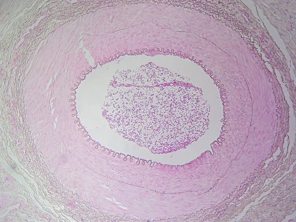
The Anatomy of Blood Vessels: Muscular Artery in High Detail
Anatomy Blood Vessels Muscular Artery High Detail — Stock Photo, Image
Abstract composition for Christmas background
Abstract composition for Christmas background — Stock Photo, Image
Liver cells of a rabit with Coccidia parasites.
Liver cells of a rabit with Coccidia parasites. — Stock Photo, Image
Microbiology, microscopic cells surface
Microbiology Microscopic Cells Surface — Stock Photo, Image
Background design textured color pattern abstract design
Background Design Textured Color Pattern Abstract Design — Stock Photo, Image
Microscopic image of onion root tip cells undergoing mitosis. Anaphases, telophases and metaphases. 1000x magnified.
Microscopic Image Onion Root Tip Cells Undergoing Mitosis Anaphases Telophases — Stock Photo, Image
Bacillary dysentery, light micrograph, photo under microscope showing presence of bacteria and accumulation of inflammatory cells in intestinal epithelium
Bacillary Dysentery Light Micrograph Photo Microscope Showing Presence Bacteria Accumulation — Stock Photo, Image
Root tip of Onion and Mitosis cell in the Root tip of Onion under a microscope.
Root Tip Onion Mitosis Cell Root Tip Onion Microscope — Stock Photo, Image
Microscopic section of Testis T.S tissue
Microscopic section of Testis T.S tissue — Stock Photo, Image
Cells of a Buttercup steam (Caltha palustris).
Cells of a Buttercup steam (Caltha palustris). — Stock Photo, Image
Abstract bright shiny color fluid background, hand drawn alcohol painting with golden streaks. Liquid ink technique texture for high resolution backdrop design
Abstract Bright Shiny Color Fluid Background Hand Drawn Alcohol Painting — Stock Photo, Image
Root tip of Onion and Mitosis cell in the Root tip of Onion under a microscope.
Root Tip Onion Mitosis Cell Root Tip Onion Microscope — Stock Photo, Image
Photomicrograph of a toxic goiter tissue sample under a microscope, revealing hypertrophy of thyroid follicular cells, increased vascularity, and colloid depletion.
Photomicrograph Toxic Goiter Tissue Sample Microscope Revealing Hypertrophy Thyroid Follicular — Stock Photo, Image
Root tip of Onion and Mitosis cell in the Root tip of Onion under a microscope.
Root Tip Onion Mitosis Cell Root Tip Onion Microscope — Stock Photo, Image
Creative and design art work, copy space wallpaper, abstract colorful texture background
Creative Design Art Work Copy Space Wallpaper Abstract Colorful Texture — Stock Photo, Image
Snake Skin Background.
Snake Skin Background. — Stock Photo, Image

Find The Best Animal Cell Images For Your Projects

If you're looking for high-quality animal cell images, you've come to the right place. Our collection of stock images features a wide variety of images that capture the intricate details of animal cells. These images are perfect for a range of projects, from educational materials to scientific presentations and publications.

Types Of Animal Cell Images Available

Our collection includes a range of animal cell images, from simple diagrams to detailed 3D renderings. You'll find images that depict various organelles, including the nucleus, mitochondria, ribosomes, and more. We also feature images that highlight the differences between animal and plant cells, including the presence of centrosomes and lysosomes.

If you're looking for a more artistic interpretation of animal cells, we also have abstract images that use color and shape to represent the various components of animal cells. These images make a striking addition to any project and can help to engage and inspire your audience.

Where To Use Animal Cell Images

Animal cell images are a valuable resource for a range of projects, from educational materials to scientific presentations. If you're creating educational materials, such as textbooks or classroom presentations, animal cell images can help students understand the structure and function of these important biological components.

If you're working on a scientific presentation or publication, animal cell images can help to illustrate your research and highlight key findings. They can also be used to compare and contrast different types of animal cells or different cellular processes, such as mitosis and meiosis.

Tips For Using Animal Cell Images

When using animal cell images, it's important to choose the right image for your project. Consider the purpose of your project and the audience you're trying to reach. If you're creating educational materials for young children, for example, you may want to use simpler, more colorful images. If you're presenting to a scientific audience, on the other hand, you may want to use more detailed and technical images.

It's also important to ensure that the images you use are accurate and up-to-date. Consult with experts in the field to make sure you're using the correct terminology and depicting the various organelles and processes correctly.

Get Your Animal Cell Images Today

Our collection of animal cell images is the perfect resource for any project that requires accurate and engaging visual aids. Browse our collection today and find the perfect image for your needs. Our images are available in JPG, AI, and EPS formats, making them easy to use in any project. Start your search today and take your project to the next level.