Human tissue Stock Photos

100,000 Human tissue pictures are available under a royalty-free license

  • Best Match
  • Fresh
  • Popular
1-100 of 100,000
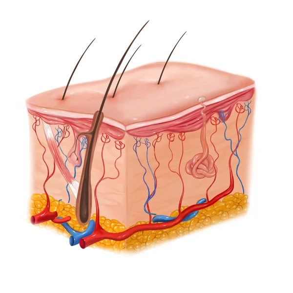
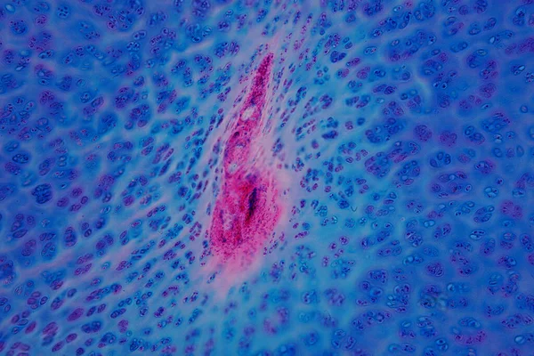
Human skin and hair structure
Human skin and hair structure — Stock Photo, Image
Pink abstract background in watercolor style
Pink Abstract Background Watercolor Style — Stock Photo, Image
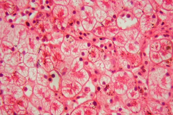
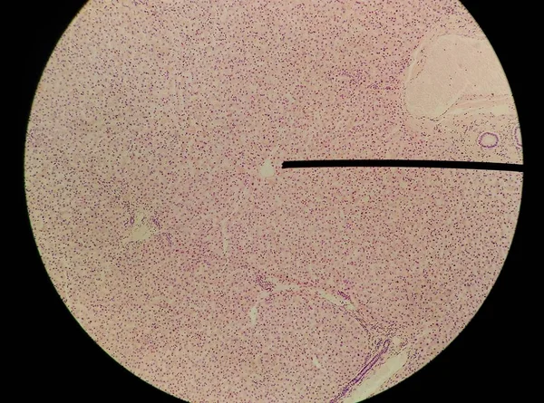
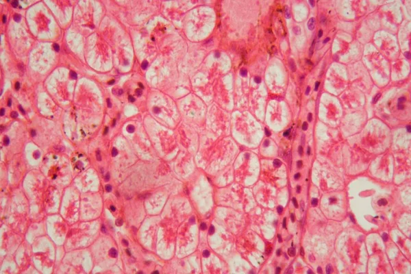
Microscope photo of a section through a frog liver.
Microscope photo of a section through a frog liver. — Stock Photo, Image
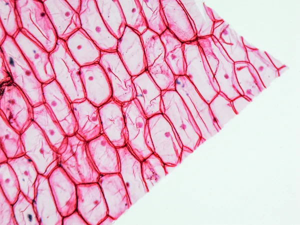
Onion epidermus micrograph
Onion epidermus micrograph — Stock Photo, Image
Swirls of alcohol ink in lilac, lavender, violet are reminiscent of marble and ripples of agate. Fluid modern Art banners, ethereal graphic design.
Swirls of alcohol ink in lilac, lavender, violet are reminiscent of marble and ripples of agate. Fluid modern Art banners, ethereal graphic design. — Stock Photo, Image
Characteristics of fungi living in wood as a group, are polyphyletic under the microscope for education.
Characteristics Fungi Living Wood Group Polyphyletic Microscope Education — Stock Photo, Image
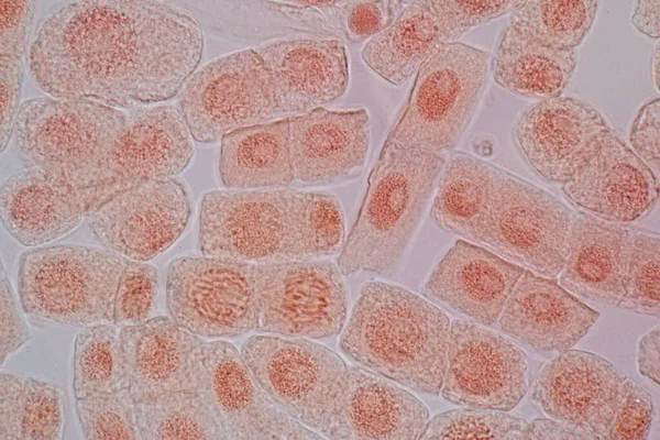
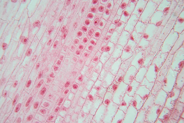
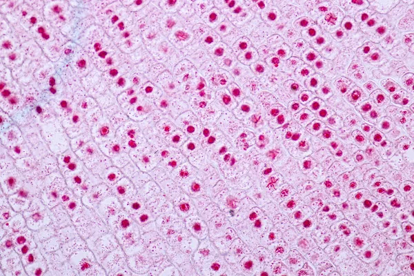
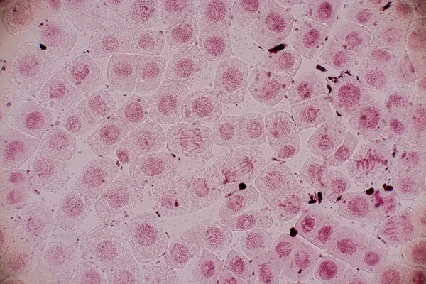
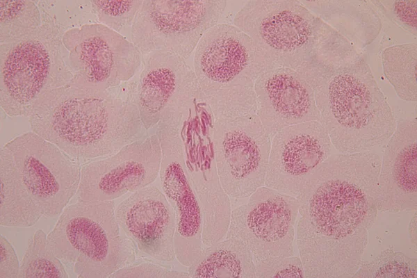
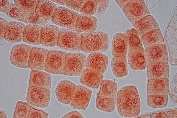
Mitosis cell in the Root tip of Onion under a microscope.
Mitosis Cell Root Tip Onion Microscope — Stock Photo, Image
Microscope view, abstract background of blue and purple surface
Microscope View Abstract Background Blue Purple Surface — Stock Photo, Image
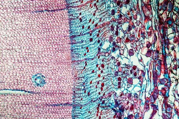
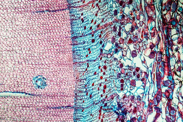
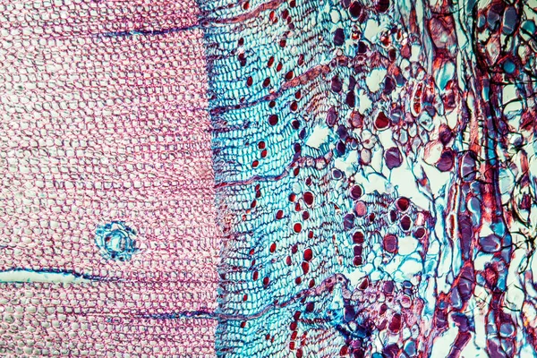
Pine trunk across 100x
Pine Trunk 100X — Stock Photo, Image
Abstract surface, microbiology cells
Abstract Surface Microbiology Cells — Stock Photo, Image
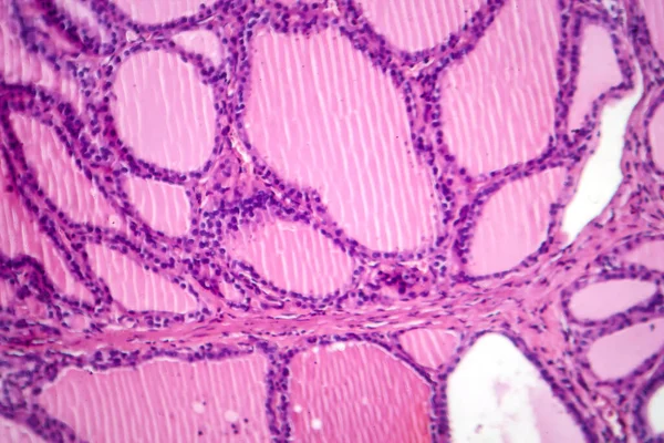
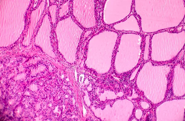
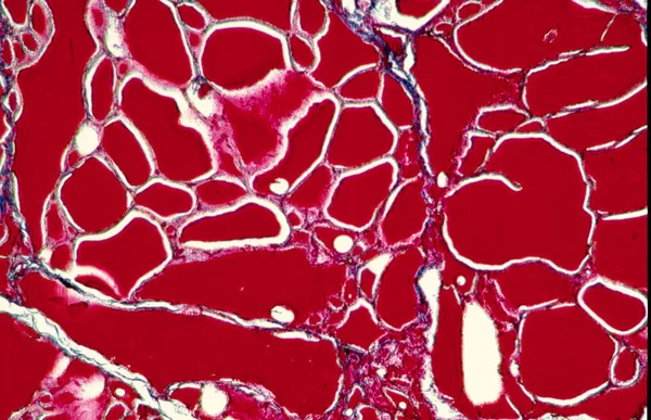
Follicles of colloid in the thyroid
Follicles Colloid Thyroid — Stock Photo, Image
Characteristics of fungi living in wood as a group, are polyphyletic under the microscope for education.
Characteristics Fungi Living Wood Group Polyphyletic Microscope Education — Stock Photo, Image
Characteristics of fungi living in wood as a group, are polyphyletic under the microscope for education.
Characteristics Fungi Living Wood Group Polyphyletic Microscope Education — Stock Photo, Image
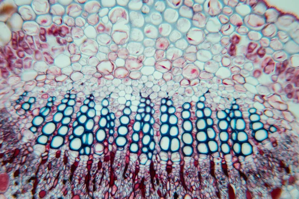
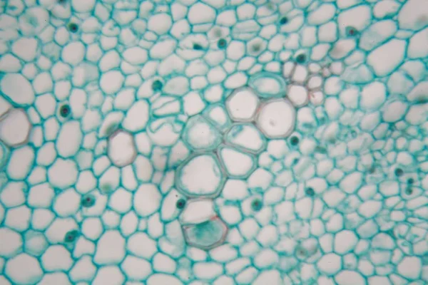
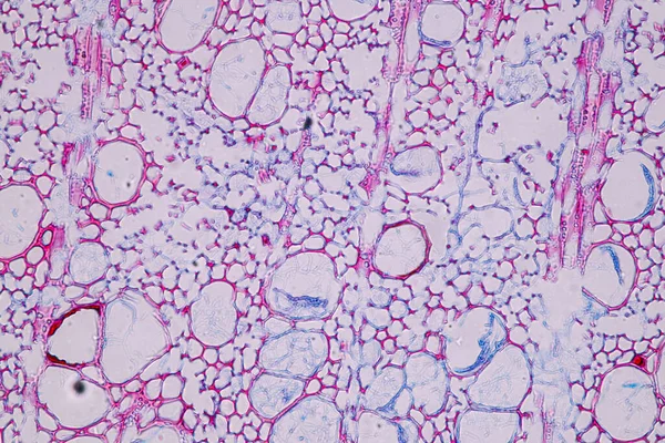
Stem with sieve cells under the microscope
Stem with sieve cells under the microscope — Stock Photo, Image
Cells of human find with microscope.
Cells of human find with microscope. — Stock Photo, Image
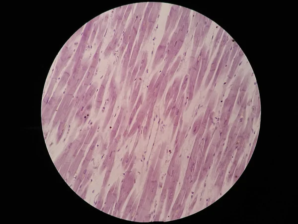
Histology of cardiac muscle under microscope view
Histology Cardiac Muscle Microscope View — Stock Photo, Image
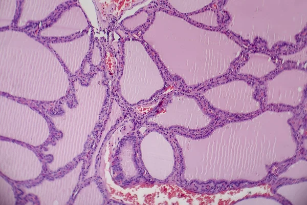
Endemic goiter, light micrograph, abnormal enlargement of the thyroid gland due to dietary iodine deficiency. Photomicrograph shows follicles of varying size, abundant colloid, lymphocytic infiltrate
Endemic Goiter Light Micrograph Abnormal Enlargement Thyroid Gland Due Dietary — Stock Photo, Image
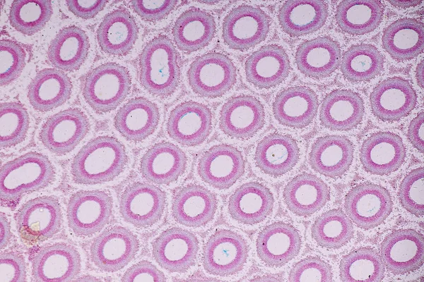
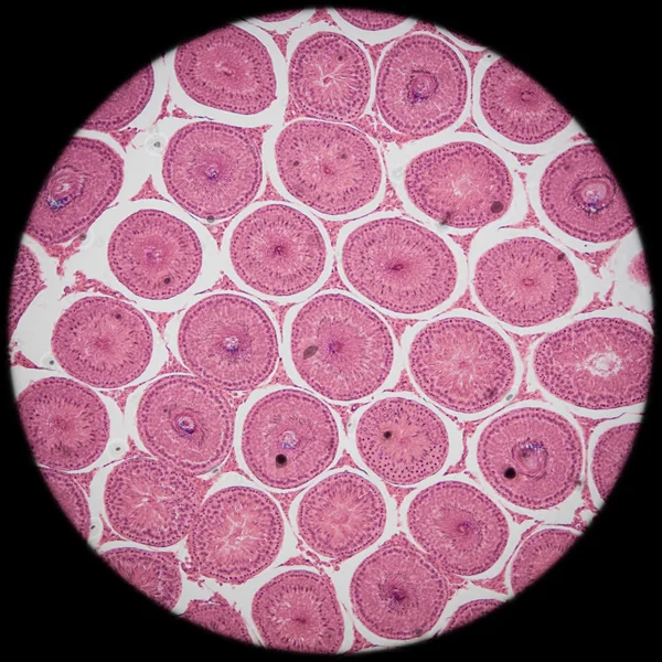
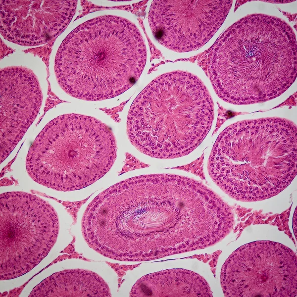
Microscopic section of Testis T.S tissue
Microscopic section of Testis T.S tissue — Stock Photo, Image
Root tip of Onion and Mitosis cell in the Root tip of Onion under a microscope.
Root Tip Onion Mitosis Cell Root Tip Onion Microscope — Stock Photo, Image
Light micrograph (LM) of a section through the fingertip.
Light Micrograph Section Fingertip — Stock Photo, Image
Pine tree trunk across 100x
Pine Tree Trunk 100X — Stock Photo, Image
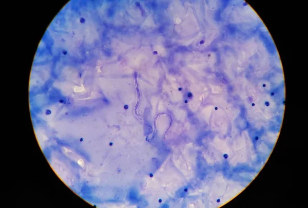
Microfilaria tissue parasite infection to human.
Microfilaria tissue parasite infection to human. — Stock Photo, Image
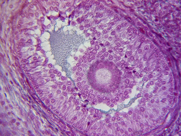
The Journey of the Egg: Histology Image in Stunning Detail
Journey Egg Histology Image Stunning Detail — Stock Photo, Image
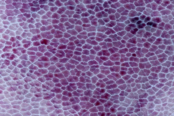
Microbiology, microscopic cells surface
Microbiology Microscopic Cells Surface — Stock Photo, Image
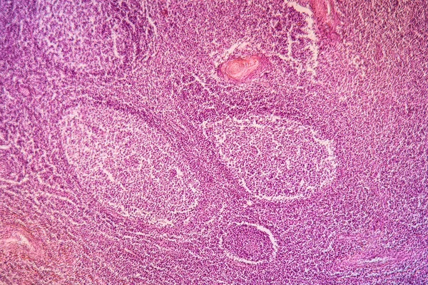
Photomicrograph of a toxic goiter tissue sample under a microscope, revealing hypertrophy of thyroid follicular cells, increased vascularity, and colloid depletion.
Photomicrograph Toxic Goiter Tissue Sample Microscope Revealing Hypertrophy Thyroid Follicular — Stock Photo, Image
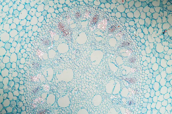
Water lily (nymphaea) stem under the microscope
Water lily (nymphaea) stem under the microscope — Stock Photo, Image
Aerial root across 100x
Aerial Root 100X — Stock Photo, Image
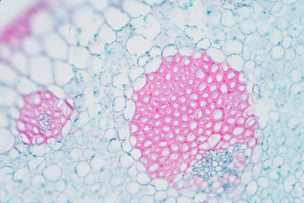
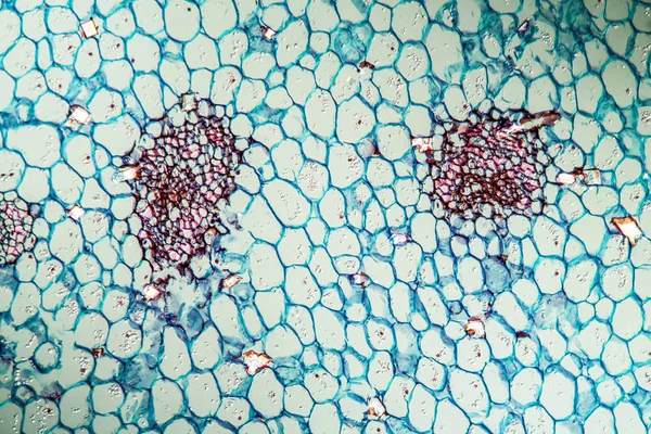
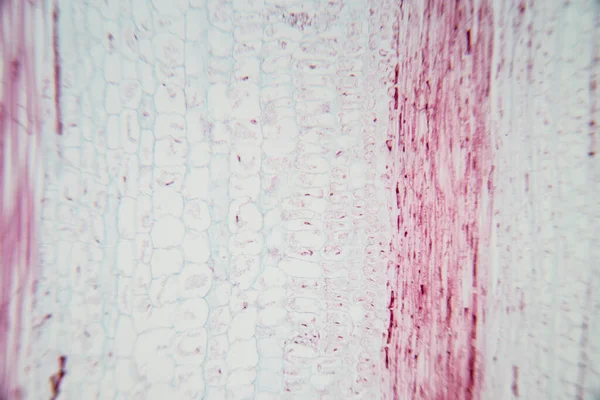
Plant vascular tissue under microscope view for education.
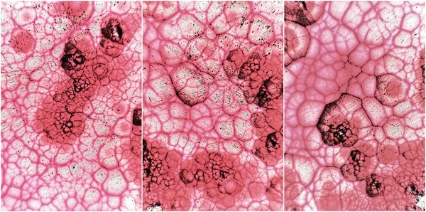
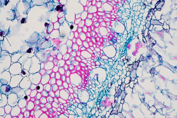
Plant Vascular Tissue Microscope View Education — Stock Photo, Image
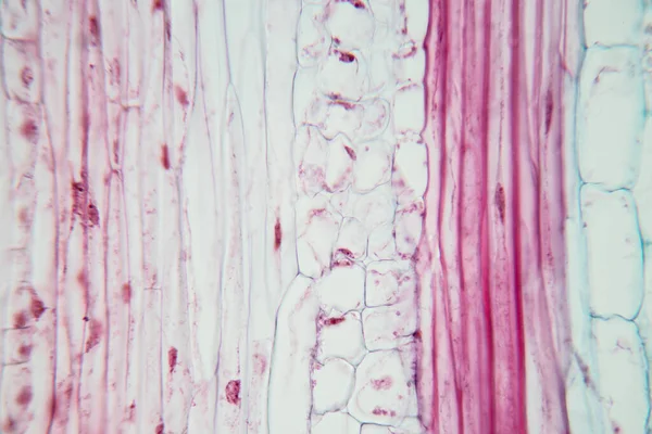
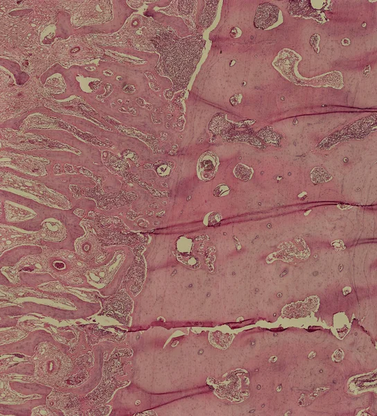
Human Hyaline cartilage bone under microscope view for education histology. Human tissue
Human Hyaline Cartilage Bone Microscope View Education Histology Human Tissue — Stock Photo, Image
Characteristics of fungi living in wood as a group, are polyphyletic under the microscope for education.
Characteristics Fungi Living Wood Group Polyphyletic Microscope Education — Stock Photo, Image
Root tip of Onion and Mitosis cell in the Root tip of Onion under a microscope.
Root Tip Onion Mitosis Cell Root Tip Onion Microscope — Stock Photo, Image
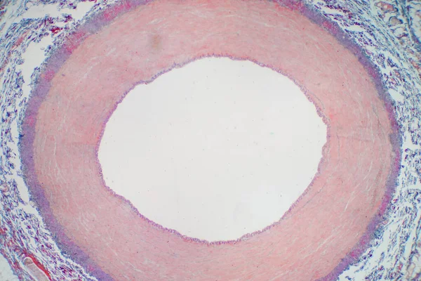
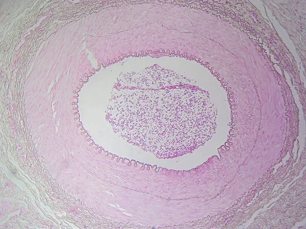
The Anatomy of Blood Vessels: Muscular Artery in High Detail
Anatomy Blood Vessels Muscular Artery High Detail — Stock Photo, Image
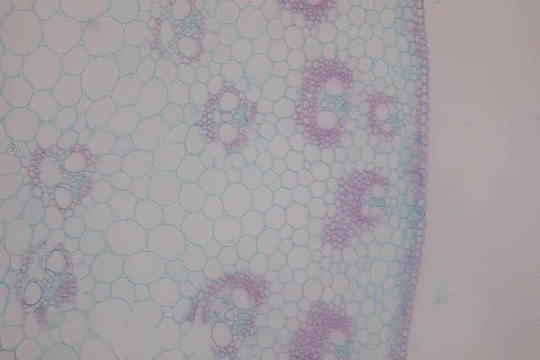
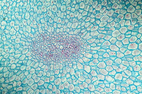
The study Plant tissue of under the microscope for classroom education.
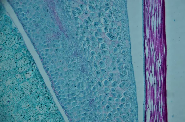
Study Plant Tissue Microscope Classroom Education — Stock Photo, Image
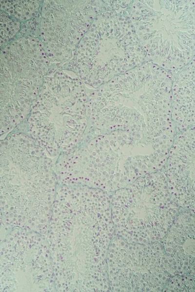
Cross section Human testis under microscope view. Shows spermatogonia, spermatocytes in meiosis, spermatids, and spermatozoa
Cross Section Human Testis Microscope View Shows Spermatogonia Spermatocytes Meiosis — Stock Photo, Image
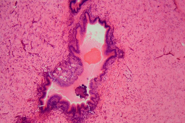
Uterus adenofibroma, light micrograph, photo under microscope. A rare benign tumor of the uterus composed of glandular and fibrous tissues
Uterus Adenofibroma Light Micrograph Photo Microscope Rare Benign Tumor Uterus — Stock Photo, Image
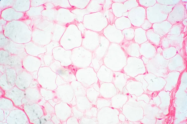
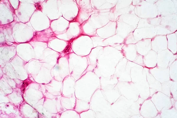
Human fat body tissue under microscope view for physiology education.
Human Fat Body Tissue Microscope View Physiology Education — Stock Photo, Image
Testicle cross-section under the microscope 100x
Testicle Cross Section Microscope 100X — Stock Photo, Image
Abstract inverse macro shot of natural stones and minerals. Set of Malachite.
Abstract Inverse Macro Shot Natural Stones Minerals Set Malachite — Stock Photo, Image
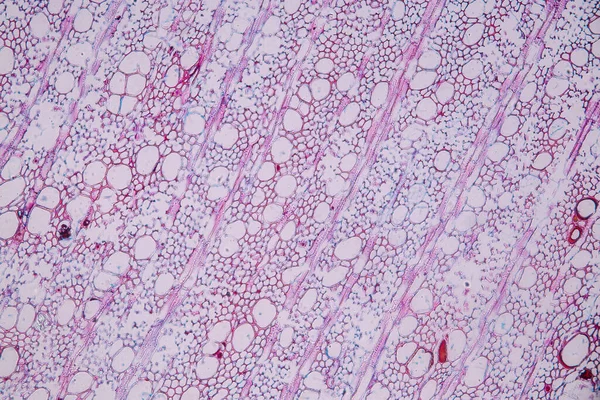
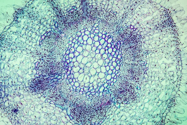
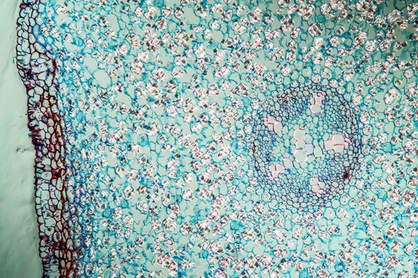
Cross sections of the plant root under the microscope view for education
Cross Sections Plant Root Microscope View Education — Stock Photo, Image
Liquid gel cosmetic smudge pink
Liquid Gel Cosmetic Smudge Pink — Stock Photo, Image
Human kidney tissue, light micrograph. Haematoxylin and eosin stain. The cuboidal epithelium of the renal tubules consists of a single layer of cuboid cells.
Human Kidney Tissue Light Micrograph Haematoxylin Eosin Stain Cuboidal Epithelium — Stock Photo, Image
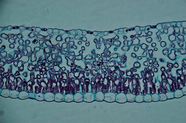
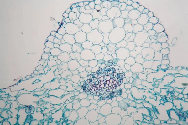
Lily leaf c.s. under microscope
Lily Leaf Microscope — Stock Photo, Image
Pink liquid ink, digital wallpaper
Pink Liquid Ink Digital Wallpaper — Stock Photo, Image
Suminagashi marble texture hand painted with pink ink Digital paper 107 performed in traditional
Suminagashi marble texture hand painted with pink ink Digital paper 107 performed in traditional — Stock Photo, Image
Red blood cells medical science background.
Red blood cells medical science background. — Stock Photo, Image
Liver cells of a rabit with Coccidia parasites.
Liver cells of a rabit with Coccidia parasites. — Stock Photo, Image
Purple texture
Purple texture — Stock Photo, Image
Mitosis cell in the Root tip of Onion under a microscope.
Mitosis Cell Root Tip Onion Microscope — Stock Photo, Image
Acute simple appendicitis, light micrograph
Acute simple appendicitis, light micrograph — Stock Photo, Image
Spinal ganglion from microscopy
Spinal Ganglion Microscopy — Stock Photo, Image
Microbiology, microscopic cells surface
Microbiology Microscopic Cells Surface — Stock Photo, Image
Root tip of Onion and Mitosis cell in the Root tip of Onion under a microscope.
Root Tip Onion Mitosis Cell Root Tip Onion Microscope — Stock Photo, Image
Microscopic section of Testis T.S tissue
Microscopic section of Testis T.S tissue — Stock Photo, Image
Onion epidermus micrograph
Onion epidermus micrograph — Stock Photo, Image
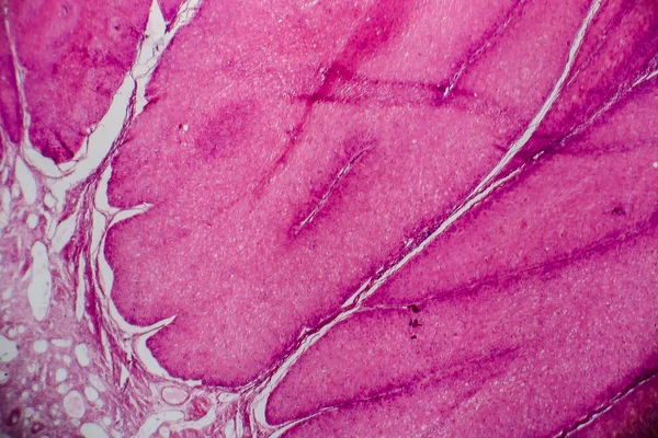
Condyloma acuminatum, also known as genital warts. Light micrograph, photo under microscope
Condyloma Acuminatum Also Known Genital Warts Light Micrograph Photo Microscope — Stock Photo, Image
Cells of a young broad bean steam (Vicia faba).
Cells of a young broad bean steam (Vicia faba). — Stock Photo, Image
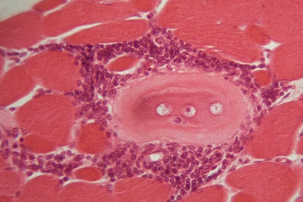
Trichinella spiralis larvae in muscle tissue under the microscope.
Trichinella spiralis larvae in muscle tissue under the microscope. — Stock Photo, Image
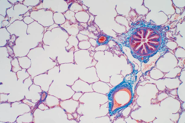
Human lung tissue under microscope, The lungs is organs of the respiratory system in humans. Human pathology education.
Human Lung Tissue Microscope Lungs Organs Respiratory System Humans Human — Stock Photo, Image
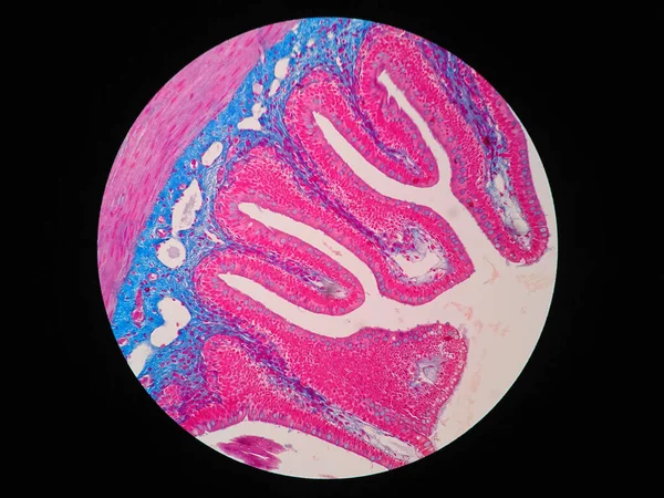
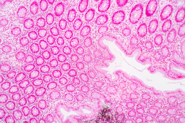
Histology of human intestine tissue under microscope view
Histology Human Intestine Tissue Microscope View — Stock Photo, Image
Shiny pink leaf gold foil texture background
Shiny Pink Leaf Gold Foil Texture Background — Stock Photo, Image
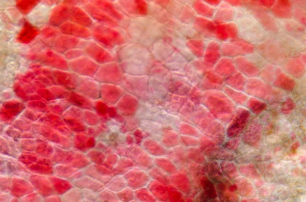
Micrograph red cherry fruit peel cell
Micrograph red cherry fruit peel cell — Stock Photo, Image
Root tip of Onion and Mitosis cell in the Root tip of Onion under a microscope.
Root Tip Onion Mitosis Cell Root Tip Onion Microscope — Stock Photo, Image
Human fat body tissue under microscope view for physiology education.
Human Fat Body Tissue Microscope View Physiology Education — Stock Photo, Image
Cells of a young broad bean steam (Vicia faba).
Cells of a young broad bean steam (Vicia faba). — Stock Photo, Image
Onion epidermus micrograph
Onion epidermus micrograph — Stock Photo, Image
Root tip of Onion and Mitosis cell in the Root tip of Onion under a microscope.
Root Tip Onion Mitosis Cell Root Tip Onion Microscope — Stock Photo, Image
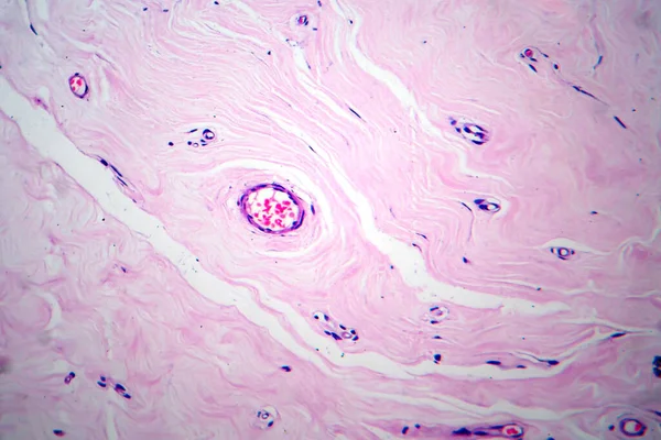
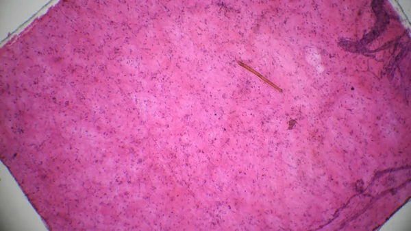
Connective tissue, light micrograph
Connective tissue, light micrograph — Stock Photo, Image
Thyroid follicular carcinoma, light micrograph, photo under microscope
Thyroid Follicular Carcinoma Light Micrograph Photo Microscope — Stock Photo, Image
Close up human cells with microscope.
Close up human cells with microscope. — Stock Photo, Image
Curves with gas bubbles are immersed in mineral water.
Curves Gas Bubbles Immersed Mineral Water — Stock Photo, Image
Abstract surface, microbiology cells
Abstract Surface Microbiology Cells — Stock Photo, Image
Microscopic image of onion root tip cells undergoing mitosis. Anaphases and metaphases. 1000x magnified
Microscopic Image Onion Root Tip Cells Undergoing Mitosis Anaphases Metaphases — Stock Photo, Image
Loose connective tissue,rattit under the microscope
Loose connective tissue,rattit under the microscope — Stock Photo, Image
Science medical anthropotomy physiology microscopic human thyroi
Science medical anthropotomy physiology microscopic human thyroi — Stock Photo, Image
Cells of a tobacco leaf (Nicotiana tabacum).
Cells of a tobacco leaf (Nicotiana tabacum). — Stock Photo, Image
Backgrounds of Characteristics Tissue of Stomach Human, Small intestine Human, Pancreas Human and Large intestine Human under the microscope in Lab.
Backgrounds Characteristics Tissue Stomach Human Small Intestine Human Pancreas Human — Stock Photo, Image
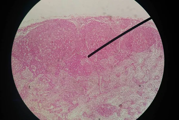
Toxic diffuse goiter, or Graves' disease, an autoimmune disease that affects the thyroid. Light micrograph shows hyperplastic thyroid follicles, decreased colloid
Toxic Diffuse Goiter Graves Disease Autoimmune Disease Affects Thyroid Light — Stock Photo, Image
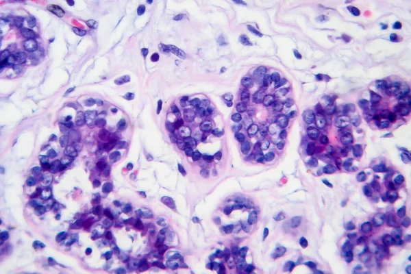
Breast cancer, light micrograph, photo under microscope
Breast Cancer Light Micrograph Photo Microscope — Stock Photo, Image
Zygomycota, or zygote fungi ,Downy mildew of cruzifers host tissue with conidia living in decaying plant on Slide under the microscope for education.
Zygomycota Zygote Fungi Downy Mildew Cruzifers Host Tissue Conidia Living — Stock Photo, Image
The study Plant tissue of under the microscope for classroom education.
Study Plant Tissue Microscope Classroom Education — Stock Photo, Image
Lily of the valley with stem 100x across
Lily Valley Stem 100X — Stock Photo, Image
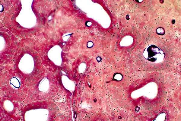
Histology of human compact bone tissue under microscope view for education, muscle bone connection and connective tissue
Histology Human Compact Bone Tissue Microscope View Education Muscle Bone — Stock Photo, Image
Lesser celandine with roots 100x across
Lesser Celandine Roots 100X — Stock Photo, Image
Abstract wallpaper, cells tissue microscope shot
Abstract Wallpaper Cells Tissue Microscope Shot — Stock Photo, Image
Texture, background, pattern. fabric lace beige sequined. Square golden mosaic music party background with stars.
Texture Background Pattern Fabric Lace Beige Sequined Square Golden Mosaic — Stock Photo, Image
The study Plant tissue of under the microscope for classroom education.
Study Plant Tissue Microscope Classroom Education — Stock Photo, Image
Mitosis cell in the Root tip of Onion under a microscope.
Mitosis Cell Root Tip Onion Microscope — Stock Photo, Image
Characteristics of fungi living in wood as a group, are polyphyletic under the microscope for education.
Characteristics Fungi Living Wood Group Polyphyletic Microscope Education — Stock Photo, Image
Abstract ink background. Marble style. Black paint stroke texture on white paper. Wallpaper for web and game design. Grunge mud art. Macro image of pen juice. Dark Smear
Abstract Ink Background Marble Style Black Paint Stroke Texture White — Stock Photo, Image
Bacillary dysentery, light micrograph, photo under microscope showing presence of bacteria and accumulation of inflammatory cells in intestinal epithelium
Bacillary Dysentery Light Micrograph Photo Microscope Showing Presence Bacteria Accumulation — Stock Photo, Image
Artery blood vessel under microscope view for education histology. Histological for human physiology.
Artery Blood Vessel Microscope View Education Histology Histological Human Physiology — Stock Photo, Image
Texture under microscope, Microscopic
Texture Microscope Microscopic — Stock Photo, Image
Colorful ebru or art of Marbling background on water surface
Colorful Ebru Art Marbling Background Water Surface — Stock Photo, Image
Microscope photo of a section through a frog liver.
Microscope photo of a section through a frog liver. — Stock Photo, Image
Cells of human find with microscope.
Cells of human find with microscope. — Stock Photo, Image
The study Plant tissue of under the microscope for classroom education.
Study Plant Tissue Microscope Classroom Education — Stock Photo, Image
Full frame image of pink abstract paint background
Full Frame Image Pink Abstract Paint Background — Stock Photo, Image
The study Plant tissue of under the microscope for classroom education.
Study Plant Tissue Microscope Classroom Education — Stock Photo, Image
Corn seed with endosperm l.s. under microscope
Corn Seed Endosperm Microscope — Stock Photo, Image
Microscopic cells texture, abstract background
Microscopic Cells Texture Abstract Background — Stock Photo, Image

Human Tissue Images: Visualize Medical Concepts with Accuracy and Realism

Human tissue images are an indispensable tool for accurately visualizing anatomy and medical concepts. In today's digital age, it is essential to use high-quality visuals that engage and educate your audience. This search page overview offers a comprehensive selection of stock images that meet the highest standards of accuracy, realism, and relevance.

Types of Images Available

Our collection of human tissue images includes a variety of formats and styles suitable for different needs. Whether you are creating educational materials, scientific publications, web pages, or marketing campaigns, we have the images you need. Our selection includes:

  • Microscopic images of cells, tissues, and organs
  • Anatomical illustrations of the human body and its systems
  • 3D renderings of medical devices, implants, and prostheses
  • Surgical and clinical photographs of patients and their conditions

All images are available in JPG, AI, and EPS formats, ensuring compatibility with most design software and publishing platforms.

Where to Use Human Tissue Images

Human tissue images can be used in a wide range of contexts, from basic education to clinical research. Here are some examples:

  • Medical textbooks and atlases
  • Online courses and tutorials
  • Doctor and patient communication materials
  • Research papers and scientific posters
  • Medical device and pharmaceutical packaging and advertising

Using high-quality images can enhance the effectiveness of your communication, provide a better understanding of complex concepts, and improve patient outcomes.

How to Choose the Right Images

Choosing the right images for your project requires careful consideration of several factors:

  • Accuracy and realism: Ensure that the images are based on current scientific knowledge and represent the subject matter as realistically as possible.
  • Relevance and context: Use images that are appropriate for your target audience and the specific topic or concept you are presenting.
  • Design and layout: Choose images that complement your overall design and layout, and do not clash with other visual or textual elements.
  • Accessibility and ethical considerations: Ensure that the images are accessible to people with different abilities and do not perpetuate negative stereotypes or biases.

By considering these factors, you can select the images that best suit your needs and communicate your message effectively.

Conclusion

Human tissue images are an essential component of medical communication and education. By using our collection of high-quality images, you can visualize medical concepts with accuracy and realism, engage your audience, and improve health outcomes. Remember to choose images that are accurate, relevant, and appropriate for your context to ensure the best results. Start browsing our collection of human tissue images now and take your medical communication to the next level.