Ligament Stock Photos

100,000 Ligament pictures are available under a royalty-free license

  • Best Match
  • Fresh
  • Popular
1-100 of 100,000
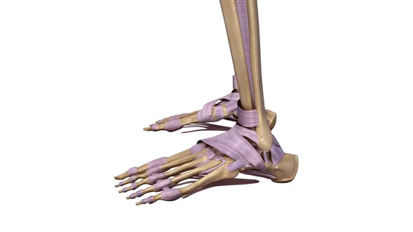
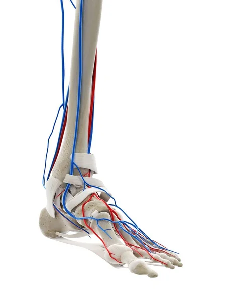
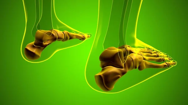
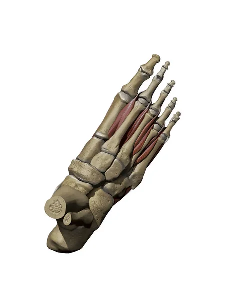
Skeleton foot with ligaments
Skeleton foot with ligaments — Stock Photo, Image
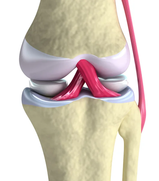
Knee joint closeup view. Isolated on white
Knee joint closeup view. Isolated on white — Stock Photo, Image
Clips. Team works
Clips. Team works — Stock Photo, Image
Female runner touching foot in pain
Female runner touching foot in pain — Stock Photo, Image
Woman Suffering From Knee Pain Sitting In Bed
Woman Suffering Knee Pain Sitting Bed — Stock Photo, Image
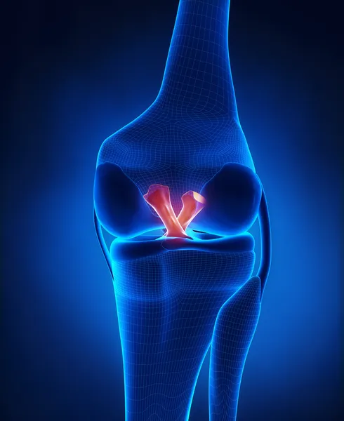
Anterior and Posterior Cruciate Ligament
Anterior and Posterior Cruciate Ligament — Stock Photo, Image
Painful Knee Close-up
Painful Knee Close-up — Stock Photo, Image
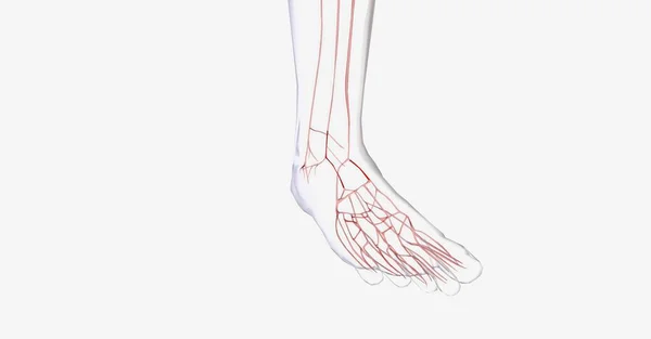
Blood vessels of foot, computer illustration
Blood Vessels Foot Computer Illustration — Stock Photo, Image
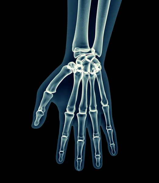
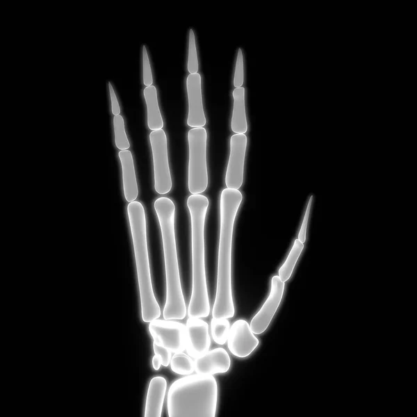
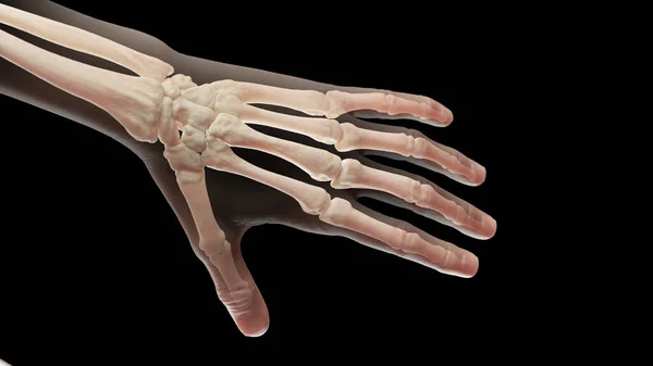
Bones of the hand, computer illustration
Bones Hand Computer Illustration — Stock Photo, Image
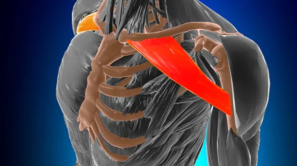
Coracobrachialis muscle. The deep muscles of the arm. Image on a black background.
Coracobrachialis muscle. The deep muscles of the arm. Image on a black background. — Stock Photo, Image
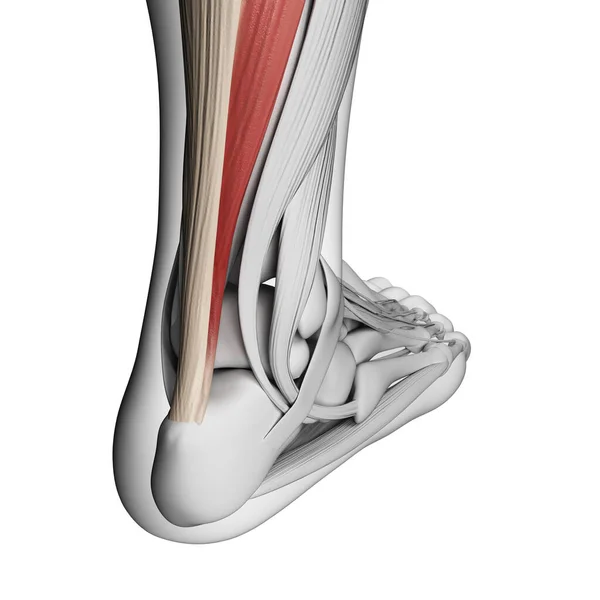
3d rendered illustration of the achilles tendon
Rendered Illustration Achilles Tendon — Stock Photo, Image
Closeup view of mother with baby and pregnant woman touching her belly on beige background, collage. Banner design
Closeup View Mother Baby Pregnant Woman Touching Her Belly Beige — Stock Photo, Image
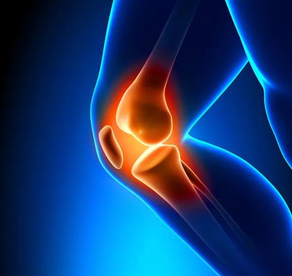
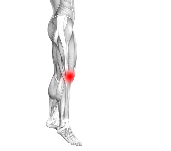
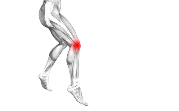
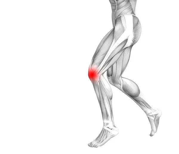
Conceptual knee human anatomy with red hot spot inflammation or articular joint pain for leg health care therapy or sport muscle concepts. 3D illustration man arthritis or bone osteoporosis disease
Conceptual knee human anatomy with red hot spot inflammation or articular joint pain for leg health care therapy or sport muscle concepts. 3D illustration man arthritis or bone osteoporosis disease — Stock Photo, Image
Focus of inflammation localized in shoulder joint
Focus of inflammation localized in shoulder joint — Stock Photo, Image
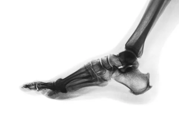
X-ray normal human foot . Lateral view . Invert color style
X-ray normal human foot . Lateral view . Invert color style — Stock Photo, Image
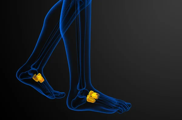
3d render medical illustration of the midfoot bone
3d render medical illustration of the midfoot bone — Stock Photo, Image
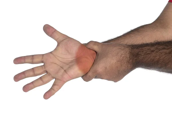
Acute pain in a woman hand isolated on white background. Clipping path on white background..
Acute pain in a woman hand isolated on white background. Clipping path on white background.. — Stock Photo, Image
Anatomy of human forearm muscles with labels
Anatomy Human Forearm Muscles Labels — Stock Photo, Image
Vascular system in my human foot. 3D rendering
Vascular System Human Foot Rendering — Stock Photo, Image
Blood vessels of foot, computer illustration
Blood Vessels Foot Computer Illustration — Stock Photo, Image
Clavicular part of deltoid Anatomy For Medical Concept 3D Illustration
Clavicular Part Deltoid Anatomy Medical Concept Illustration — Stock Photo, Image
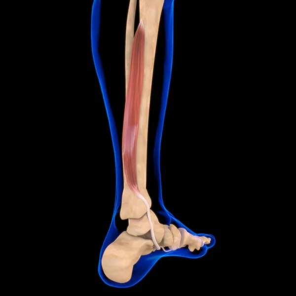
Lower arm musculature
Lower arm musculature — Stock Photo, Image
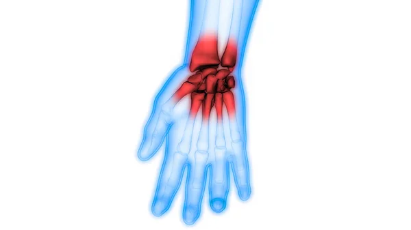
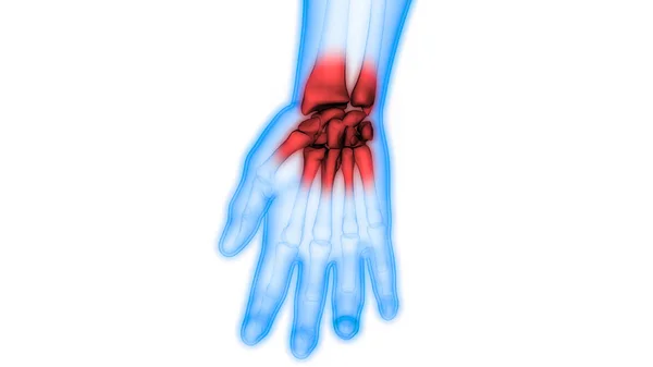
Human Body Bone Joint Pains (Hand Joint) - Illustration
Human Body Bone Joint Pains Hand Joint Illustration — Stock Photo, Image
3d illustration of bones, human organs system anatomy banner
Illustration Bones Human Organs System Anatomy Banner — Stock Photo, Image
Conceptual knee human anatomy with red hot spot inflammation or articular joint pain for leg health care therapy or sport muscle concepts. 3D illustration man arthritis or bone osteoporosis disease
Conceptual Knee Human Anatomy Red Hot Spot Inflammation Articular Joint — Stock Photo, Image
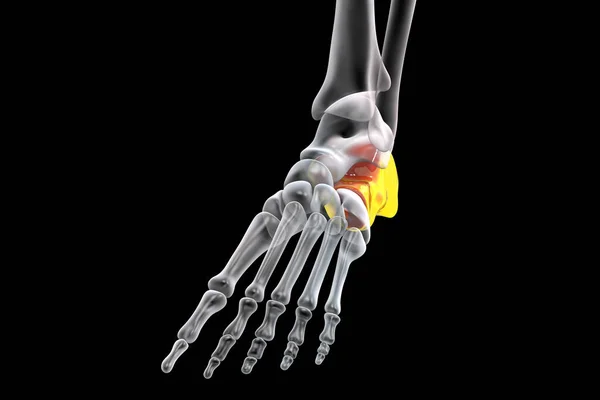
Illustration of the calcaneus, or heel, bone of the foot.
Illustration Calcaneus Heel Bone Foot — Stock Photo, Image
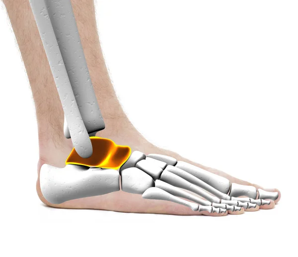
Talus Anklebone Astragalus Bones - Anatomy Male - Studio photo i
Talus Anklebone Astragalus Bones - Anatomy Male - Studio photo i — Stock Photo, Image
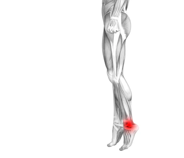
Conceptual ankle human anatomy with red hot spot inflammation or articular joint pain for leg health care therapy or sport muscle concepts. 3D illustration man arthritis or bone osteoporosis disease
Conceptual Ankle Human Anatomy Red Hot Spot Inflammation Articular Joint — Stock Photo, Image
Illustration of wrist replacement on black background.
Illustration Wrist Replacement Black Background — Stock Photo, Image
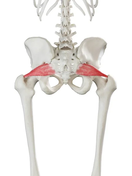
Human skeleton model with detailed Piriformis muscle, digital illustration.
Human Skeleton Model Detailed Piriformis Muscle Digital Illustration — Stock Photo, Image
Illustration of human knee anatomy on white background.
Illustration Human Knee Anatomy White Background — Stock Photo, Image
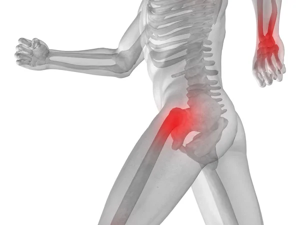
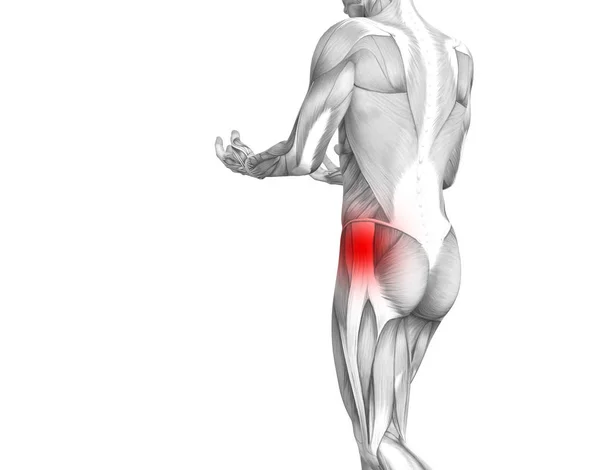
Conceptual hip human anatomy with red hot spot inflammation articular joint pain for leg health care therapy or sport muscle concepts. 3D illustration man arthritis or bone sore osteoporosis disease
Conceptual hip human anatomy with red hot spot inflammation articular joint pain for leg health care therapy or sport muscle concepts. 3D illustration man arthritis or bone sore osteoporosis disease — Stock Photo, Image
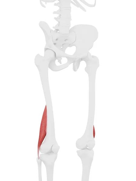
Human skeleton part with detailed red Short biceps femoris muscle, digital illustration.
Human Skeleton Part Detailed Red Short Biceps Femoris Muscle Digital — Stock Photo, Image
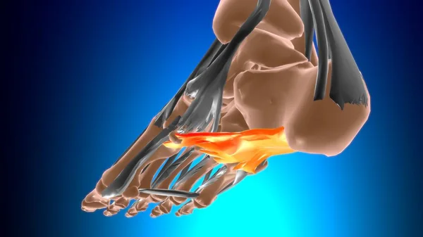
Long plantar ligament Anatomy For Medical Concept 3D Illustration
Long Plantar Ligament Anatomy Medical Concept Illustration — Stock Photo, Image
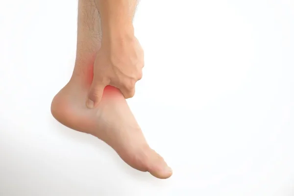
Man with foot pain and copyspace
Man Foot Pain Copyspace — Stock Photo, Image
Bones of the left hand, illustration.
Bones Left Hand Illustration — Stock Photo, Image
Anatomy of the nerves of the upper limb (arm) in palmar view.
Anatomy Nerves Upper Limb Arm Palmar View — Stock Photo, Image
Conceptual knee human anatomy with red hot spot inflammation or articular joint pain for leg health care therapy or sport muscle concepts. 3D illustration man arthritis or bone osteoporosis disease
Conceptual Knee Human Anatomy Red Hot Spot Inflammation Articular Joint — Stock Photo, Image
Adductor magnu Muscle Anatomy For Medical Concept 3D Illustration
Adductor Magnu Muscle Anatomy Medical Concept Illustration — Stock Photo, Image
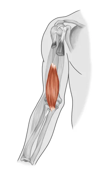
Human arm musculature
Human arm musculature — Stock Photo, Image
Bones of foot, computer illustration
Bones Foot Computer Illustration — Stock Photo, Image
Back view of human lumbar vertebra 3d render
Back View Human Lumbar Vertebra Render — Stock Photo, Image
The muscles of the upper right limb in anterior view.
Muscles Upper Right Limb Anterior View — Stock Photo, Image
X-ray image of a man isolated on black
X-ray image of a man isolated on black — Stock Photo, Image
Medical illustration of the palmaris longus
Medical Illustration Palmaris Longus — Stock Photo, Image
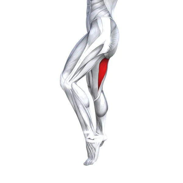
Human upper legs anatomy
Human upper legs anatomy — Stock Photo, Image
Ankle with exercise injuries.
Ankle Exercise Injuries — Stock Photo, Image
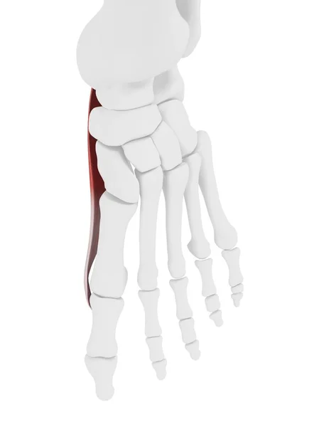
Human skeleton with red colored Abductor hallucis muscle, computer illustration.
Human Skeleton Red Colored Abductor Hallucis Muscle Computer Illustration — Stock Photo, Image
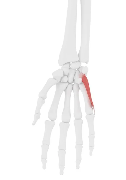
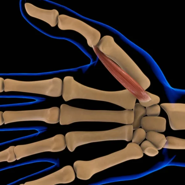
Human skeleton part with detailed Opponens digiti minimi muscle, digital illustration.
Human Skeleton Part Detailed Opponens Digiti Minimi Muscle Digital Illustration — Stock Photo, Image
Anatomical Concept 3D illustration
Anatomical Concept 3D illustration — Stock Photo, Image
Calcaneus bone of the foot. Heel bone, the largest of the tarsal bones in the foot. Human foot anatomy. 3D illustration
Calcaneus Bone Foot Heel Bone Largest Tarsal Bones Foot Human — Stock Photo, Image
Conceptual knee human anatomy with red hot spot inflammation or articular joint pain for leg health care therapy or sport muscle concepts. 3D illustration man arthritis or bone osteoporosis disease
Conceptual Knee Human Anatomy Red Hot Spot Inflammation Articular Joint — Stock Photo, Image
3D Illustration, Muscle is a soft tissue, Muscle cells contain proteins , producing a contraction that changes both the length and the shape of the cell. Muscles function to produce force and motion.
Illustration Muscle Soft Tissue Muscle Cells Contain Proteins Producing Contraction — Stock Photo, Image
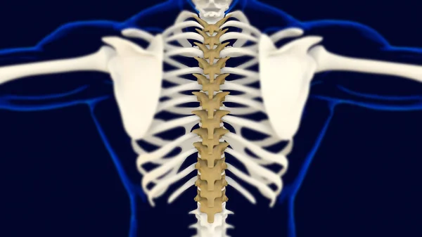
Thoracic Vertebrae Anatomy for medical concept 3D Illustration
Thoracic Vertebrae Anatomy Medical Concept Illustration — Stock Photo, Image
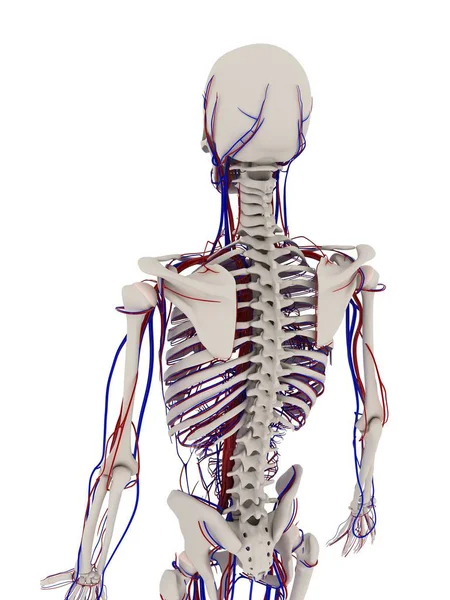
Skeletal system and cardiovascular system
Skeletal system and cardiovascular system — Stock Photo, Image
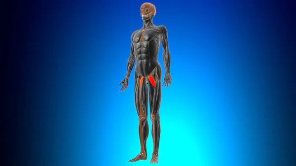
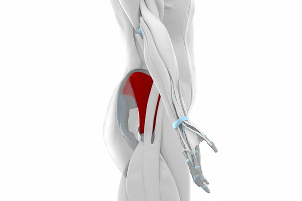
Pectineus Muscle Anatomy For Medical Concept 3D Illustration
Pectineus Muscle Anatomy Medical Concept Illustration — Stock Photo, Image
Shoulder and the bones that constitute it: the scapula, the humerus. .
Shoulder Bones Constitute Scapula Humerus — Stock Photo, Image
MRI L-S spine myelogram
MRI L-S  spine myelogram — Stock Photo, Image
Illustration of human anatomy
Illustration of human anatomy — Stock Photo, Image
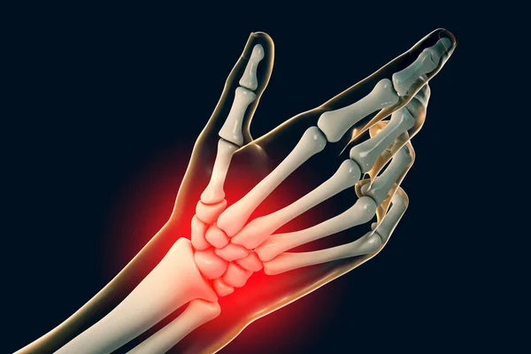
Illustration of a painful wrist.
Illustration Painful Wrist — Stock Photo, Image
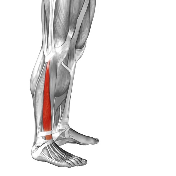
Illustration human lower leg anatomy
Illustration human lower leg anatomy — Stock Photo, Image
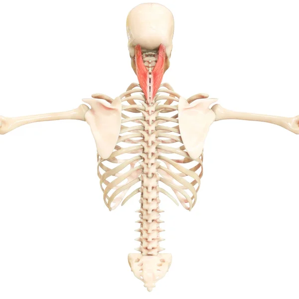
Human Muscular System Torso Muscles Capitus Muscle Anatomy. 3D
Human Muscular System Torso Muscles Capitus Muscle Anatomy — Stock Photo, Image
Biceps femoris Muscle Anatomy For Medical Concept 3D Illustration
Biceps Femoris Muscle Anatomy Medical Concept Illustration — Stock Photo, Image
Biceps, Brachii, Brachioradial, Brachioradial muscles - Black White
Biceps, Brachii, Brachioradial, Brachioradial muscles - Black White — Stock Photo, Image
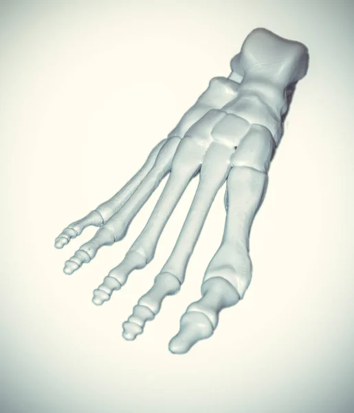
White prototype of the human foot skeleton printed on 3d printer
White prototype of the human foot skeleton printed on 3d printer — Stock Photo, Image
Human Body Bone Joint Pains Anatomy (Palm Hand). 3D
Human Body Bone Joint Pains Anatomy Palm Hand — Stock Photo, Image
3D Illustration, Muscle is a soft tissue, Muscle cells contain proteins , producing a contraction that changes both the length and the shape of the cell. Muscles function to produce force and motion.
Illustration Muscle Soft Tissue Muscle Cells Contain Proteins Producing Contraction — Stock Photo, Image
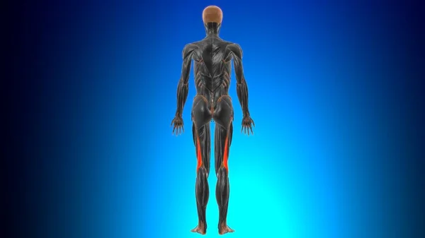
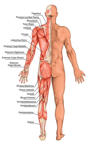
Anatomical board, male anatomy, man's anatomical body, human muscular system, surface anatomy, body shapes, posterior view, full body
Anatomical board, male anatomy, man's anatomical body, human muscular system, surface anatomy, body shapes, posterior view, full body — Stock Photo, Image
Human skeleton part with detailed Interosseous dorsal muscle, digital illustration.
Human Skeleton Part Detailed Interosseous Dorsal Muscle Digital Illustration — Stock Photo, Image
Conceptual human anatomy with red inflammation
Conceptual Human Anatomy Red Inflammation — Stock Photo, Image
Fantasy chaotic colorful fractal pattern. Abstract fractal shapes. 3D rendering illustration background or wallpaper.
Fantasy Chaotic Colorful Fractal Pattern Abstract Fractal Shapes Rendering Illustration — Stock Photo, Image
Biceps, Brachii, Brachioradial, Brachialis muscles - Black White
Biceps, Brachii, Brachioradial, Brachialis muscles - Black White — Stock Photo, Image
Human Body Bones Joint Pains (Feet joints and Bones). 3D - Illustration
Human Body Bones Joint Pains Feet Joints Bones Illustration — Stock Photo, Image
Human Skeleton Foot bones Anatomy For Medical Concept 3D Illustration
Human Skeleton Foot Bones Anatomy Medical Concept Illustration — Stock Photo, Image
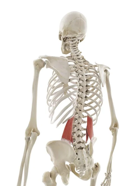
Quadratus lumborum muscle, computer illustration
Quadratus Lumborum Muscle Computer Illustration — Stock Photo, Image
Human Skeleton System Bones (Hands) Anatomy. 3D
Human Skeleton System Bones Hands Anatomy — Stock Photo, Image
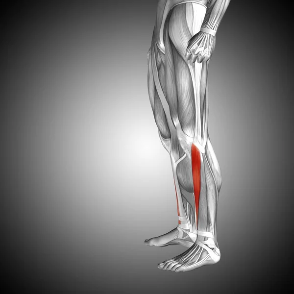
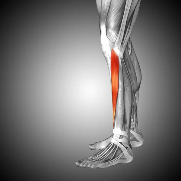
Human lower legs anatomy
Human lower legs anatomy — Stock Photo, Image
Medial head of gastrocnemius Muscle Anatomy For Medical Concept 3D Illustration
Medial Head Gastrocnemius Muscle Anatomy Medical Concept Illustration — Stock Photo, Image
3d render illustration of the talus bone
3d render illustration of the talus bone — Stock Photo, Image
Human Skeleton System Bone Joints Anatomy. 3D
Human Skeleton System Bone Joints Anatomy — Stock Photo, Image
Concept conceptual 3D illustration fit strong back lower leg human anatomy, anatomical muscle isolated white background for body medical health tendon foot and biological gym fitness muscular system
Concept Conceptual Illustration Fit Strong Back Lower Leg Human Anatomy — Stock Photo, Image
Nerves of foot, computer illustration
Nerves Foot Computer Illustration — Stock Photo, Image
Anatomy of hand, computer illustration
Anatomy Hand Computer Illustration — Stock Photo, Image
Medical accurate 3d illustration of the hand bones
Medical accurate 3d illustration of the hand bones — Stock Photo, Image
Knee ligaments, tendons, x-ray
Knee ligaments, tendons, x-ray — Stock Photo, Image
Young man suffering from pain in leg on white background
Young Man Suffering Pain Leg White Background — Stock Photo, Image
Gluteus minimus Muscles
Gluteus minimus Muscles — Stock Photo, Image
Illustration of fingers bones in human skeleton.
Illustration Fingers Bones Human Skeleton — Stock Photo, Image
Conceptual knee human anatomy with red hot spot inflammation or articular joint pain for leg health care therapy or sport muscle concepts
Conceptual Knee Human Anatomy Red Hot Spot Inflammation Articular Joint — Stock Photo, Image
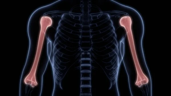
Humerus Bone Joints of Human Skeleton System Anatomy 3d rendering
Humerus Bone Joints Human Skeleton System Anatomy Rendering — Stock Photo, Image
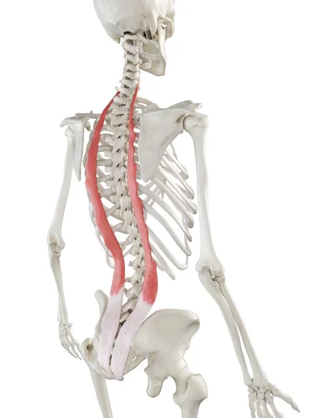
Human skeleton with red colored Iliocostalis muscle, computer illustration.
Human Skeleton Red Colored Iliocostalis Muscle Computer Illustration — Stock Photo, Image
Close up joint x-ray
Close up joint  x-ray — Stock Photo, Image
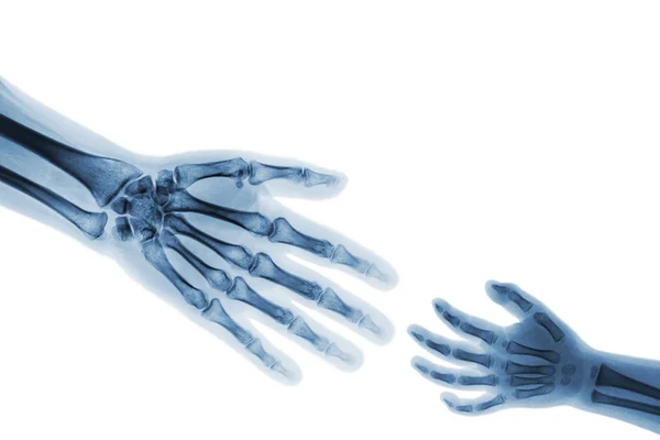
Adult help child .
Adult help child . — Stock Photo, Image
Foot with dorsal muscles and bone structures
Foot Dorsal Muscles Bone Structures — Stock Photo, Image
Man with wrist pain
Man with wrist pain — Stock Photo, Image
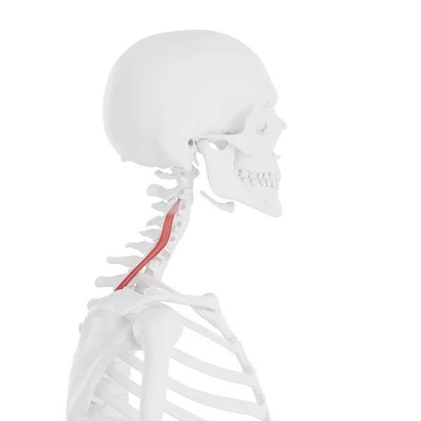
Human skeleton with red colored Longissimus cervicis muscle, digital illustration.
Human Skeleton Red Colored Longissimus Cervicis Muscle Digital Illustration — Stock Photo, Image
3D Illustration, Muscle is a soft tissue, Muscle cells contain proteins , producing a contraction that changes both the length and the shape of the cell. Muscles function to produce force and motion.
Illustration Muscle Soft Tissue Muscle Cells Contain Proteins Producing Contraction — Stock Photo, Image
Iliotibial tract or band Anatomy For Medical Concept 3D Illustration
Iliotibial Tract Band Anatomy Medical Concept Illustration — Stock Photo, Image

Explore High-Quality Ligament Images for Your Medical Projects

When it comes to medical projects and presentations, visual aids are essential in conveying the right message. Ligament images are particularly useful in explaining injuries, surgeries, and medical procedures. We offer a vast collection of top-quality ligament images that are perfect for medical professionals, students, and researchers.

Types of Ligament Images

Our collection consists of a wide range of ligament images, including anatomical illustrations and 3D renderings. These images can be useful in demonstrating the different types of ligaments in the body, their functions, and the injuries that can result in their damage.

Where to Use Ligament Images

Ligament images can be used in a variety of medical projects, such as presentations, brochures, textbooks, and websites. Medical professionals can use these images to discuss injuries and treatment options with their patients, while students can use them in their research papers and presentations.

Choosing the Right Ligament Image

Choosing the right ligament image for your project is crucial in conveying the right message. It's important to select an image that is relevant to the topic and accurately represents the information you're trying to convey. For instance, if you're discussing an injury to the knee ligament, you'll need an image that specifically shows the knee area.

Moreover, you should consider the quality of the image, its resolution, and the format. We offer high-resolution JPG, AI, and EPS files that can be easily customized to fit your project's needs.

Get the Best Ligament Images for Your Project

Our collection of ligament images is perfect for medical professionals, students, and researchers who require top-quality visuals to emphasize their message. With our vast collection of anatomical illustrations and 3D renderings, you're sure to find the perfect image for your project.

So why wait? Browse our collection today and get the best ligament images for your next medical project!