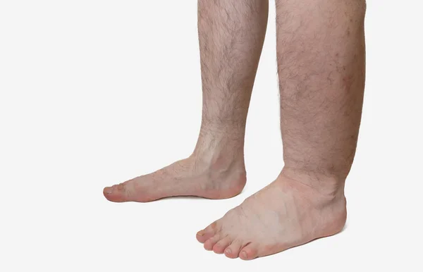
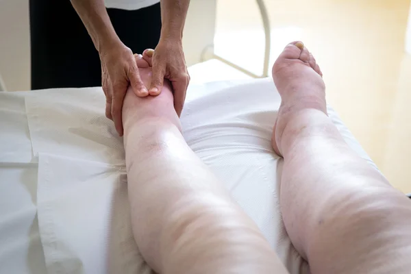
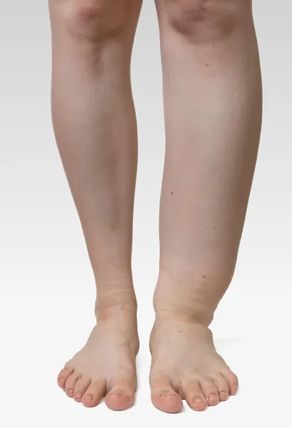
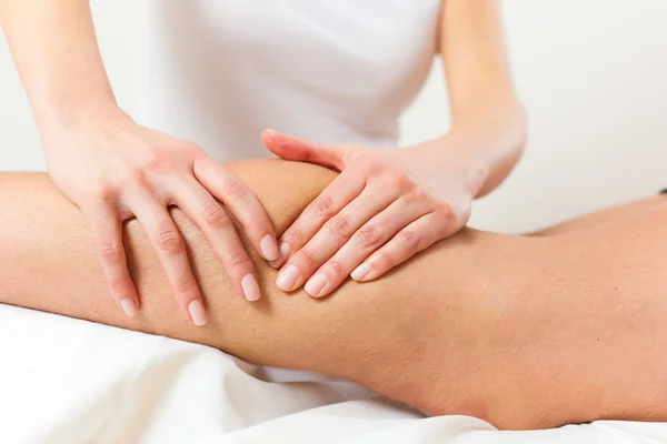
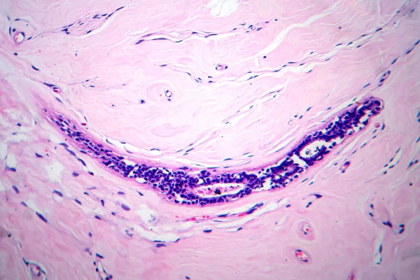
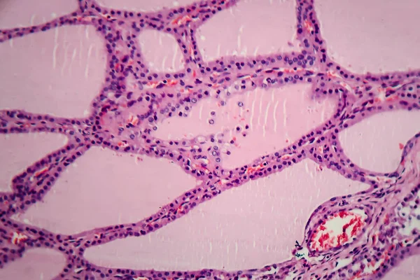
Leg of diseased patient who suffers from Edema.
Leg of diseased patient who suffers from Edema. — Stock Photo, Image
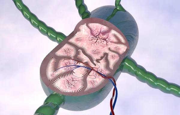
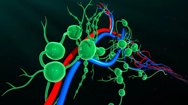
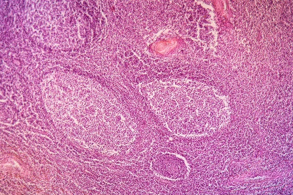
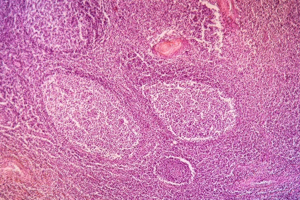
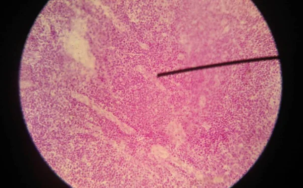
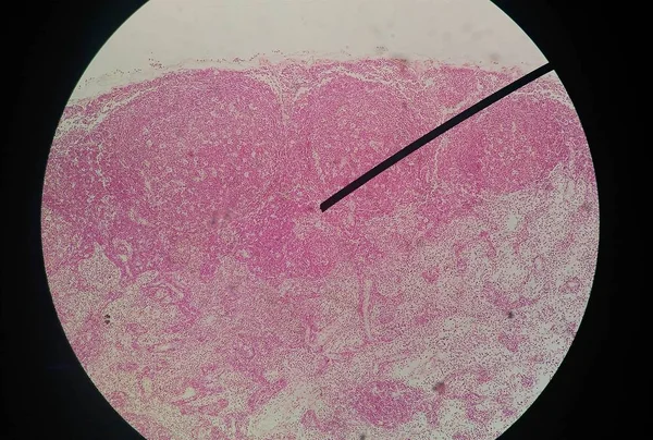
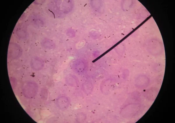
Lymph node section
Lymph node section — Stock Photo, Image
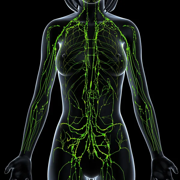
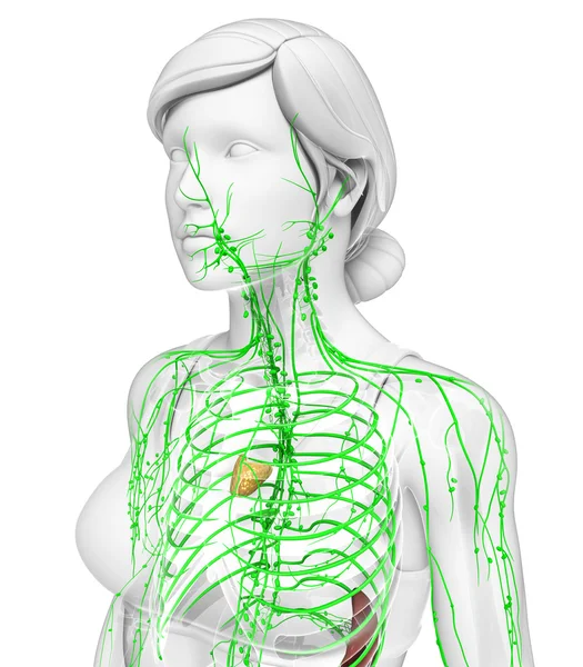
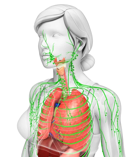
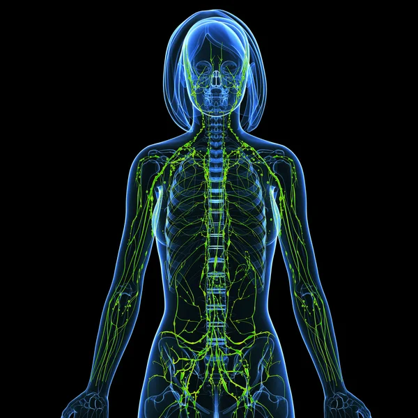
3d art illustration of lymphatic system of female
3d art illustration of lymphatic system of female — Stock Photo, Image
Lymph system amatomy
Lymph system amatomy — Stock Photo, Image
Lymph nodes in organism
Lymph nodes in organism — Stock Photo, Image
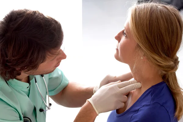
Doctor checking lymph nodes
Doctor checking lymph nodes — Stock Photo, Image
Lymphatic system of female body
Lymphatic system of  female body — Stock Photo, Image
Male doctor checks little girl lymph nodes
Male doctor checks little girl lymph nodes — Stock Photo, Image
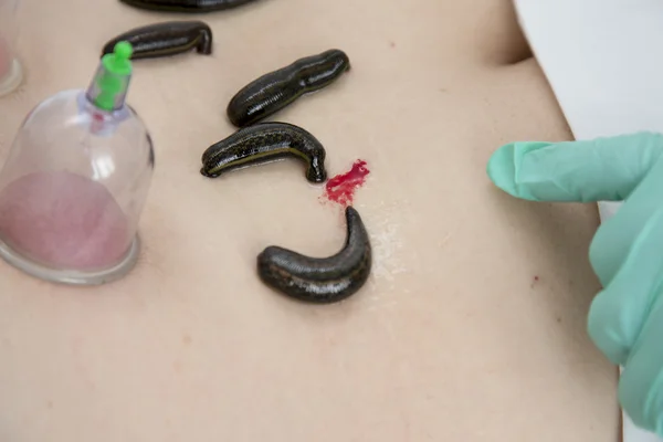
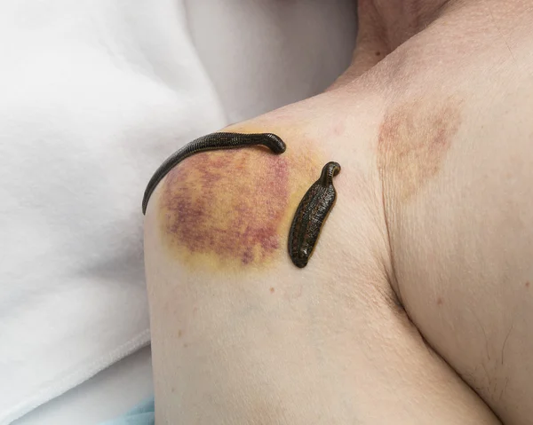
Treatment of people of medical leeches.
Treatment of people of medical leeches. — Stock Photo, Image
Doctor is showing Edema diagnosis on tablet. View from top.
Doctor is showing Edema diagnosis on tablet. View from top. — Stock Photo, Image
Pediatrician checking a baby
Pediatrician checking a baby — Stock Photo, Image
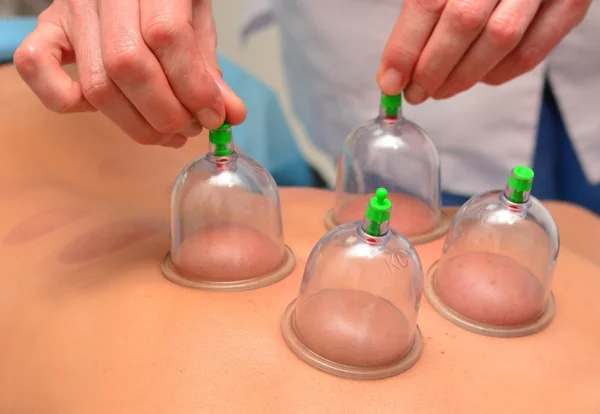
Cupping therapy, woman doctor removes cup from the patient's back
Cupping therapy, woman doctor removes cup from the patient's back — Stock Photo, Image
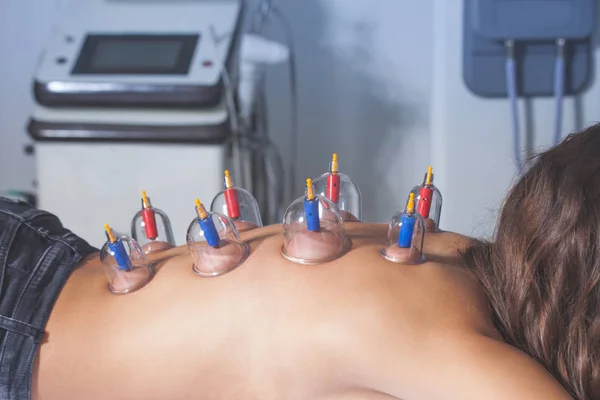
Woman on vacuum cupping therapy
Woman on vacuum cupping therapy — Stock Photo, Image
Women doing Fitness on Mini Trampoline
Women doing Fitness on Mini Trampoline — Stock Photo, Image
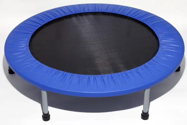
Low impact exercise equipment you can bounce on used to increase lymph flow, tone muscles, and more.
Low Impact Exercise Equipment You Can Bounce Used Increase Lymph — Stock Photo, Image
Checking lymph nodes
Checking lymph nodes — Stock Photo, Image
Thyroid Disease - Text on Chalkboard. 3D Illustration.
Thyroid Disease - Text on Chalkboard. 3D Illustration. — Stock Photo, Image
Treatment of medical leeches people and acupuncture.
Treatment of medical leeches people and acupuncture. — Stock Photo, Image
WOMAN PALPATING LYMPH NODE
WOMAN PALPATING LYMPH NODE — Stock Photo, Image
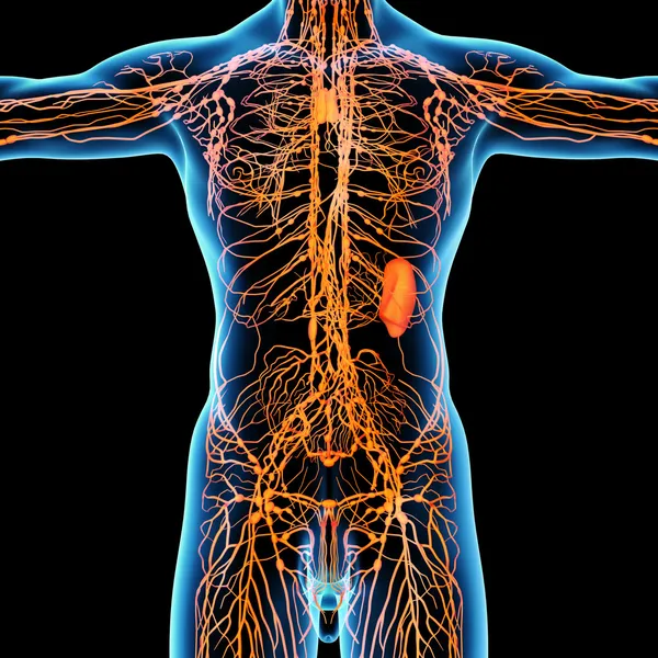
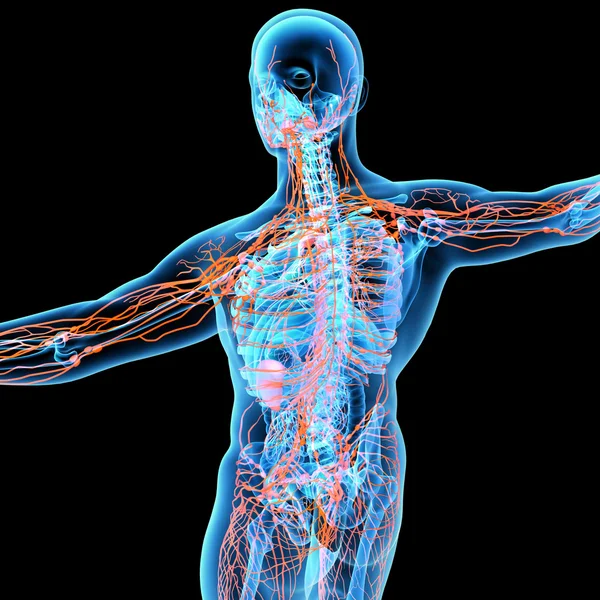
3d art illustration of lymphatic system of male
3d art illustration of lymphatic system of male — Stock Photo, Image
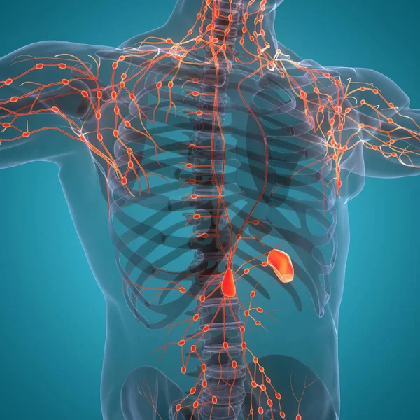
Human Internal system Lymph Nodes Anatomy. 3D
Human Internal System Lymph Nodes Anatomy — Stock Photo, Image
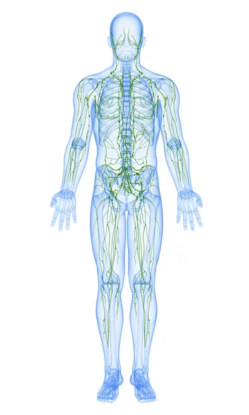
Lymphatic system - front view
Lymphatic system - front view — Stock Photo, Image
Lymph nodes anatomy
Lymph nodes anatomy — Stock Photo, Image
Woman doing lymphatic drainage massage
Woman doing lymphatic drainage massage — Stock Photo, Image
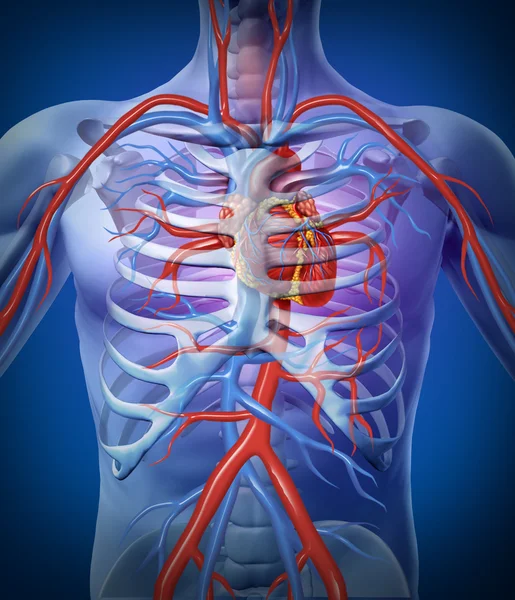
Human Heart Circulation In a Skeleton
Human Heart Circulation In a Skeleton — Stock Photo, Image
Female muscles anatomy
Female muscles anatomy — Stock Photo, Image
Treatment of people of medical leeches.
Treatment of people of medical leeches. — Stock Photo, Image
Lymphoma background concept
Lymphoma background concept — Stock Photo, Image
Businessman showing paper
Businessman showing paper — Stock Photo, Image
Beautiful young woman touching her face and neck
Beautiful young woman touching her face and neck — Stock Photo, Image
Thyroid function examination
Thyroid function examination — Stock Photo, Image
Legs pressotherapy machine on woman patient in hospital
Legs pressotherapy machine on woman patient in hospital — Stock Photo, Image
Woman visit a doctor
Woman visit a doctor — Stock Photo, Image
Doctor osteopath massage therapist is doing massage to a little boy. Hands massage the back and cervical-collar zone. Close-up view without face. Mom gives her baby a massage.
Doctor Osteopath Massage Therapist Doing Massage Little Boy Hands Massage — Stock Photo, Image
Treatment of medical leeches people and acupuncture.
Treatment of medical leeches people and acupuncture. — Stock Photo, Image
Lymph node section
Lymph node section — Stock Photo, Image
Young woman having her head massaged at the beauty salon
Young Woman Having Her Head Massaged Beauty Salon — Stock Photo, Image
3d render medical illustration of the lymphatic system
3d render medical illustration of the lymphatic system — Stock Photo, Image
Lymphatic system of female with half body
Lymphatic system of female with half body — Stock Photo, Image
Male doctor checking lymph nodes of pregnant woman
Male doctor checking lymph nodes of pregnant woman — Stock Photo, Image
Young woman having her head massaged at the beauty salon
Young Woman Having Her Head Massaged Beauty Salon — Stock Photo, Image
Hand writing Lymphedema with marker
Hand writing Lymphedema with marker — Stock Photo, Image
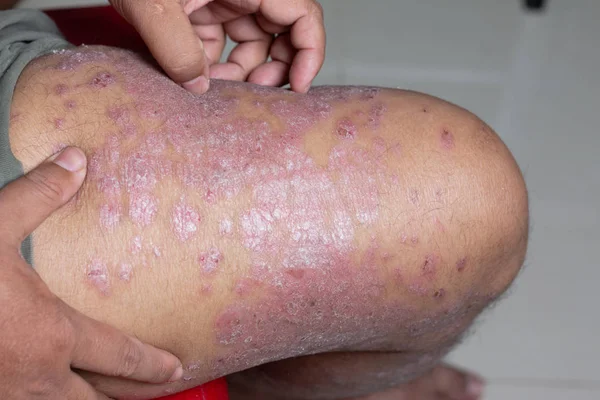
Men are using his hands to scratch the leg ulcers. Diseases caused by abnormalities of the lymph. Psoriasis is a skin disease. Select focus shallow depth of field.
Men Using His Hands Scratch Leg Ulcers Diseases Caused Abnormalities — Stock Photo, Image
Woman bandaging patient's legs
Woman bandaging patient's legs — Stock Photo, Image
Leg of diseased patient who suffers from Edema.
Leg of diseased patient who suffers from Edema. — Stock Photo, Image
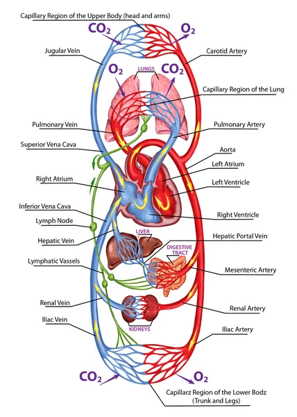
Human bloodstream - didactic board of anatomy of blood system of human circulation, sanguine and cardiovascular system
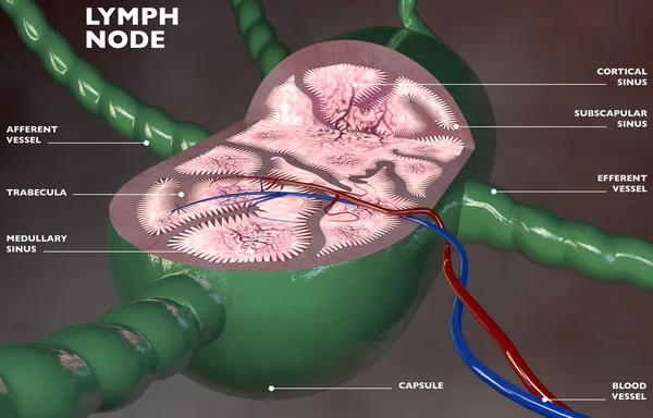
Human bloodstream - didactic board of anatomy of blood system of human circulation, sanguine and cardiovascular system — Stock Photo, Image

Sick woman touching her lymph nodes at home
Sick woman touching her lymph nodes at home — Stock Photo, Image
Lymphatic and digestive system of Female body artwork
Lymphatic and digestive system of Female body artwork — Stock Photo, Image
Senior man hand holding he neck and massaging in pain area.
Senior man hand holding he neck and massaging in pain area. — Stock Photo, Image
Treatment with leeches shoulder and neck area, back area in the
Treatment with leeches shoulder and neck area, back area in the — Stock Photo, Image
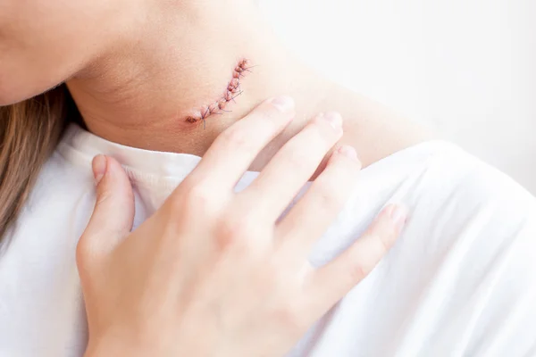
Scar mole on woman's neck
Scar mole on woman's neck — Stock Photo, Image
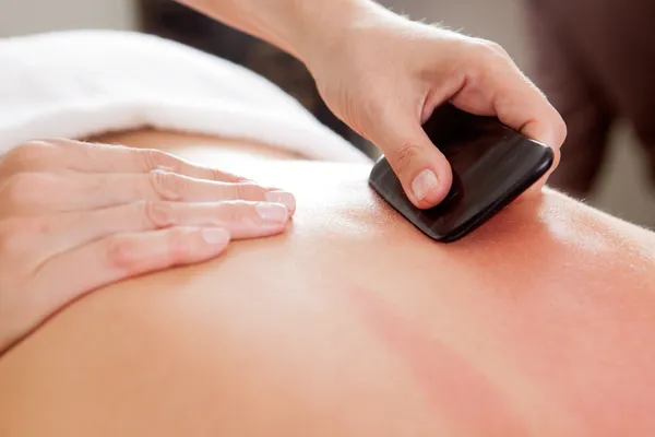
Gua-Sha Acupuncture Treatment on Back
Gua-Sha Acupuncture Treatment on Back — Stock Photo, Image
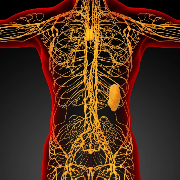
Lymphatic system - back view
Lymphatic system - back view — Stock Photo, Image
Patient at the physiotherapy - massage
Patient at the physiotherapy - massage — Stock Photo, Image
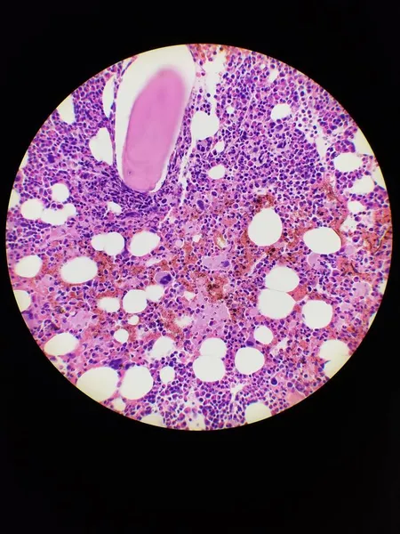
Bone marrow biopsy H and E stain showing eosinophilia
Bone marrow biopsy H and E stain showing eosinophilia — Stock Photo, Image
Microbiology, microscopic cells surface
Microbiology Microscopic Cells Surface — Stock Photo, Image
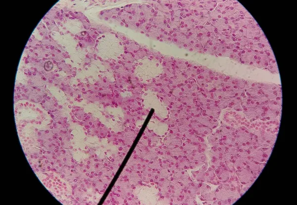
Close up human cells with microscope.
Close up human cells with microscope. — Stock Photo, Image
Inflamed tissue under the microscope 100x
Inflamed Tissue Microscope 100X — Stock Photo, Image
Microscopic cells texture, abstract background
Microscopic Cells Texture Abstract Background — Stock Photo, Image
Endometrial adenocarcinoma, light micrograph, photo under microscope
Endometrial Adenocarcinoma Light Micrograph Photo Microscope — Stock Photo, Image
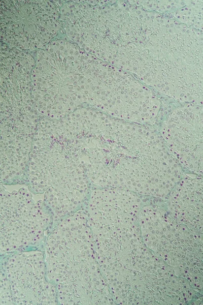
Testicle cross-section under the microscope 100x
Testicle Cross Section Microscope 100X — Stock Photo, Image
Cells of human find with microscope.
Cells of human find with microscope. — Stock Photo, Image
Cells of human find with microscope.
Cells of human find with microscope. — Stock Photo, Image
The image of the wall, for use as a background.
Image Wall Use Background — Stock Photo, Image
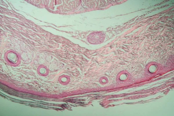
Tail of the rat tissue across 100x
Tail Rat Tissue 100X — Stock Photo, Image
Photomicrograph of fibroadenoma, showcasing benign glandular and fibrous tissue growth within the breast, a common noncancerous tumor.
Photomicrograph Fibroadenoma Showcasing Benign Glandular Fibrous Tissue Growth Breast Common — Stock Photo, Image
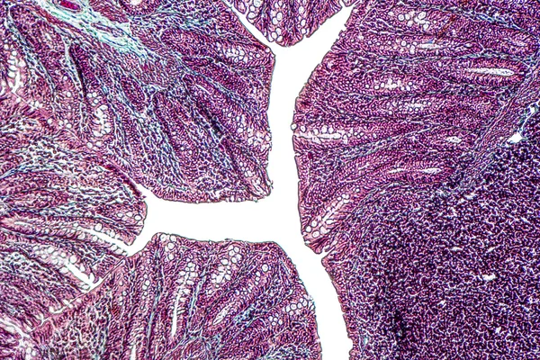
Close up human cells with microscope.
Close up human cells with microscope. — Stock Photo, Image
Microbiology, microscopic cells surface
Microbiology Microscopic Cells Surface — Stock Photo, Image
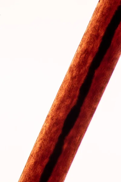
Characteristics of Hair cell of human under microscope view for education in laboratory.
Characteristics Hair Cell Human Microscope View Education Laboratory — Stock Photo, Image
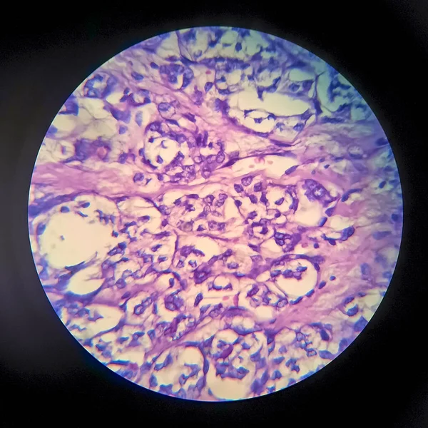
Thyroid follicular carcinoma, light micrograph, photo under microscope
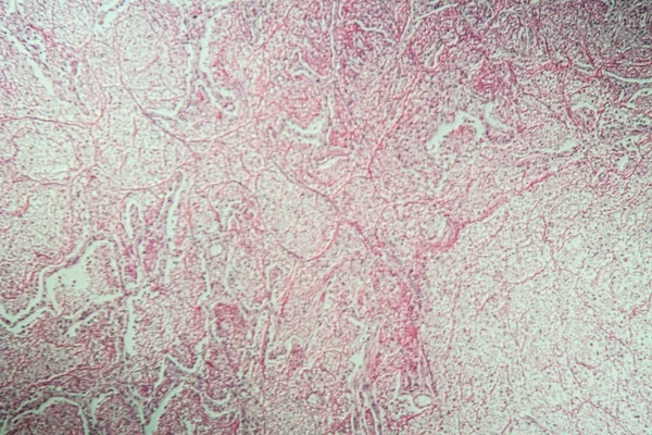
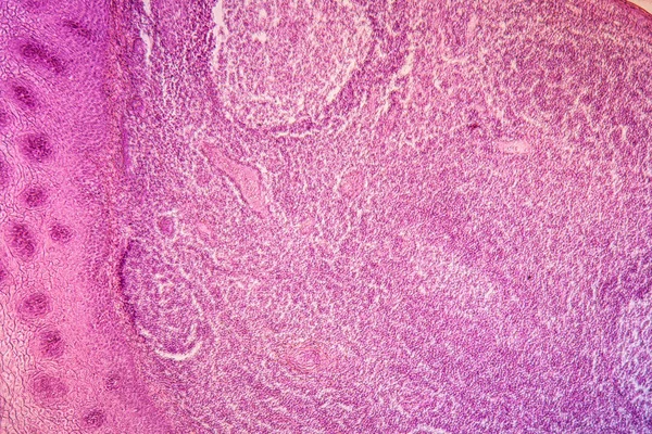
Thyroid Follicular Carcinoma Light Micrograph Photo Microscope — Stock Photo, Image
Background made of paper. Abstract background. Texture. Mixing colors. Color mix
Background Made Paper Abstract Background Texture Mixing Colors Color Mix — Stock Photo, Image
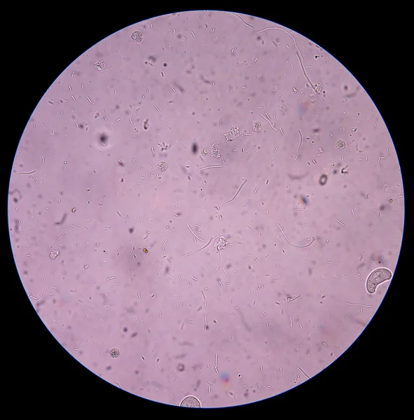
Bacteria in urine
Bacteria in urine — Stock Photo, Image
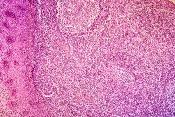
Spinal ganglion from microscopy
Spinal Ganglion Microscopy — Stock Photo, Image
Salty pink lake.
Salty pink lake. — Stock Photo, Image
Abstract backdrop in pink and black tones in grunge style, colorful background for poster, site, interior design, stickers,
Abstract Backdrop Pink Black Tones Grunge Style Colorful Background Poster — Stock Photo, Image
Thyroid follicular carcinoma, light micrograph, photo under microscope
Thyroid Follicular Carcinoma Light Micrograph Photo Microscope — Stock Photo, Image
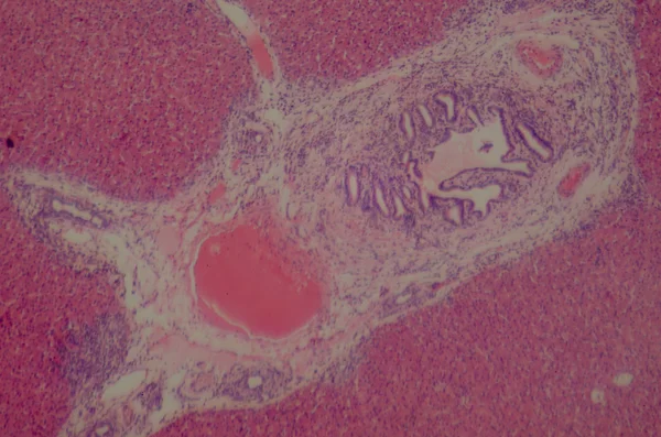
Microscopic section of human liver
Microscopic section of human liver — Stock Photo, Image
Thyroid follicular carcinoma, light micrograph, photo under microscope
Thyroid Follicular Carcinoma Light Micrograph Photo Microscope — Stock Photo, Image
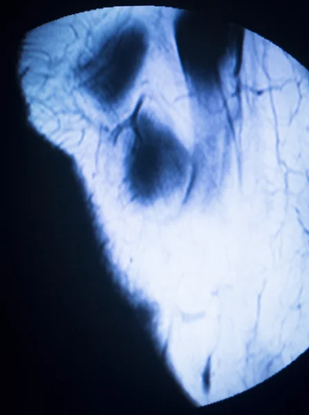
MRI magnetic resonance imaging medical scan
MRI magnetic resonance imaging medical scan — Stock Photo, Image
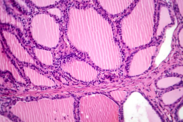
Endemic goiter, light micrograph, abnormal enlargement of the thyroid gland due to dietary iodine deficiency. Photomicrograph shows follicles of varying size, abundant colloid, lymphocytic infiltrate
Endemic Goiter Light Micrograph Abnormal Enlargement Thyroid Gland Due Dietary — Stock Photo, Image
Abstract ink background. Marble style. Black paint stroke texture on white paper. Wallpaper for web and game design. Grunge mud art. Macro image of pen juice. Dark Smear
Abstract Ink Background Marble Style Black Paint Stroke Texture White — Stock Photo, Image
Cells of human find with microscope.
Cells of human find with microscope. — Stock Photo, Image
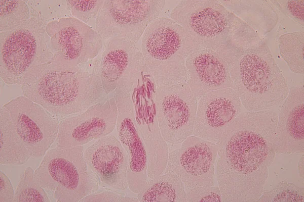
Root tip of Onion and Mitosis cell in the Root tip of Onion under a microscope.
Root Tip Onion Mitosis Cell Root Tip Onion Microscope — Stock Photo, Image
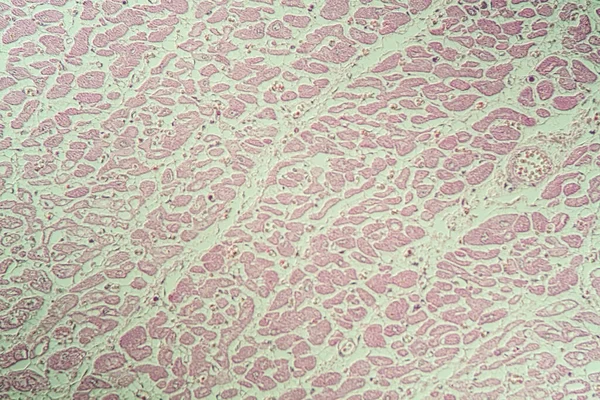
Inflamed heart muscle sick tissue under the microscope 200x
Inflamed Heart Muscle Sick Tissue Microscope 200X — Stock Photo, Image
Photomicrograph of a toxic goiter tissue sample under a microscope, revealing hypertrophy of thyroid follicular cells, increased vascularity, and colloid depletion.
Photomicrograph Toxic Goiter Tissue Sample Microscope Revealing Hypertrophy Thyroid Follicular — Stock Photo, Image
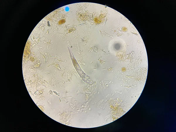
Demodex mange from a microscope view. The parasite causing a skin disease -Demodecosis.
Demodex Mange Microscope View Parasite Causing Skin Disease Demodecosis — Stock Photo, Image
Dramatic black, monochrome abstraction, reminiscent of dragon scales or caked lava, shimmering. Fragment of a modern artwork. Macro photography of the smallest details of a panting. Contemporary art. Artistic background image.
Dramatic Black Monochrome Abstraction Reminiscent Dragon Scales Caked Lava Shimmering — Stock Photo, Image
Cells in reproductive female cytology and histology concept.
Cells in reproductive female cytology and histology concept. — Stock Photo, Image
The study Plant tissue of under the microscope for classroom education.
Study Plant Tissue Microscope Classroom Education — Stock Photo, Image
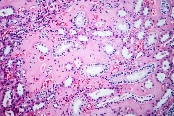
Histopathology of hypertensive renal disease, light micrograph, photo under microscope
Histopathology Hypertensive Renal Disease Light Micrograph Photo Microscope — Stock Photo, Image
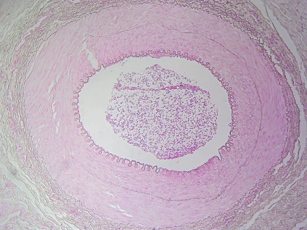
The Anatomy of Blood Vessels: Muscular Artery in High Detail
Anatomy Blood Vessels Muscular Artery High Detail — Stock Photo, Image
Palate tonsils inflammation diseased tissue 100x
Palate Tonsils Inflammation Diseased Tissue 100X — Stock Photo, Image
Actinomyces in the jaw Diseased tissue 200x
Actinomyces Jaw Diseased Tissue 200X — Stock Photo, Image
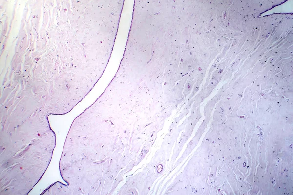
Uterus adenofibroma, light micrograph, photo under microscope. A rare benign tumor of the uterus composed of glandular and fibrous tissues
Uterus Adenofibroma Light Micrograph Photo Microscope Rare Benign Tumor Uterus — Stock Photo, Image
Schistosoma mansoni parasites under the microscope.
Schistosoma mansoni parasites under the microscope. — Stock Photo, Image
Tissue diseased with alcoholism 100x
Tissue Diseased Alcoholism 100X — Stock Photo, Image
Large intestine of a rat
Large intestine of a rat — Stock Photo, Image
Inflamed tissue under the microscope 100x
Inflamed Tissue Microscope 100X — Stock Photo, Image
Abstract surface, microbiology cells
Abstract Surface Microbiology Cells — Stock Photo, Image
Endemic goiter, light micrograph, abnormal enlargement of the thyroid gland due to dietary iodine deficiency. Photomicrograph shows follicles of varying size, abundant colloid, lymphocytic infiltrate
Endemic Goiter Light Micrograph Abnormal Enlargement Thyroid Gland Due Dietary — Stock Photo, Image

Find the Best Lymph Images for Your Next Project

Looking for lymph images to use in your medical projects? You've come to the right place. Our stock image collection offers a wide range of high-quality lymph-related images that can be used for a variety of purposes. With files in JPG, AI, and EPS formats, you'll find the perfect image to fit your needs.

Explore Our Collection

Our lymph image collection is extensive and includes a diverse range of imagery. Whether you need images of anatomical structures, medical procedures or diseases, we have them all. Our collection is continuously expanding to ensure that you have easy access to all the best images for your projects.

Our images are perfect for use in medical journals, textbooks, online articles, and presentations. Wherever your project takes you, we have the perfect image to help you communicate your message effectively and professionally.

Choose the Right Image for Your Project

Choosing the right image is essential for creating a professional and high-quality project. When picking an image, consider the message that you're trying to convey, the intended audience, and the context in which the image will be used. Using the right image can engage your audience and communicate your message more effectively.

Be sure to choose images that are high-quality, clear, and relevant to your project. Avoid using images with low resolutions or images that are unclear or difficult to interpret. Additionally, ensure the images are well-cited and that you have the necessary rights and permissions to use them in your project.

Get the Best Value for Your Money

Our lymph image collection is designed to provide you with the best value for your money. Our images are available for purchase individually or as part of a package, making it easy to get the images you need without breaking the bank. We also offer free previews before you make a purchase, so you can see exactly what you're getting.

With our high-quality images and affordable prices, you'll be able to take your project to the next level and impress your audience without spending a fortune.

Conclusion

Our collection of lymph images is the perfect source for a wide range of medical projects. With high-quality images available in a variety of formats and sizes, our collection is perfect for medical textbooks, articles, reports, presentations, and more.

Remember to choose images that are clear, relevant, and appropriate for your project, and be sure to cite them accurately. Don't settle for low-quality images or images that don't work for your project. Take advantage of our extensive lymph image collection and create the perfect project today!