Optic nerve Stock Photos

100,000 Optic nerve pictures are available under a royalty-free license

  • Best Match
  • Fresh
  • Popular
1-100 of 100,000
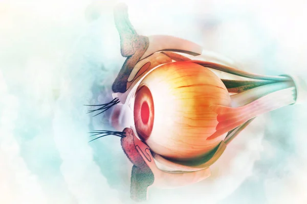
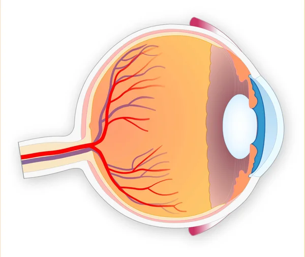
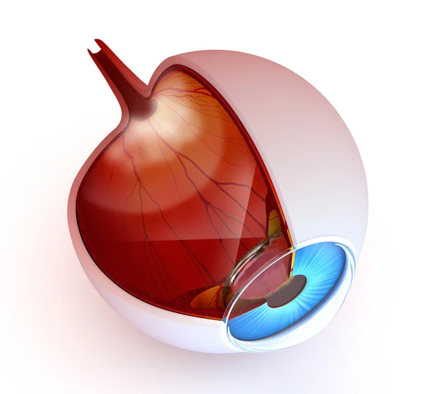
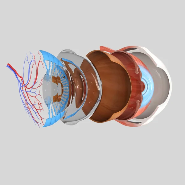
Human eye anatomy. Inner structure. 3d illustration
Human Eye Anatomy Inner Structure Illustration — Stock Photo, Image
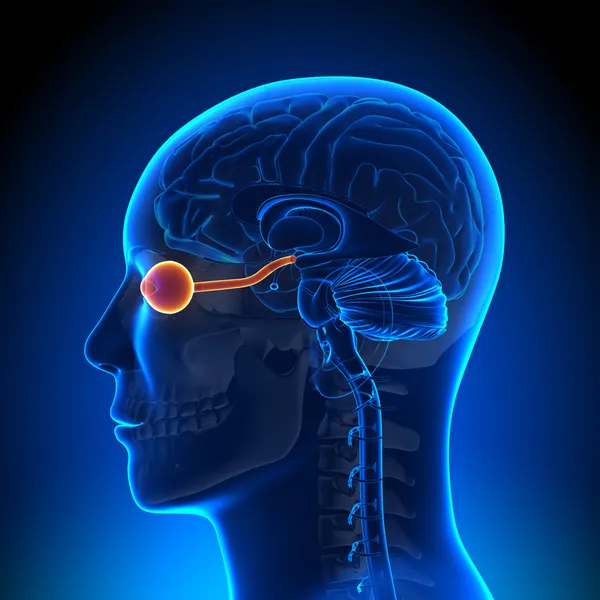
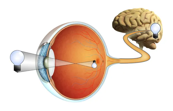
Brain Anatomy - Optical Nerve Eye
Brain Anatomy - Optical Nerve Eye — Stock Photo, Image
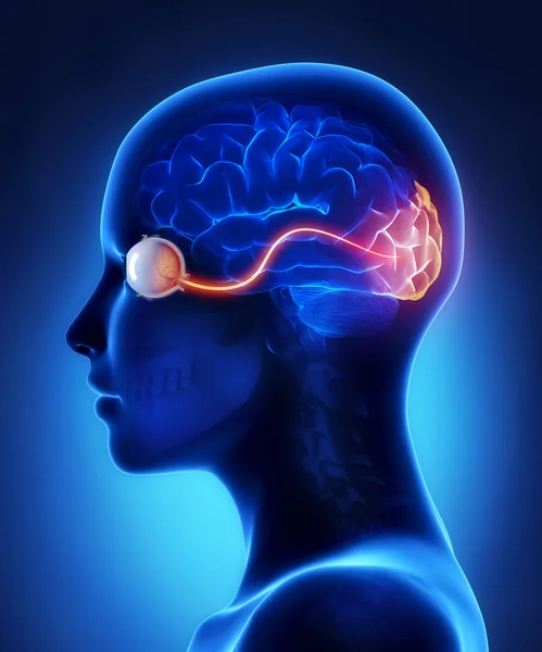
Eye and visual cortex nerves
Eye and visual cortex nerves — Stock Photo, Image
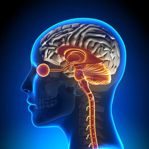
Brain Stem, Cerebellum, Optical Nerve, Female Brain Anatomy
Brain Stem, Cerebellum, Optical Nerve, Female Brain Anatomy — Stock Photo, Image
Human Eye
Human Eye — Stock Photo, Image
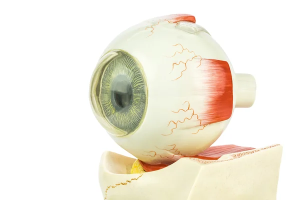
Artificial model of human eye
Artificial model of human eye — Stock Photo, Image
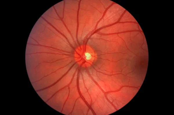
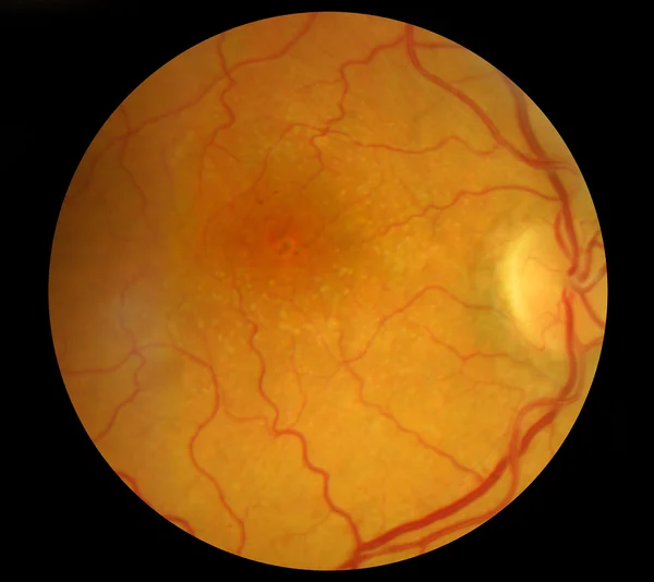
Human Retina - Optic Nerve
Human Retina - Optic Nerve — Stock Photo, Image
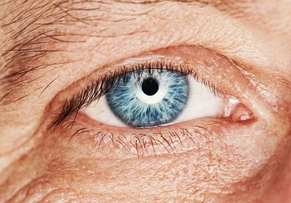
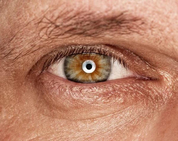
Picture of a human eye close up
Picture Human Eye Close — Stock Photo, Image
Eye Cross Section Real Human Anatomy - isolated on white
Eye Cross Section Real Human Anatomy - isolated on white — Stock Photo, Image
Eye anatomy - inner structure isolated on white
Eye anatomy - inner structure isolated on white — Stock Photo, Image
3d rendered medically accurate illustration of a male brain anatomy
Rendered Medically Accurate Illustration Male Brain Anatomy — Stock Photo, Image
3d rendered medically accurate illustration of a female brain anatomy
Rendered Medically Accurate Illustration Female Brain Anatomy — Stock Photo, Image
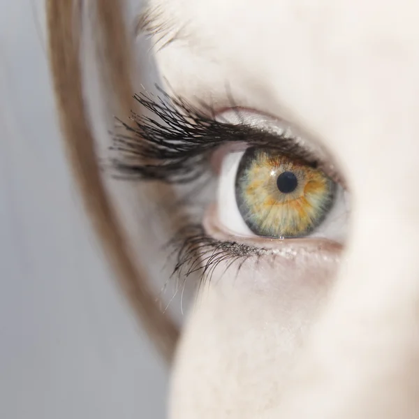
Beautiful woman eye close up
Beautiful woman eye close up — Stock Photo, Image
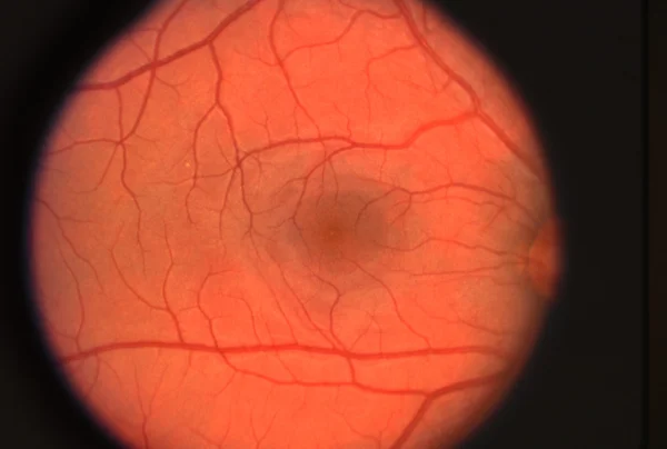
Retina
Retina — Stock Photo, Image
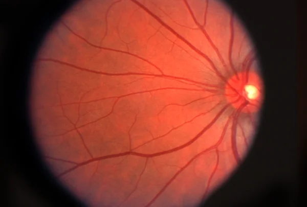
Retina
Retina — Stock Photo, Image
Vision
Vision — Stock Photo, Image
Medical Fundus photo of retinal pathology, hemmorhages, vein occ
Medical Fundus photo of retinal pathology, hemmorhages, vein occ — Stock Photo, Image
Picture of a human eye
Picture Human Eye — Stock Photo, Image
Human eye anatomy
Human eye anatomy — Stock Photo, Image
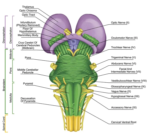
Brainstem, brain stem, ventral view, posterior part of the brain, adjoining and structurally continuous with the spinal cord, motor and sensory innervation to the face and neck via thecranial nerves
Brainstem, brain stem, ventral view, posterior part of the brain, adjoining and structurally continuous with the spinal cord, motor and sensory innervation to the face and neck via thecranial nerves — Stock Photo, Image
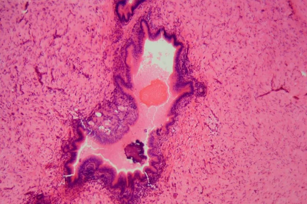
Liver cells of a rabit with Coccidia parasites.
Liver cells of a rabit with Coccidia parasites. — Stock Photo, Image
Black and green color oil pastel textured background
Black Green Color Oil Pastel Textured Background — Stock Photo, Image
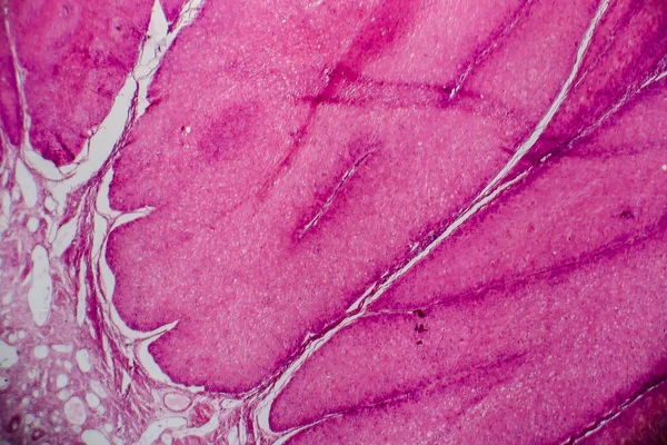
Uterus adenofibroma, light micrograph, photo under microscope. A rare benign tumor of the uterus composed of glandular and fibrous tissues
Uterus Adenofibroma Light Micrograph Photo Microscope Rare Benign Tumor Uterus — Stock Photo, Image
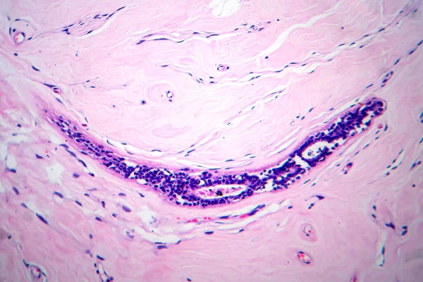
Photomicrograph of fibroadenoma, showcasing benign glandular and fibrous tissue growth within the breast, a common noncancerous tumor.
Photomicrograph Fibroadenoma Showcasing Benign Glandular Fibrous Tissue Growth Breast Common — Stock Photo, Image
Trachea, light micrograph. Hyaline cartilage from trachea showing chondrocytes, collagen fibers and matrix.
Trachea Light Micrograph Hyaline Cartilage Trachea Showing Chondrocytes Collagen Fibers — Stock Photo, Image
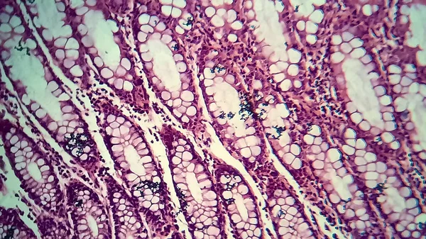
Bacillary dysentery, light micrograph, photo under microscope showing presence of bacteria and accumulation of inflammatory cells in intestinal epithelium
Bacillary Dysentery Light Micrograph Photo Microscope Showing Presence Bacteria Accumulation — Stock Photo, Image
Bright color wall background
Bright color wall background — Stock Photo, Image
Neurofibromatosis, light micrograph, photo under microscope. A group of genetic disorders with formation of tumors in the nervous system
Neurofibromatosis Light Micrograph Photo Microscope Group Genetic Disorders Formation Tumors — Stock Photo, Image
3D rendering abstract round light background
Rendering Abstract Light Background — Stock Photo, Image
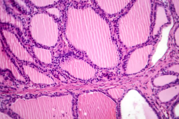
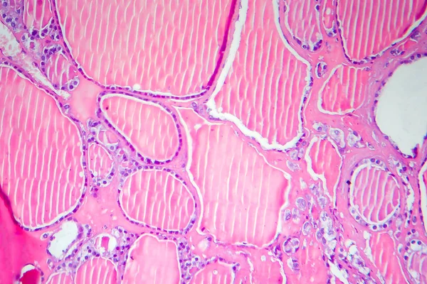
Photomicrograph of a toxic goiter tissue sample under a microscope, revealing hypertrophy of thyroid follicular cells, increased vascularity, and colloid depletion.
Photomicrograph Toxic Goiter Tissue Sample Microscope Revealing Hypertrophy Thyroid Follicular — Stock Photo, Image
Characteristics of fungi living in wood as a group, are polyphyletic under the microscope for education.
Characteristics Fungi Living Wood Group Polyphyletic Microscope Education — Stock Photo, Image
Colorful abstract background for copy space
Colorful Abstract Background Copy Space — Stock Photo, Image
Red blood cells medical science background.
Red blood cells medical science background. — Stock Photo, Image
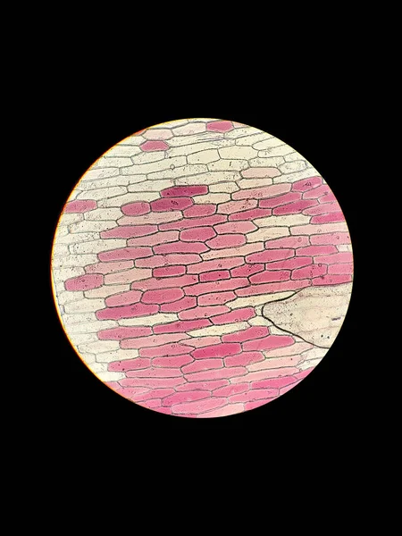
Biological preparation of onion plant cells under a microscope with the phenomenon of plasmolysis
Biological Preparation Onion Plant Cells Microscope Phenomenon Plasmolysis — Stock Photo, Image
Motor nerves in spinal tissue.
Motor Nerves Spinal Tissue — Stock Photo, Image
Abstract bright shiny color fluid background, hand drawn alcohol painting with golden streaks. Liquid ink technique texture for high resolution backdrop design
Abstract Bright Shiny Color Fluid Background Hand Drawn Alcohol Painting — Stock Photo, Image
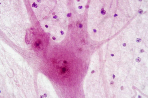
Education Spinal cord and Motor Neuron under the microscope in Lab.
Education Spinal Cord Motor Neuron Microscope Lab — Stock Photo, Image
Cliated epithelium of human under the microscope in Lab.
Cliated Epithelium Human Microscope Lab — Stock Photo, Image
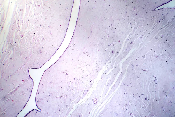
Purulent meningitis, light micrograph, photo under microscope
Purulent Meningitis Light Micrograph Photo Microscope — Stock Photo, Image
Cerebellum, Thalamus, Medulla oblongata, Spinal cord and Motor Neuron human under the microscope in Lab.
Cerebellum Thalamus Medulla Oblongata Spinal Cord Motor Neuron Human Microscope — Stock Photo, Image
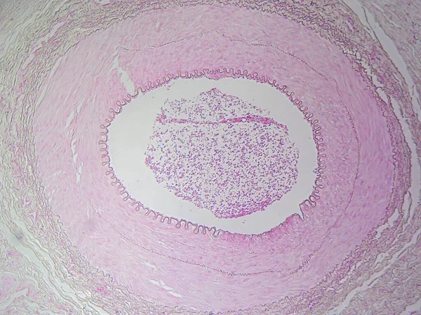
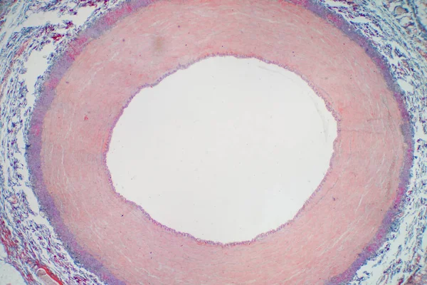
The Anatomy of Blood Vessels: Muscular Artery in High Detail
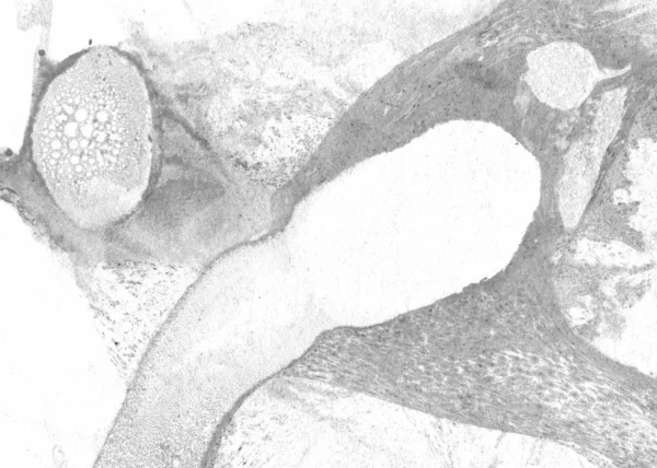
Anatomy Blood Vessels Muscular Artery High Detail — Stock Photo, Image
Copy space wallpaper, grunge background, abstract colorful texture background
Copy Space Wallpaper Grunge Background Abstract Colorful Texture Background — Stock Photo, Image
Artery blood vessel under microscope view for education histology. Histological for human physiology.
Artery Blood Vessel Microscope View Education Histology Histological Human Physiology — Stock Photo, Image
Abstract green texture background, copy space wallpaper
Abstract Green Texture Background Copy Space Wallpaper — Stock Photo, Image
Condyloma acuminatum, also known as genital warts. Light micrograph, photo under microscope
Condyloma Acuminatum Also Known Genital Warts Light Micrograph Photo Microscope — Stock Photo, Image
Space Flight Into A Star Field In Galaxy Clouds And Lightning Nebula
Space Flight Star Field Galaxy Clouds Lightning Nebula — Stock Photo, Image
Condyloma acuminatum, also known as genital warts. Light micrograph, photo under microscope
Condyloma Acuminatum Also Known Genital Warts Light Micrograph Photo Microscope — Stock Photo, Image
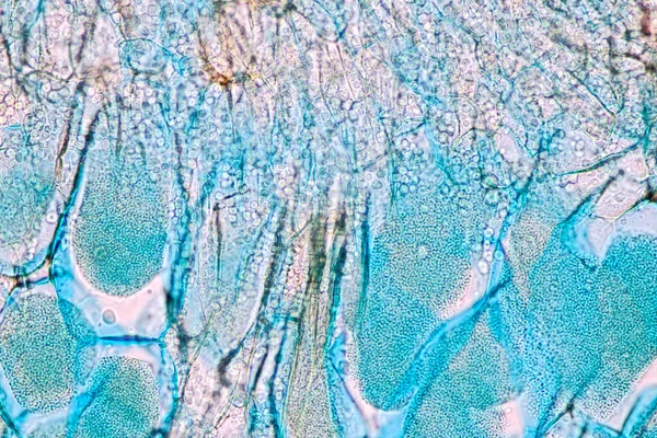
Host cells with spores (mold) are inside wood under the microscope for education.
Host Cells Spores Mold Wood Microscope Education — Stock Photo, Image
3D rendering abstract multicolor fractal light background
Rendering Abstract Multicolor Fractal Light Background — Stock Photo, Image
Rustic stones, corroded textures, colorful and interesting backgrounds and charming textures, used for web design and abstract backgrounds
Rustic Stones Corroded Textures Colorful Interesting Backgrounds Charming Textures Used — Stock Photo, Image
Creative illustrations in life. Colorful and beautiful background patterns for designers.
Creative Illustrations Life Colorful Beautiful Background Patterns Designers — Stock Photo, Image
Very nice paint.
Very nice paint. — Stock Photo, Image
Areolar connective tissue under the microscope view. Histological for human physiology.
Areolar Connective Tissue Microscope View Histological Human Physiology — Stock Photo, Image
Creative and design art work, copy space wallpaper, abstract colorful texture background
Creative Design Art Work Copy Space Wallpaper Abstract Colorful Texture — Stock Photo, Image
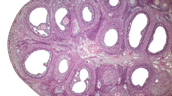
Light micrograph of the Cat's ovarian. Micrograph of ovary showing primordial, primary and secondary follicles isolated on white background. Hematoxylin and eosin stain.
Light Micrograph Cat Ovarian Micrograph Ovary Showing Primordial Primary Secondary — Stock Photo, Image
Characteristics of fungi living in wood as a group, are polyphyletic under the microscope for education.
Characteristics Fungi Living Wood Group Polyphyletic Microscope Education — Stock Photo, Image
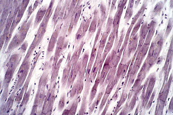
Human cardiac tissue, light micrograph. Haematoxylin and eosin stain.
Human Cardiac Tissue Light Micrograph Haematoxylin Eosin Stain — Stock Photo, Image
Endometrial adenocarcinoma, light micrograph, photo under microscope
Endometrial Adenocarcinoma Light Micrograph Photo Microscope — Stock Photo, Image
Slime molds, as a group, are polyphyletic under the microscope for education.
Slime Molds Group Polyphyletic Microscope Education — Stock Photo, Image
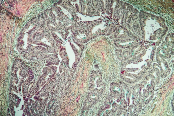
Colon carcinoma tissue thin section 100x
Colon Carcinoma Tissue Thin Section 100X — Stock Photo, Image
Vertical social template for backgrounds web banner posters and your creative design works etc
Vertical Social Template Backgrounds Web Banner Posters Your Creative Design — Stock Photo, Image
3D rendering abstract circle light background
Rendering Abstract Circle Light Background — Stock Photo, Image
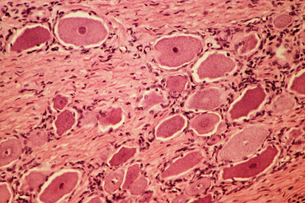
Spinal ganglion view from microscope
Spinal Ganglion View Microscope — Stock Photo, Image
Characteristics of fungi living in wood as a group, are polyphyletic under the microscope for education.
Characteristics Fungi Living Wood Group Polyphyletic Microscope Education — Stock Photo, Image
3D rendering abstract multicolor fractal light background
Rendering Abstract Multicolor Fractal Light Background — Stock Photo, Image
Follicular adenoma of thyroid gland, light micrograph. Histopathology of thydoid adenoma. Photo under microscope
Follicular Adenoma Thyroid Gland Light Micrograph Histopathology Thydoid Adenoma Photo — Stock Photo, Image
The study Plant tissue of under the microscope for classroom education.
Study Plant Tissue Microscope Classroom Education — Stock Photo, Image
Spinal ganglion from microscopy
Spinal Ganglion Microscopy — Stock Photo, Image
Micrograph of small intestinum tenue tissue
Micrograph of small intestinum tenue tissue — Stock Photo, Image
Abstract wooden texture background
Abstract Wooden Texture Background — Stock Photo, Image
Traditional marbling artwork patterns as colorful abstract background
Traditional Marbling Artwork Patterns Colorful Abstract Background — Stock Photo, Image
Abstract marbling art patterns as colorful background
Abstract marbling art patterns as colorful background — Stock Photo, Image
Texture or background wall of shabby paint and plaster cracks
Texture or background wall of shabby paint and plaster cracks — Stock Photo, Image
Electric lighting spark
Electric lighting spark — Stock Photo, Image
Dramatic black, monochrome abstraction, reminiscent of dragon scales or caked lava, shimmering. Fragment of a modern artwork. Macro photography of the smallest details of a panting. Contemporary art. Artistic background image.
Dramatic Black Monochrome Abstraction Reminiscent Dragon Scales Caked Lava Shimmering — Stock Photo, Image
Multicolored hypnotic swirl of spiral fractal art design background
Multicolored hypnotic swirl of spiral fractal art design background — Stock Photo, Image
Swirls of alcohol ink in lilac, lavender, violet are reminiscent of marble and ripples of agate. Fluid modern Art banners, ethereal graphic design.
Swirls of alcohol ink in lilac, lavender, violet are reminiscent of marble and ripples of agate. Fluid modern Art banners, ethereal graphic design. — Stock Photo, Image
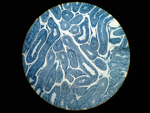
Cross section Human testis under microscope view. Shows spermatogonia, spermatocytes in meiosis, spermatids, and spermatozoa
Cross Section Human Testis Microscope View Shows Spermatogonia Spermatocytes Meiosis — Stock Photo, Image
The study Plant tissue of under the microscope for classroom education.
Study Plant Tissue Microscope Classroom Education — Stock Photo, Image
Abstract color texture, background, copy space wallpaper
Abstract Color Texture Background Copy Space Wallpaper — Stock Photo, Image
Abstract green texture background, copy space wallpaper
Abstract Green Texture Background Copy Space Wallpaper — Stock Photo, Image
3D rendering abstract red fractal light background
Rendering Abstract Red Fractal Light Background — Stock Photo, Image
Uterus adenofibroma, light micrograph, photo under microscope. A rare benign tumor of the uterus composed of glandular and fibrous tissues
Uterus Adenofibroma Light Micrograph Photo Microscope Rare Benign Tumor Uterus — Stock Photo, Image
Green textured grungy background
Green Textured Grungy Background — Stock Photo, Image
Fantasy chaotic colorful fractal pattern. Abstract fractal shapes. 3D rendering illustration background or wallpaper.
Fantasy Chaotic Colorful Fractal Pattern Abstract Fractal Shapes Rendering Illustration — Stock Photo, Image
Characteristics of fungi living in wood as a group, are polyphyletic under the microscope for education.
Characteristics Fungi Living Wood Group Polyphyletic Microscope Education — Stock Photo, Image
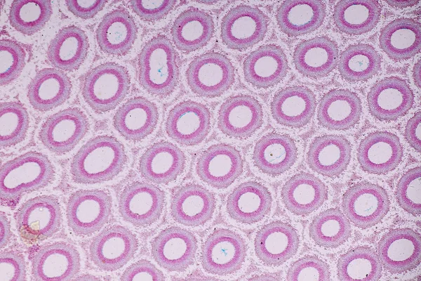
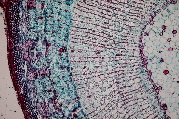
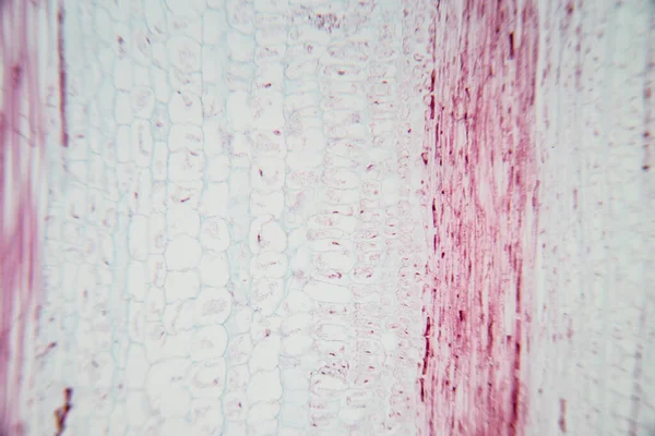
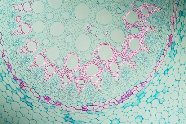
Philodendron aerial root in cross section 100x
Philodendron Aerial Root Cross Section 100X — Stock Photo, Image
Abstract color texture, grunge background, copy space wallpaper
Abstract Color Texture Grunge Background Copy Space Wallpaper — Stock Photo, Image
Abstract Colorful Backdrop wallpaper
Abstract Colorful Backdrop Wallpaper — Stock Photo, Image
Marbling. Marble texture. Paint splash. Colorful fluid. Abstract colored background. Raster illustration. Colorful abstract painting background. Highly-textured oil paint. High quality details.
Marbling Marble Texture Paint Splash Colorful Fluid Abstract Colored Background — Stock Photo, Image
Fibrous thyroiditis, light micrograph, photo under microscope. A disease characterized by chronic inflammation and fibrosis of the thyroid gland
Fibrous Thyroiditis Light Micrograph Photo Microscope Disease Characterized Chronic Inflammation — Stock Photo, Image
Imaginatory fractal abstract background Image
Imaginatory fractal abstract background Image — Stock Photo, Image
Imaginatory fractal abstract background Image
Imaginatory fractal abstract background Image — Stock Photo, Image
Green texture, abstract background
Green Texture Abstract Background — Stock Photo, Image
Microscopic cells texture, abstract background
Microscopic Cells Texture Abstract Background — Stock Photo, Image
Curves with gas bubbles are immersed in mineral water.
Curves Gas Bubbles Immersed Mineral Water — Stock Photo, Image
Copy space wallpaper, grunge background, abstract colorful texture background
Copy Space Wallpaper Grunge Background Abstract Colorful Texture Background — Stock Photo, Image
Abstract ink background. Marble style. Black paint stroke texture on white paper. Wallpaper for web and game design. Grunge mud art. Macro image of pen juice. Dark Smear
Abstract Ink Background Marble Style Black Paint Stroke Texture White — Stock Photo, Image
Spinal cord c.s. under microscope
Spinal Cord Microscope — Stock Photo, Image
Microorganism,on small leaf-mold
Microorganism Small Leaf Mold — Stock Photo, Image

Find High-Quality Optic Nerve Images for Your Project

When it comes to visualizing the human anatomy, the optic nerve is one of the most complex and critical parts of the body. As an information pathway from the eyes to the brain, it plays an essential role in vision and perception. Whether you are a medical professional, a graphic designer, or a health educator, finding the right optic nerve images can make all the difference in communicating your message effectively. With our extensive collection of high-quality stock images, you can bring clarity and precision to your work.

Types of Optic Nerve Images Available

Our collection includes a wide variety of optic nerve images, ranging from diagrams and illustrations to detailed photographs and scans. You can choose from images that showcase the anatomy of the optic nerve, its connection to the eyes and the brain, and its functional pathways. We offer images that feature different angles, magnifications, and color schemes, so you can find the perfect visual for your project. Whether you need images for a medical journal article, a website, or a patient brochure, we have you covered.

Where You Can Use Optic Nerve Images

The versatility of our optic nerve images makes them useful for a wide range of applications. Medical professionals can use them to educate patients on eye diseases, injuries, and conditions related to the optic nerve. Scientists and researchers can use them to study the structure and function of the optic nerve and develop new treatments and therapies. Graphic designers and illustrators can use them to create eye-catching visuals for books, websites, and presentations. No matter the context, our optic nerve images are designed to inform, educate, and engage your audience.

How to Choose the Right Optic Nerve Images

Choosing the right optic nerve images for your project can seem daunting, especially if you are not a medical expert or an experienced designer. However, with a few simple tips, you can make sure that your visuals are accurate, compelling, and relevant. First, consider your audience and your message. Who are you trying to reach, and what do you want to convey? Second, select images that are clear, well-lit, and in focus. Third, verify the source and the copyright status of the image to avoid legal issues. With these guidelines in mind, you can create visuals that stand out and make an impact.

Get Started with Our Optic Nerve Images Today

Are you ready to take your project to the next level with high-quality optic nerve images? Browse our collection today and discover the possibilities. With our easy-to-use search tool and flexible licensing options, you can find the perfect images for your needs and budget. Whether you need a single image or a bulk package, we are here to help you succeed.