Tonsil Stock Photos

100,000 Tonsil pictures are available under a royalty-free license

  • Best Match
  • Fresh
  • Popular
1-100 of 100,000
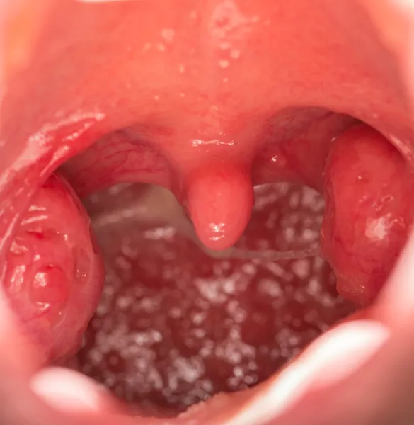
Tonsil throat, Oral tonsil
Tonsil throat, Oral tonsil — Stock Photo, Image
Child with doctor in the office of a pediatrician, medical examination
Child with doctor in the office of a pediatrician, medical examination — Stock Photo, Image
Open mouth view of tonsils
Open mouth view of tonsils — Stock Photo, Image
Sick young woman having pain in her throat
Sick young woman having pain in her throat — Stock Photo, Image
Pediatrician examining little child throat
Pediatrician examining little child throat — Stock Photo, Image
Young female having sore throat
Young female having sore throat — Stock Photo, Image
Doctor examining little child boy
Doctor examining little child boy — Stock Photo, Image
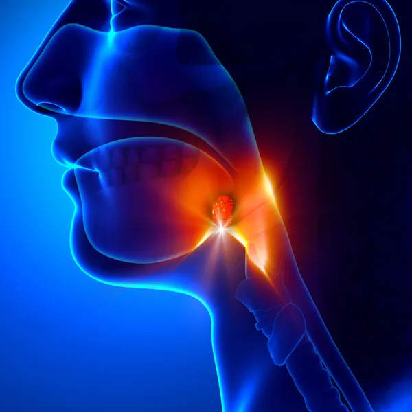
Tonsils - Angina Pectoris
Tonsils - Angina Pectoris — Stock Photo, Image
Young little girl being examinated by a doctor
Young little girl being examinated by a doctor — Stock Photo, Image
Female doctor touch male patient's throat because she thinking about the tonsillitis.
Female Doctor Touch Male Patient Throat Because She Thinking Tonsillitis — Stock Photo, Image
Throat check
Throat check — Stock Photo, Image
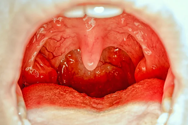
Chronic tonsillitis. Herpes with throat
Chronic Tonsillitis Herpes Throat — Stock Photo, Image
Macro opened mouth with throat tonsil
Macro opened mouth with throat tonsil — Stock Photo, Image
Woman with thyroid gland problem on the blue background
Woman Thyroid Gland Problem Blue Background — Stock Photo, Image
Woman with sore throat touching her neck colored in red
Woman with sore throat touching her neck colored in red — Stock Photo, Image
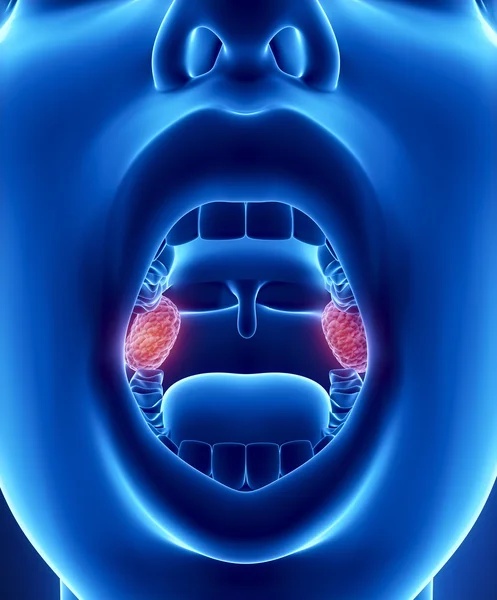
Tonsils anatomy
Tonsils anatomy — Stock Photo, Image
Medical Exam
Medical Exam — Stock Photo, Image
Child have sore throat sick
Child have sore throat sick — Stock Photo, Image
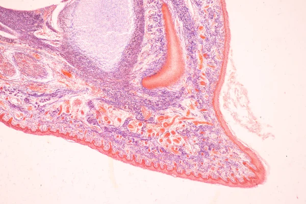
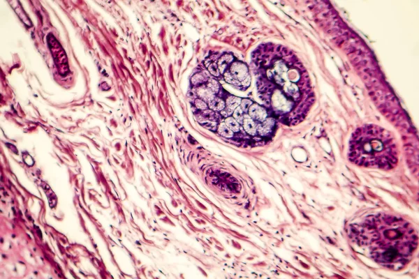
Under high magnification microscopic view of labium minus
High Magnification Microscopic View Labium — Stock Photo, Image
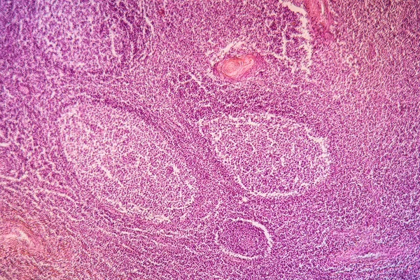
Lymph node in tuberculosis
Lymph node in tuberculosis — Stock Photo, Image
Liver cirrhosis diseased tissue under the microscope 100x
Liver Cirrhosis Diseased Tissue Microscope 100X — Stock Photo, Image
Microscopic cells texture, abstract background
Microscopic Cells Texture Abstract Background — Stock Photo, Image
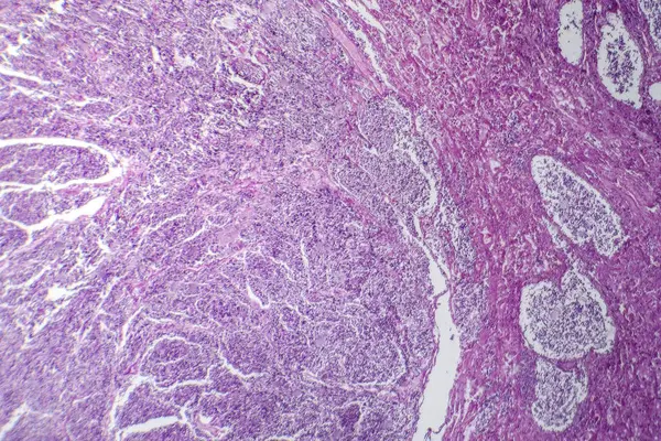
Endometrial adenocarcinoma, light micrograph, photo under microscope
Endometrial Adenocarcinoma Light Micrograph Photo Microscope — Stock Photo, Image
Human liver cells with tuberculosis under the microscope.
Human liver cells with tuberculosis under the microscope. — Stock Photo, Image
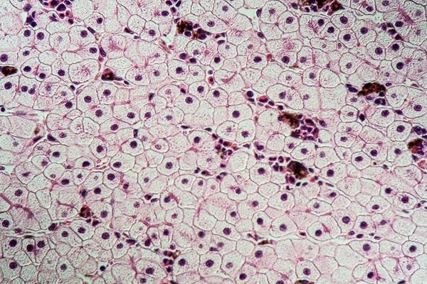
Axolot amphibians with liver cells 100x
Axolot Amphibians Liver Cells 100X — Stock Photo, Image
Tooth development from human under microscope view for education.
Tooth Development Human Microscope View Education — Stock Photo, Image
Epithelial cells of the oral mucosa with cell nuclei and bacteria
Epithelial Cells Oral Mucosa Cell Nuclei Bacteria — Stock Photo, Image
Pink abstract background in watercolor style
Pink Abstract Background Watercolor Style — Stock Photo, Image
Cells of human uterus tissue with inoffensive tumor cells
Cells of human uterus tissue with inoffensive tumor cells — Stock Photo, Image
Microbiology, microscopic cells surface
Microbiology Microscopic Cells Surface — Stock Photo, Image
Pink soap bubbles background
Pink soap bubbles background — Stock Photo, Image
Close up human cells with microscope.
Close up human cells with microscope. — Stock Photo, Image
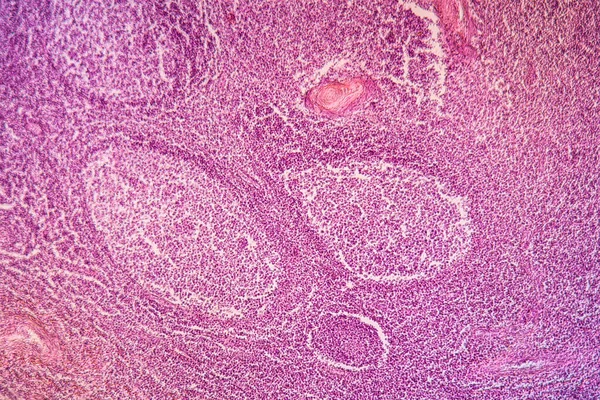
Lymph node structure
Lymph node structure — Stock Photo, Image
Neurofibromatosis, light micrograph, photo under microscope. A group of genetic disorders with formation of tumors in the nervous system
Neurofibromatosis Light Micrograph Photo Microscope Group Genetic Disorders Formation Tumors — Stock Photo, Image
Bacillary dysentery, light micrograph, photo under microscope showing presence of bacteria and accumulation of inflammatory cells in intestinal epithelium
Bacillary Dysentery Light Micrograph Photo Microscope Showing Presence Bacteria Accumulation — Stock Photo, Image
Liver mouse,schistosomiasis diseased tissue 100x
Liver Mouse Schistosomiasis Diseased Tissue 100X — Stock Photo, Image
Close up human cells with microscope.
Close up human cells with microscope. — Stock Photo, Image
Photomicrograph of primary particulate contracted kidney, illustrating shrunken renal tissue and abnormal nephron structures.
Photomicrograph Primary Particulate Contracted Kidney Illustrating Shrunken Renal Tissue Abnormal — Stock Photo, Image
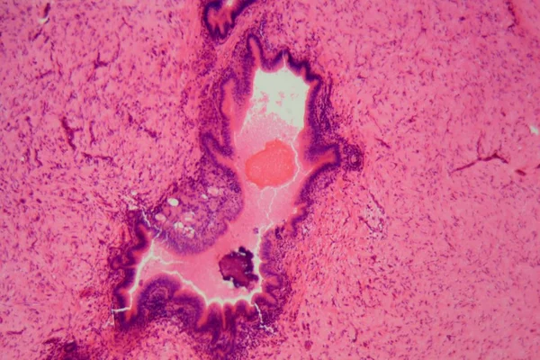
Photomicrograph of chronic bronchitis, showing inflamed bronchial lining with excess mucus production, characteristic of airway disease.
Photomicrograph Chronic Bronchitis Showing Inflamed Bronchial Lining Excess Mucus Production — Stock Photo, Image
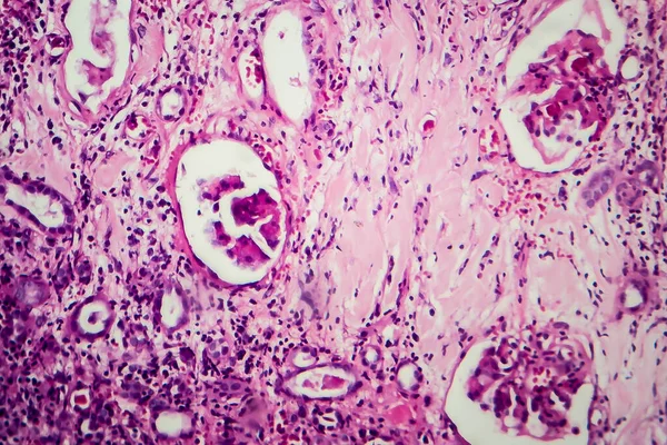
Histopathology of diffuse sclerosing glomerulonephritis, light micrograph, photo under microscope
Histopathology Diffuse Sclerosing Glomerulonephritis Light Micrograph Photo Microscope — Stock Photo, Image
Vintage pink background. Rough painted wall of fiesta color. Imperfect plane of rosy colored. Uneven old decorative toned backdrop of rouge tint. Texture of pink hue. Ornamental stony surface.
Vintage Pink Background Rough Painted Wall Fiesta Color Imperfect Plane — Stock Photo, Image
Acute simple appendicitis, light micrograph
Acute simple appendicitis, light micrograph — Stock Photo, Image
Histopathology of diffuse sclerosing glomerulonephritis, light micrograph, photo under microscope
Histopathology Diffuse Sclerosing Glomerulonephritis Light Micrograph Photo Microscope — Stock Photo, Image
Bazex syndrome or paraneoplastic acrokeratosis is a psoriasiform eruption that is seen more frequently in adult Caucasian males and is characterized by a predominantly acral distribution with nail dystrophy.
Bazex Syndrome Paraneoplastic Acrokeratosis Psoriasiform Eruption Seen More Frequently Adult — Stock Photo, Image
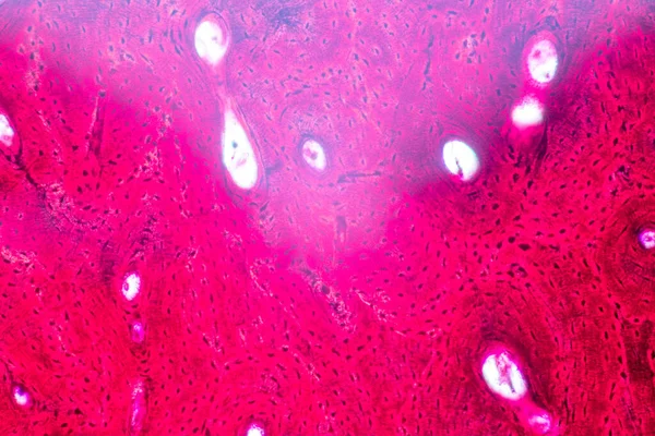
Anatomy and Histological Bone, Elastic cartilage human and Joint of human foetus under the microscope for education.
Anatomy Histological Bone Elastic Cartilage Human Joint Human Foetus Microscope — Stock Photo, Image
Colorful old paper texture background
Colorful Old Paper Texture Background — Stock Photo, Image
Human liver tissue under microscope view
Human Liver Tissue Microscope View — Stock Photo, Image
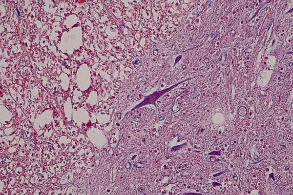
Neurons cells from the spinal cord under the microscope view
Neurons Cells Spinal Cord Microscope View — Stock Photo, Image
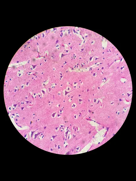
Nerve cells under light microscope
Nerve cells under light microscope — Stock Photo, Image
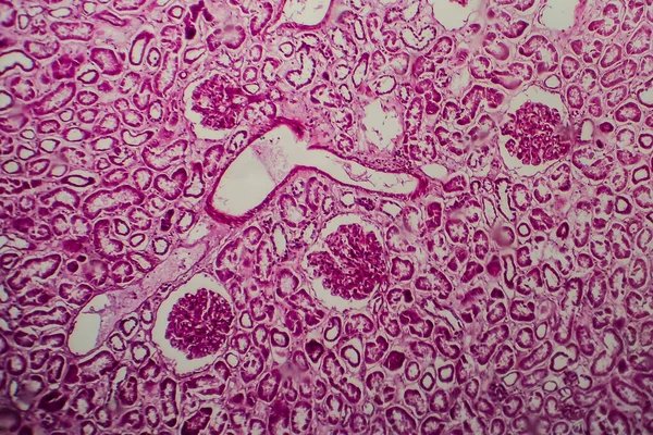
Kidney. Cortex and medulla
Kidney. Cortex and medulla — Stock Photo, Image
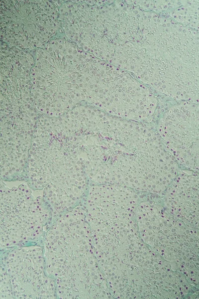
Testicle cross-section under the microscope 100x
Testicle Cross Section Microscope 100X — Stock Photo, Image
Microscope photo of a large intestine section with inflammation (Colitis).
Microscope photo of a large intestine section with inflammation (Colitis). — Stock Photo, Image
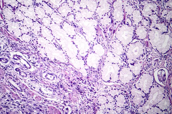
Photomicrograph of mucinous carcinoma in the stomach, displaying malignant mucin-producing cells, characteristic of an aggressive stomach cancer.
Photomicrograph Mucinous Carcinoma Stomach Displaying Malignant Mucin Producing Cells Characteristic — Stock Photo, Image
Photomicrograph of mucinous carcinoma in the stomach, displaying malignant mucin-producing cells, characteristic of an aggressive stomach cancer.
Photomicrograph Mucinous Carcinoma Stomach Displaying Malignant Mucin Producing Cells Characteristic — Stock Photo, Image
Bowen's Disease Tumor under the microscope 100x
Bowen Disease Tumor Microscope 100X — Stock Photo, Image
Abstract watercolor marble texture background. natural pink pattern
Abstract Watercolor Marble Texture Background Natural Pink Pattern — Stock Photo, Image
Liver cells of a rabit with Coccidia parasites.
Liver cells of a rabit with Coccidia parasites. — Stock Photo, Image
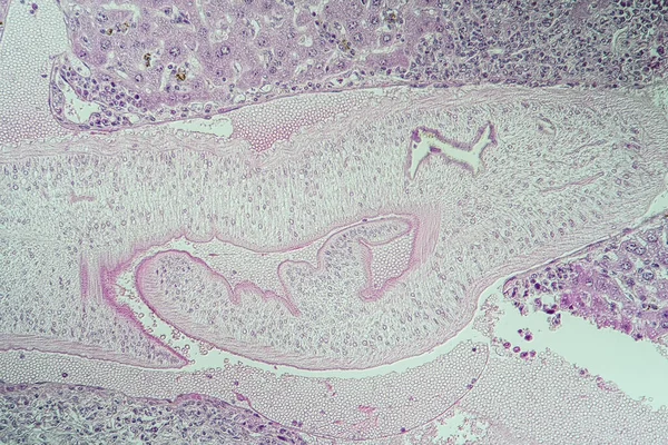
Hydra is a genus of small, fresh-water animals of the phylum Cnidaria and class Hydrozoa under the microscope for education.
Hydra Genus Small Fresh Water Animals Phylum Cnidaria Class Hydrozoa — Stock Photo, Image
Watercolor painted paper background texture
Watercolor painted paper background texture — Stock Photo, Image
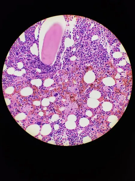
Bone marrow biopsy H and E stain showing eosinophilia
Bone marrow biopsy H and E stain showing eosinophilia — Stock Photo, Image
Neurofibromatosis, light micrograph, photo under microscope. A group of genetic disorders with formation of tumors in the nervous system
Neurofibromatosis Light Micrograph Photo Microscope Group Genetic Disorders Formation Tumors — Stock Photo, Image
Axolot amphibians with liver cells 100x
Axolot Amphibians Liver Cells 100X — Stock Photo, Image
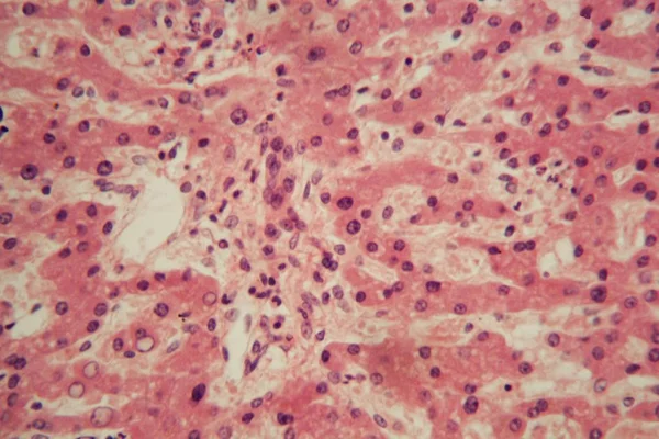
Viral hepatitis, light micrograph
Viral hepatitis, light micrograph — Stock Photo, Image
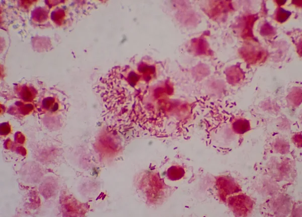
Bacteria cell with white blood cells.Gram negative.
Bacteria Cell White Blood Cells Gram Negative — Stock Photo, Image
Swirls of alcohol ink in lilac, lavender, violet are reminiscent of marble and ripples of agate. Fluid modern Art banners, ethereal graphic design.
Swirls of alcohol ink in lilac, lavender, violet are reminiscent of marble and ripples of agate. Fluid modern Art banners, ethereal graphic design. — Stock Photo, Image
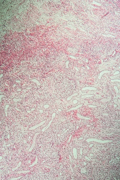
Poly nephritis, tissue under the microscope 100x
Poly Nephritis Tissue Microscope 100X — Stock Photo, Image
Photomicrograph of small cell lung cancer, revealing densely packed malignant cells characteristic of an aggressive lung malignancy.
Photomicrograph Small Cell Lung Cancer Revealing Densely Packed Malignant Cells — Stock Photo, Image
Neurofibromatosis, light micrograph, photo under microscope. A group of genetic disorders with formation of tumors in the nervous system
Neurofibromatosis Light Micrograph Photo Microscope Group Genetic Disorders Formation Tumors — Stock Photo, Image
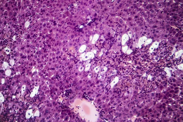
Photomicrograph of fatty liver showing lipid steatosis. Photomicrograph showing large vacuoles of triglyceride fat accumulated inside liver cells, it occurs in alcohol overuse, toxins, in diabetes.
Photomicrograph Fatty Liver Showing Lipid Steatosis Photomicrograph Showing Large Vacuoles — Stock Photo, Image
Tissue with schistosomiasis sick tissue 100x
Tissue Schistosomiasis Sick Tissue 100X — Stock Photo, Image
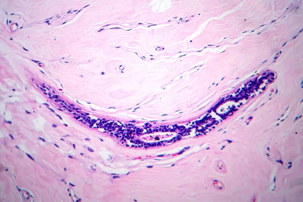
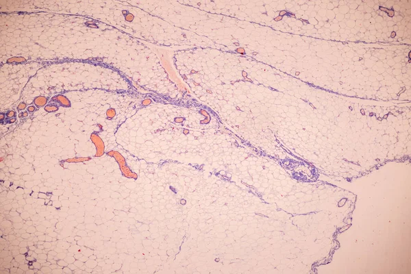
Connective tissue, light micrograph
Connective tissue, light micrograph — Stock Photo, Image
Red blood cells medical science background.
Red blood cells medical science background. — Stock Photo, Image
Hyalinosis of lien
Hyalinosis of lien — Stock Photo, Image
Histopathology of interstitial nephritis, light micrograph, photo under microscope
Histopathology Interstitial Nephritis Light Micrograph Photo Microscope — Stock Photo, Image
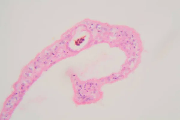
Section of a common liver fluke (Fasciola) under the microscope
Section of a common liver fluke (Fasciola) under the microscope — Stock Photo, Image
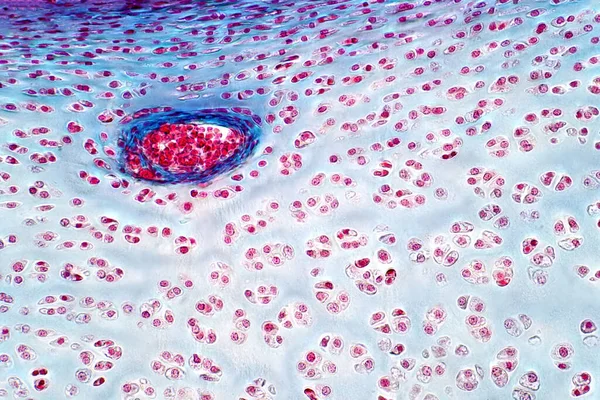
Human Hyaline cartilage bone under light microscope view for histology education. Haematoxylin and eosin staining technique for histology.
Human Hyaline Cartilage Bone Light Microscope View Histology Education Haematoxylin — Stock Photo, Image
Pattern with Alligator Skin.
Pattern with Alligator Skin. — Stock Photo, Image
Muscle composite cells on a microscope slide.
Muscle Composite Cells Microscope Slide — Stock Photo, Image
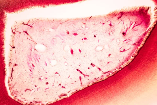
Tooth development from human under microscope view for education.
Tooth Development Human Microscope View Education — Stock Photo, Image
The image of the wall, for use as a background.
Image Wall Use Background — Stock Photo, Image
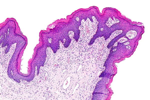
Skin papilloma of a human
Skin papilloma of a human — Stock Photo, Image
Schistosoma mansoni parasites under the microscope.
Schistosoma mansoni parasites under the microscope. — Stock Photo, Image
Gray liquid ink, digital wallpaper
Gray Liquid Ink Digital Wallpaper — Stock Photo, Image
Uterus adenofibroma, light micrograph, photo under microscope. A rare benign tumor of the uterus composed of glandular and fibrous tissues
Uterus Adenofibroma Light Micrograph Photo Microscope Rare Benign Tumor Uterus — Stock Photo, Image
Anatomy and Histological Bone, Elastic cartilage human and Joint of human foetus under the microscope for education.
Anatomy Histological Bone Elastic Cartilage Human Joint Human Foetus Microscope — Stock Photo, Image
Mitosis cell in the Root tip of Onion under a microscope.
Mitosis Cell Root Tip Onion Microscope — Stock Photo, Image
Pink abstract background in watercolor style
Pink Abstract Background Watercolor Style — Stock Photo, Image
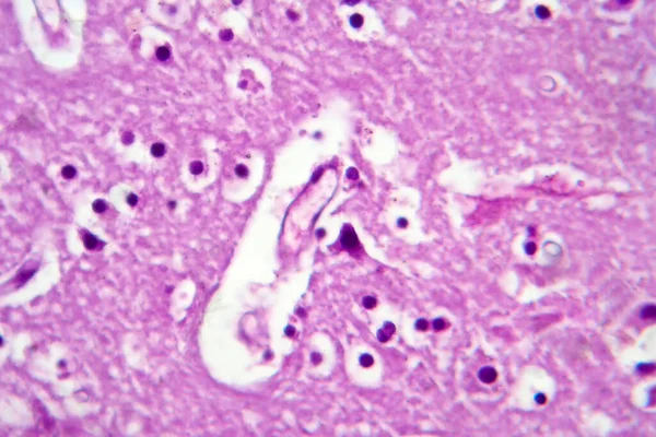
Histopathology of Japanese encephalitis, light micrograph, photo under microscope
Histopathology Japanese Encephalitis Light Micrograph Photo Microscope — Stock Photo, Image
Tissue of Blood human, Vein human, Artery human and Heart muscle human under the microscope in Lab.
Tissue Blood Human Vein Human Artery Human Heart Muscle Human — Stock Photo, Image
Tissue of Blood human, Vein human, Artery human and Heart muscle human under the microscope in Lab.
Tissue Blood Human Vein Human Artery Human Heart Muscle Human — Stock Photo, Image
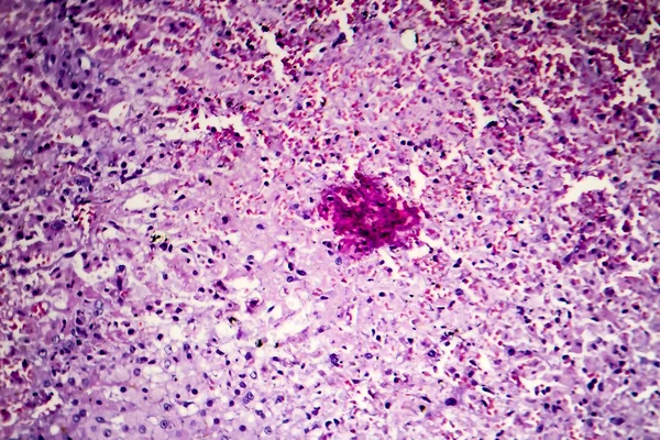
Hepatic congestion, light micrograph
Hepatic congestion, light micrograph — Stock Photo, Image
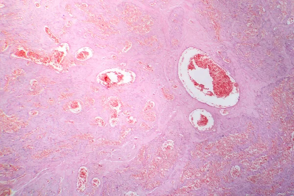
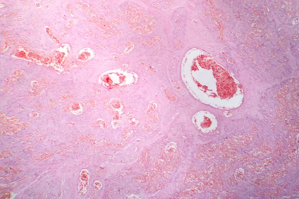
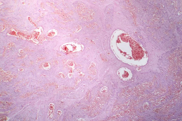
Photomicrograph of capillary hemangioma, illustrating abnormal proliferation of capillaries, characteristic of a benign vascular tumor.
Photomicrograph Capillary Hemangioma Illustrating Abnormal Proliferation Capillaries Characteristic Benign Vascular — Stock Photo, Image
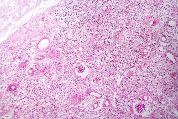
Photomicrograph of capillary hemangioma, illustrating abnormal proliferation of capillaries, characteristic of a benign vascular tumor.
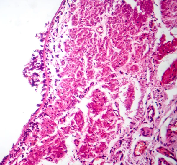
Photomicrograph Capillary Hemangioma Illustrating Abnormal Proliferation Capillaries Characteristic Benign Vascular — Stock Photo, Image
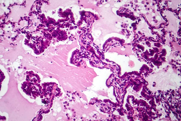
Photomicrograph of capillary hemangioma, illustrating abnormal proliferation of capillaries, characteristic of a benign vascular tumor.
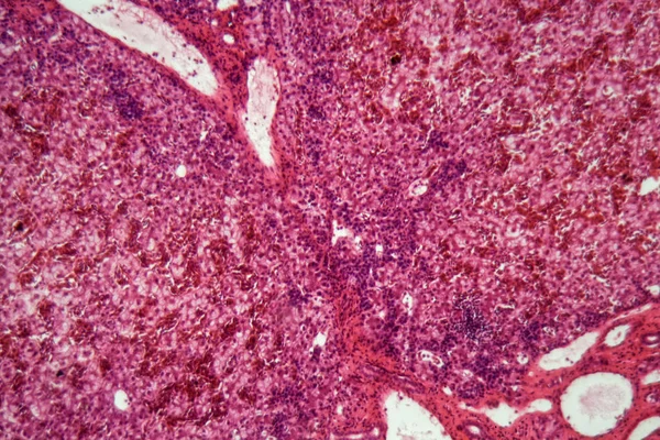
Photomicrograph Capillary Hemangioma Illustrating Abnormal Proliferation Capillaries Characteristic Benign Vascular — Stock Photo, Image
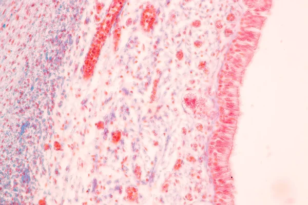
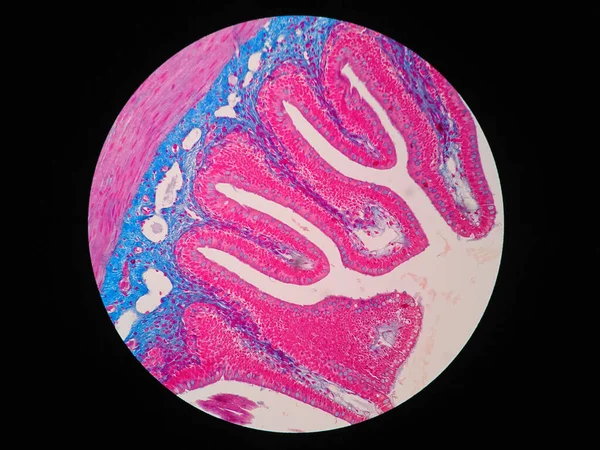
Ciliated Epithelium sec. under microscope
Ciliated Epithelium Sec Microscope — Stock Photo, Image
Histology of human intestine tissue under microscope view
Histology Human Intestine Tissue Microscope View — Stock Photo, Image
Human liver cells with cancer
Human liver cells with cancer — Stock Photo, Image
Photomicrograph of lung adenocarcinoma, displaying malignant glandular cells indicative of the most common type of lung cancer.
Photomicrograph Lung Adenocarcinoma Displaying Malignant Glandular Cells Indicative Most Common — Stock Photo, Image
Histopathology of diffuse sclerosing glomerulonephritis, light micrograph, photo under microscope
Histopathology Diffuse Sclerosing Glomerulonephritis Light Micrograph Photo Microscope — Stock Photo, Image
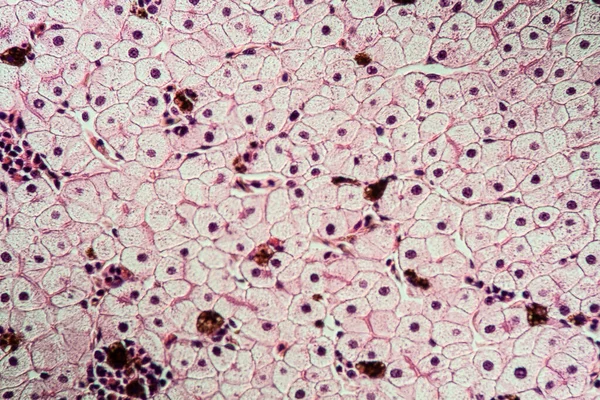
Hemosiderosis liver 100x under a microscope
Hemosiderosis Liver 100X Microscope — Stock Photo, Image

Tonsil Images: Exploring Our Library of Stock Images

When it comes to finding high-quality images of tonsils, our library has everything that you need to make your project stand out. Our collection includes a range of different image formats, including JPG, AI, and EPS, making it easy for you to find the perfect image for your purposes.

Our Selection of Tonsil Images

We offer a wide range of tonsil images that are perfect for use in educational materials, medical websites, and even advertising campaigns. From detailed graphics to high-resolution photographs, our library has everything that you need to make your project a success.

Our images are available in a range of different formats, making them easy to use in a variety of different contexts. Whether you are looking for a simple graphic to use in a brochure or a detailed photograph for a medical paper, our collection of tonsil images has something for everyone.

Choosing the Right Image for Your Project

When choosing an image for your project, it is important to think carefully about the context in which it will be used. Are you aiming to educate your audience about the anatomy of the tonsils, or are you trying to create a more visceral impact? Do you want your image to stand out or blend in with the surrounding text?

By carefully considering all of these factors, you can make an informed decision about which image to use. Additionally, our library of tonsil images offers a range of different visual styles, making it easy to find an image that perfectly matches your specific needs.

Using Images to Enhance Your Project

When used correctly, images can be an incredibly powerful tool for enhancing the impact of your project. By choosing the right image, you can grab your audience's attention and make your message more memorable.

With our collection of tonsil images, you can rest assured that you are getting high-quality visuals that will help you achieve your goals. So why wait? Browse our collection today and find the perfect image for your project!