Ulna Stock Photos

100,000 Ulna pictures are available under a royalty-free license

  • Best Match
  • Fresh
  • Popular
1-100 of 100,000
Palm Anatomy - Anatomy Bones
Palm Anatomy - Anatomy Bones — Stock Photo, Image
Male bones anatomy
Male bones anatomy — Stock Photo, Image
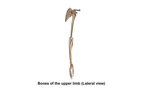
Bones of upper limb
Bones of upper limb — Stock Photo, Image
Human Skeleton Finger Joints
Human Skeleton Finger Joints — Stock Photo, Image
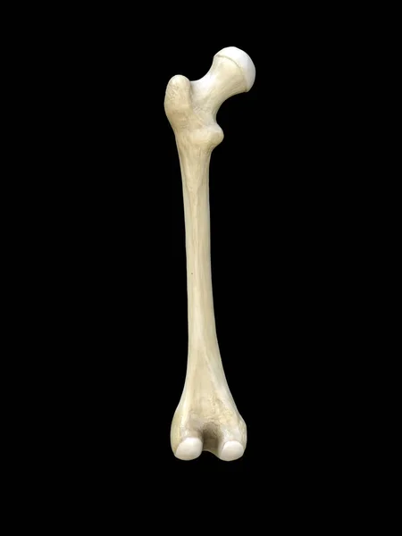
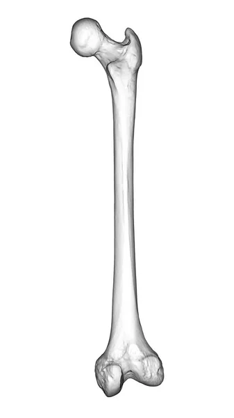
A femur bone, the largest bone in the human body located in the thigh, 3D illustration showcasing its shape and structure in back view
Femur Bone Largest Bone Human Body Located Thigh Illustration Showcasing — Stock Photo, Image
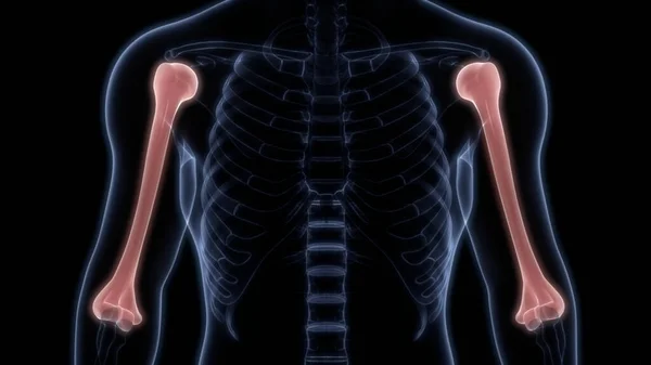
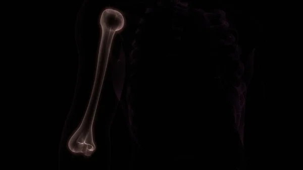
Humerus Bone Joints of Human Skeleton System Anatomy 3d rendering
Humerus Bone Joints Human Skeleton System Anatomy Rendering — Stock Photo, Image
Human Skeleton System Palm Hand Bone Joints Anatomy. 3D
Human Skeleton System Palm Hand Bone Joints Anatomy — Stock Photo, Image
Human Skeleton System Hand Bone Joints Anatomy. 3D
Human Skeleton System Hand Bone Joints Anatomy — Stock Photo, Image
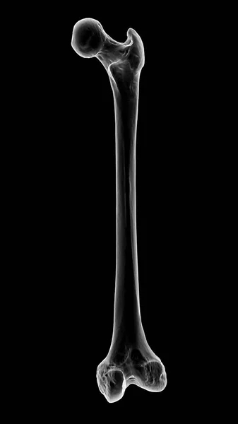
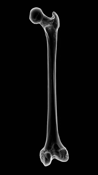
Left human femur bone, posterior view, close up isolated on black background, 3d rendering
Left Human Femur Bone Posterior View Close Isolated Black Background — Stock Photo, Image
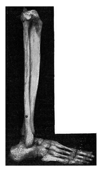
Skeleton of the lower thigh and foot of a Japanese seen externally, vintage engraved illustration. From the Universe and Humanity, 1910..
Skeleton Lower Thigh Foot Japanese Seen Externally Vintage Engraved Illustration — Stock Photo, Image
Tracheal and partial laryngeal stenosis following cicatrization of a gumma, vintage engraved illustration..
Tracheal Partial Laryngeal Stenosis Cicatrization Gumma Vintage Engraved Illustration — Stock Photo, Image
Brachioradialis muscle. Pain and trigger points in the hand.
Brachioradialis muscle. Pain and trigger points in the hand. — Stock Photo, Image
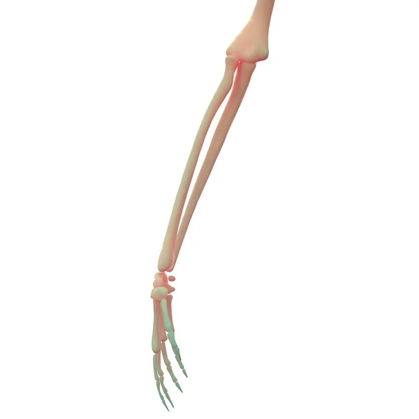
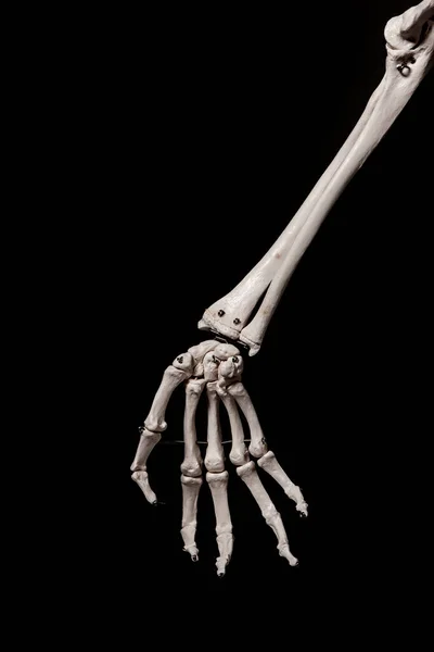
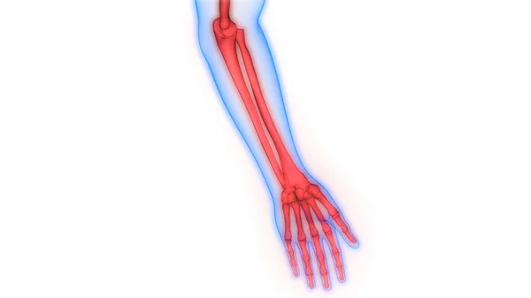
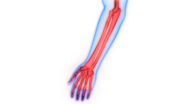
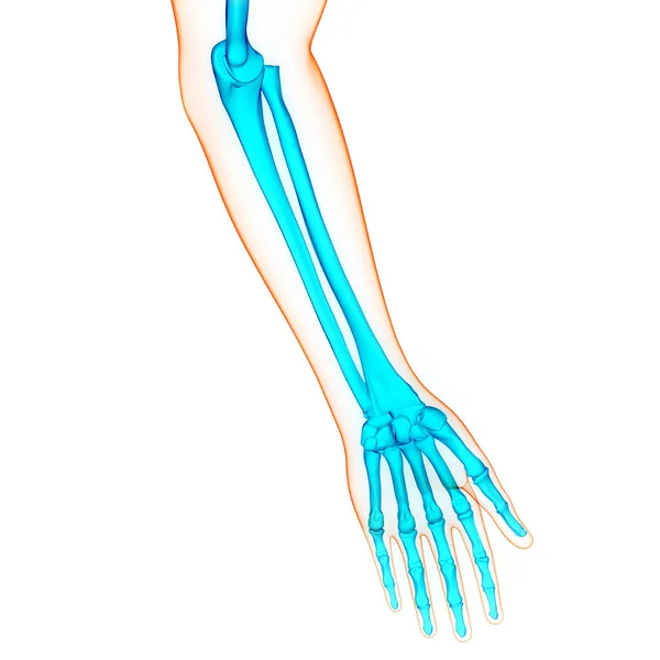
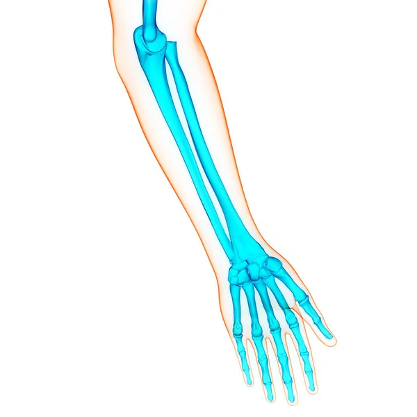
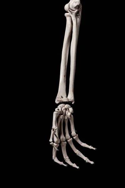
Human forearm skeleton anatomy bone
Human Forearm Skeleton Anatomy Bone — Stock Photo, Image
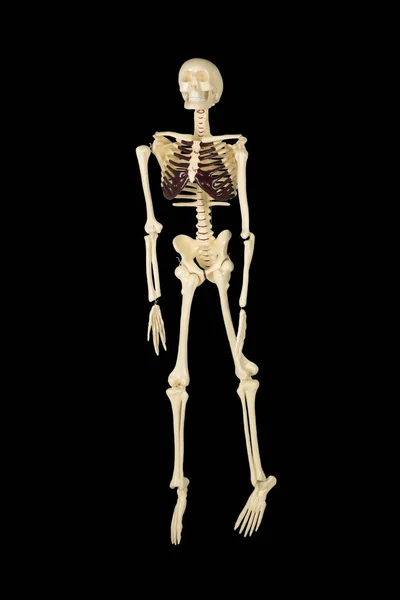
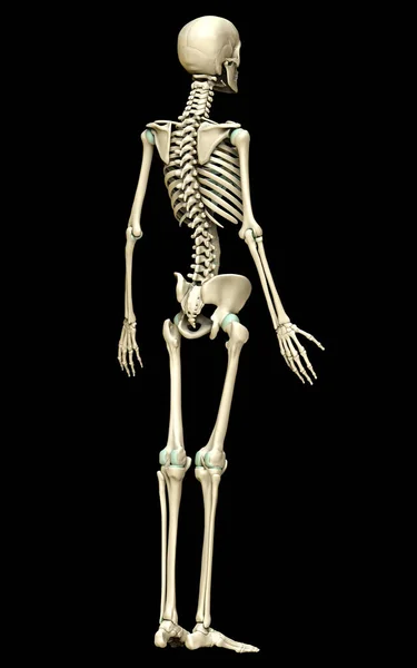
Full length of human skeleton in the studio with dark background
Full Length Human Skeleton Studio Dark Background — Stock Photo, Image
Black watercolor ink abstract shape background.
Black Watercolor Ink Abstract Shape Background — Stock Photo, Image
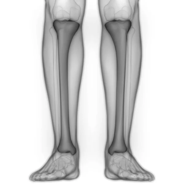
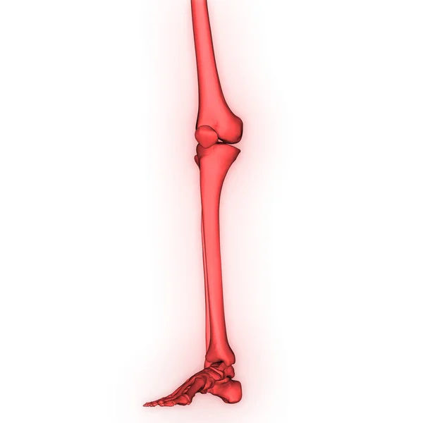
Human leg bones anatomy medical background
Human Leg Bones Anatomy Medical Background — Stock Photo, Image
Bones of the hand, computer illustration
Bones Hand Computer Illustration — Stock Photo, Image
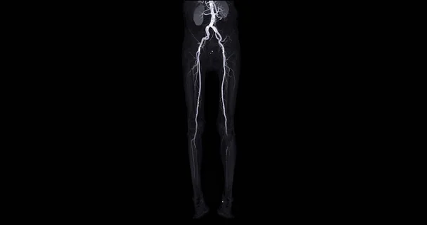
CTA femoral artery run off image of femoral artery for diagnostic Acute or Chronic Peripheral Arterial Disease.
Cta Femoral Artery Run Image Femoral Artery Diagnostic Acute Chronic — Stock Photo, Image
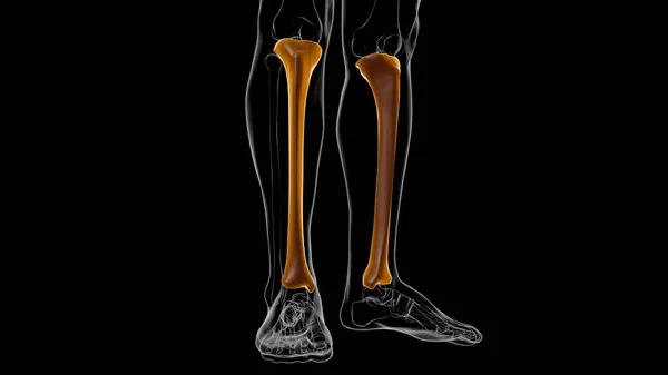
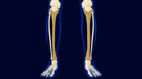
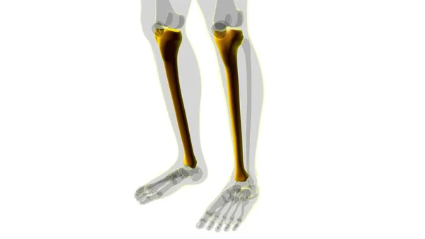
Human skeleton anatomy Tibia Bone 3D Rendering For Medical Concept
Human Skeleton Anatomy Tibia Bone Rendering Medical Concept — Stock Photo, Image
Human Skeleton System Hand Bone Joints Anatomy. 3D
Human Skeleton System Hand Bone Joints Anatomy — Stock Photo, Image
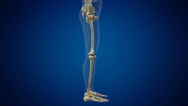
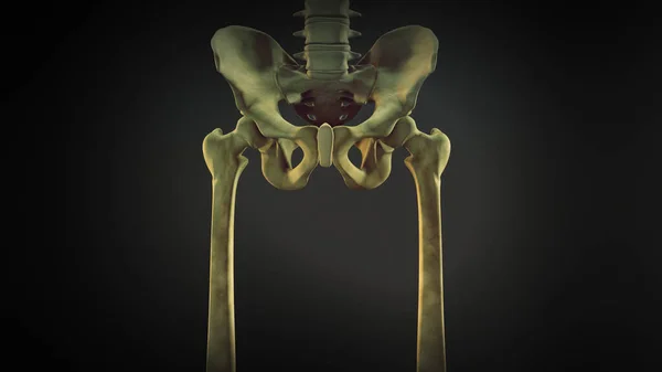
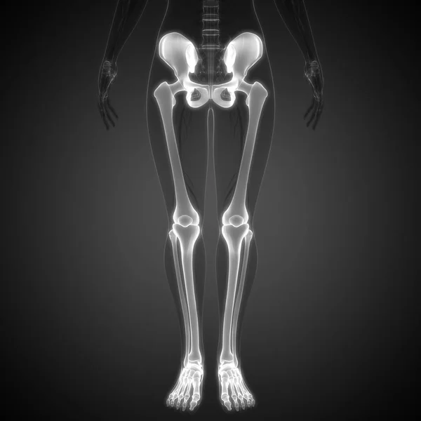
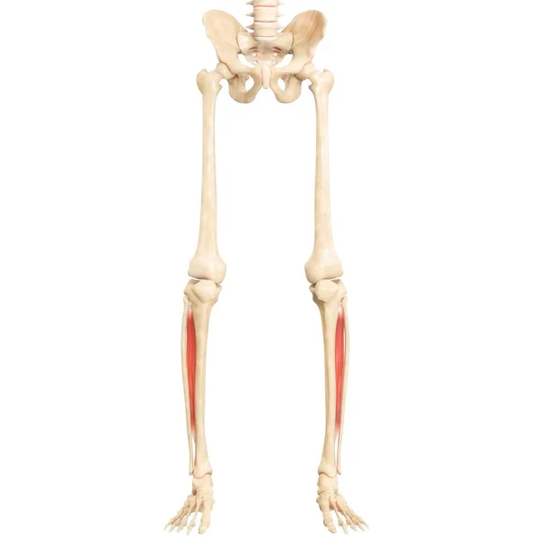
Pelvis and legs skeleton of human body
Pelvis Legs Skeleton Human Body — Stock Photo, Image
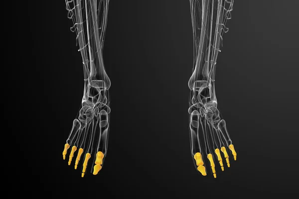
3d render medical illustration of the phalanges foot
3d render medical illustration of the phalanges foot — Stock Photo, Image
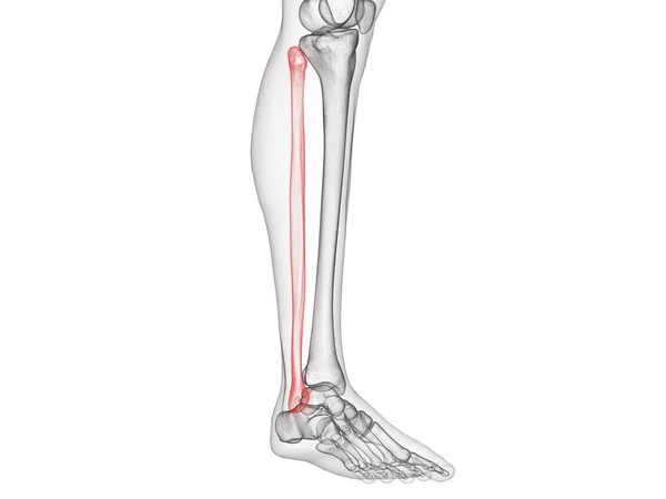
Fibula Bone Human skeleton anatomy 3D Rendering For Medical Concept
Fibula Bone Human Skeleton Anatomy Rendering Medical Concept — Stock Photo, Image
Human Skeleton System Hand Bone Joints Anatomy. 3D
Human Skeleton System Hand Bone Joints Anatomy — Stock Photo, Image
Human Skeleton System Palm Hand Bone Joints Anatomy. 3D
Human Skeleton System Palm Hand Bone Joints Anatomy — Stock Photo, Image
Human Skeleton System Hand Bone Joints Anatomy. 3D
Human Skeleton System Hand Bone Joints Anatomy — Stock Photo, Image
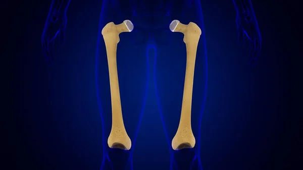
Human skeleton anatomy Femur Bone 3D Rendering For Medical Concept
Human Skeleton Anatomy Femur Bone Rendering Medical Concept — Stock Photo, Image
Human skeleton anatomy Tibia Bone 3D Rendering For Medical Concept
Human Skeleton Anatomy Tibia Bone Rendering Medical Concept — Stock Photo, Image
3D Illustration of Human Skeleton System Bone Joints Anatomy
Illustration Human Skeleton System Bone Joints Anatomy — Stock Photo, Image
Finger Joints with Knee Joint
Finger Joints with Knee Joint — Stock Photo, Image
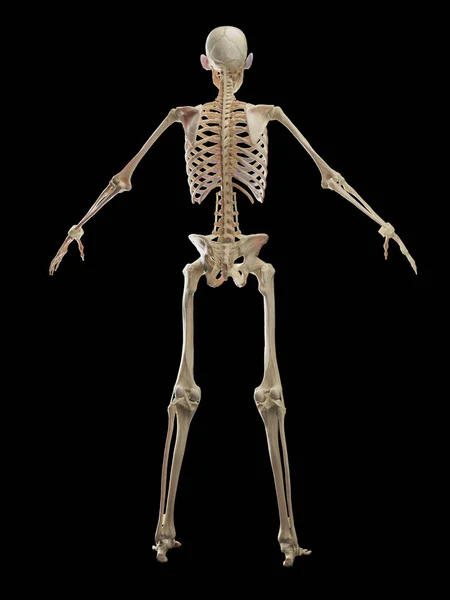
3d rendered, medically accurate illustration of the skeleton system
Rendered Medically Accurate Illustration Skeleton System — Stock Photo, Image
Muscle hand
Muscle hand — Stock Photo, Image
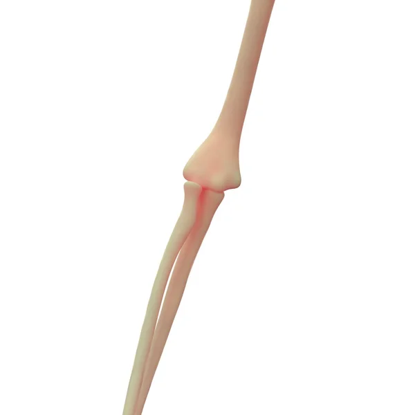
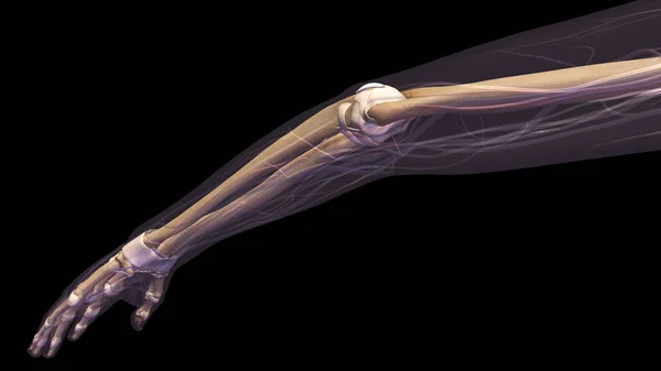
Human Skeleton Elbow Joint
Human Skeleton Elbow Joint — Stock Photo, Image
Broken arm xray
Broken arm xray — Stock Photo, Image
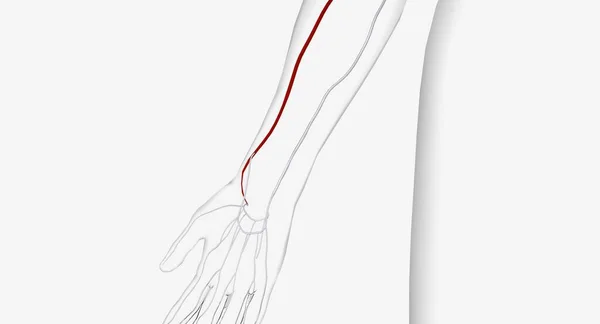
The radial artery is a major artery in the forearm that supplies blood to the lower arm and hand. 3D Render
Radial Artery Major Artery Forearm Supplies Blood Lower Arm Hand — Stock Photo, Image
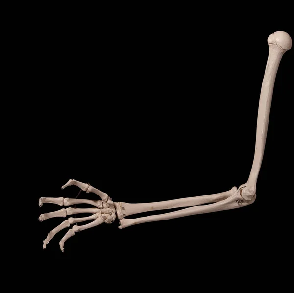
Human arm bones anatomy
Human arm bones anatomy — Stock Photo, Image
Human anatomy. The skeleton of the leg on a white background. 3D illustration
Human Anatomy Skeleton Leg White Background Illustration — Stock Photo, Image
Abstract ink background. Marble style. Black paint stroke texture on white paper. Wallpaper for web and game design. Grunge mud art. Macro image of pen juice. Dark Smear
Abstract Ink Background Marble Style Black Paint Stroke Texture White — Stock Photo, Image
3d rendered, medically accurate illustration of the skeleton system
Rendered Medically Accurate Illustration Skeleton System — Stock Photo, Image
Human forearm skeleton anatomy bone
Human Forearm Skeleton Anatomy Bone — Stock Photo, Image
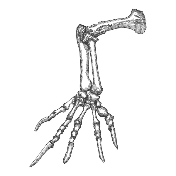
Bird foot sketch
Bird foot sketch — Stock Photo, Image
Computer illustration of a femur (thigh bone), the largest bone in the human body, showing its shape and structure in back view.
Computer Illustration Femur Thigh Bone Largest Bone Human Body Showing — Stock Photo, Image
Stylized drawing lizard bones
Stylized drawing lizard bones — Stock Photo, Image
2d cartoon illustration of arm bones
2d cartoon illustration of arm bones — Stock Photo, Image
Human Skeleton System Bone Joints Anatomy. 3D
Human Skeleton System Bone Joints Anatomy — Stock Photo, Image
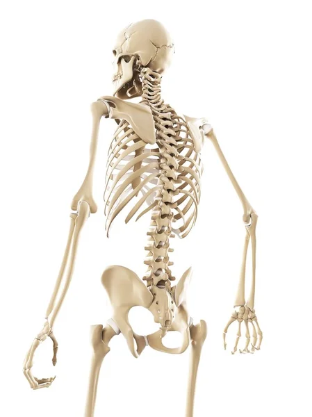
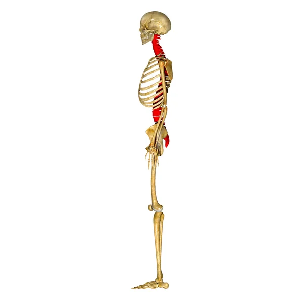
Human body, skeleton
Human body, skeleton — Stock Photo, Image
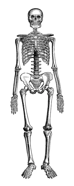
The Human Skeleton
The Human Skeleton — Stock Photo, Image
Anatomy of the nerves of the upper limb (arm) in palmar view.
Anatomy Nerves Upper Limb Arm Palmar View — Stock Photo, Image
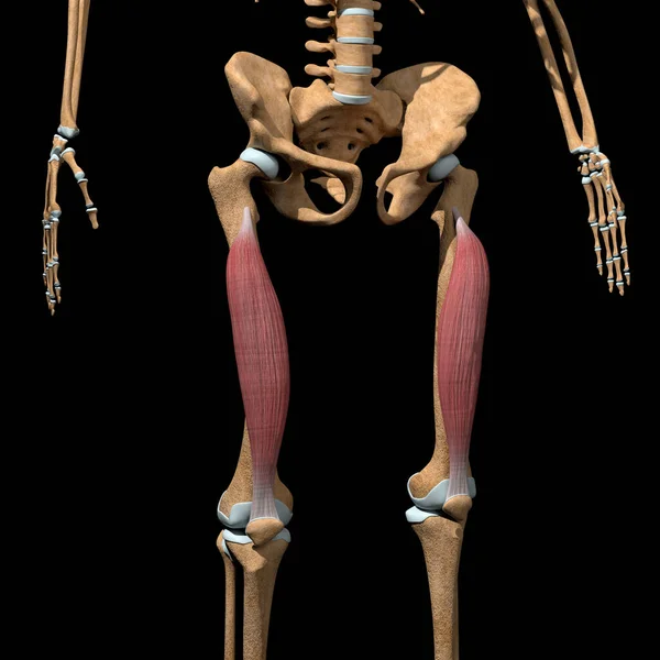
This 3d illustration shows the vastus intermedius muscles on skeleton
Illustration Shows Vastus Intermedius Muscles Skeleton — Stock Photo, Image
Tibia Bones Human skeleton anatomy 3D Rendering For Medical Concept
Tibia Bones Human Skeleton Anatomy Rendering Medical Concept — Stock Photo, Image
Human skeleton anatomy Fibula Bone 3D Rendering For Medical Concept
Human Skeleton Anatomy Fibula Bone Rendering Medical Concept — Stock Photo, Image
3D Illustration of Human Skeleton System Anatomy
Illustration Human Skeleton System Anatomy — Stock Photo, Image
Bamboo Bird Whistle on Black Background
Bamboo Bird Whistle on Black Background — Stock Photo, Image
Abstract black and white background. Monochrome texture.
Abstract Black White Background Monochrome Texture — Stock Photo, Image
Spooky 3d human skeleton screaming
Spooky Human Skeleton Screaming — Stock Photo, Image
Right human femur bone, posterior view, bone anatomy, white background, 3d rendering
Right Human Femur Bone Posterior View Bone Anatomy White Background — Stock Photo, Image
3D Illustration, Muscle is a soft tissue, Muscle cells contain proteins , producing a contraction that changes both the length and the shape of the cell. Muscles function to produce force and motion.
Illustration Muscle Soft Tissue Muscle Cells Contain Proteins Producing Contraction — Stock Photo, Image
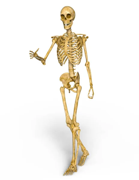
3D CG rendering of a skeleton
3D CG rendering of a skeleton — Stock Photo, Image
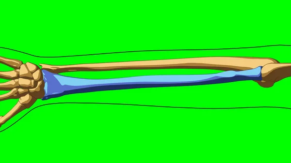
Human skeleton Radius bone anatomy for medical concept 3D illustration with green matte
Human Skeleton Radius Bone Anatomy Medical Concept Illustration Green Matte — Stock Photo, Image
Human Muscular System Muscles Anatomy. 3D
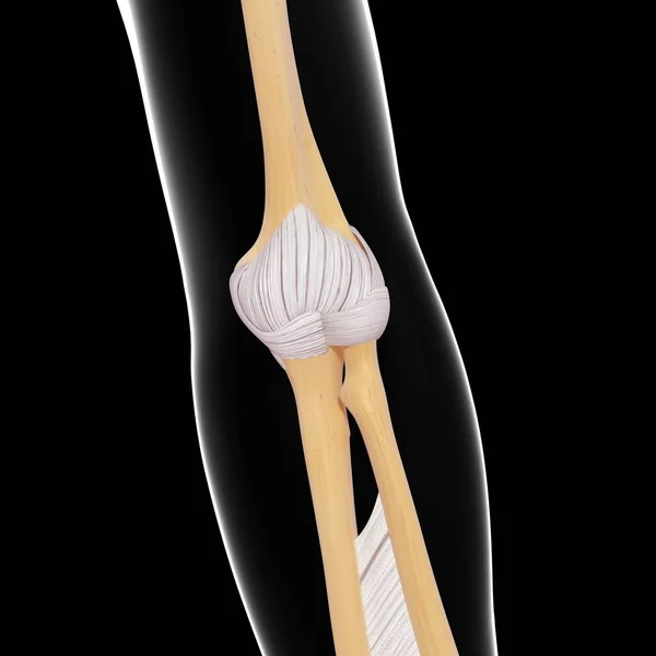
Human Muscular System Muscles Anatomy — Stock Photo, Image
Humerus Bone Joints of Human Skeleton System Anatomy 3d rendering
Humerus Bone Joints Human Skeleton System Anatomy Rendering — Stock Photo, Image
Human skeletal system
Human skeletal system — Stock Photo, Image
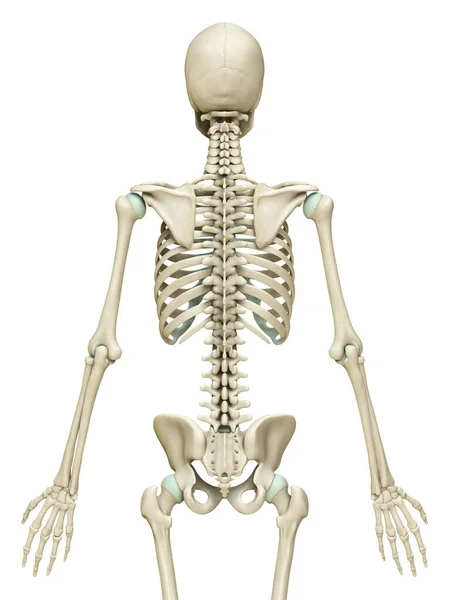
Male skeletal system, illustration
Male Skeletal System Illustration — Stock Photo, Image
3d rendered, medically accurate illustration of the skeleton system
Rendered Medically Accurate Illustration Skeleton System — Stock Photo, Image
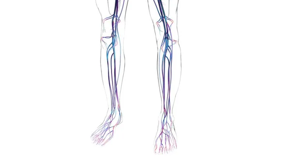
Human Arteries And Veins For Medical Concept 3D Illustration
Human Arteries Veins Medical Concept Illustration — Stock Photo, Image
3D Illustration of Human Skeleton System Bone Joints Anatomy
Illustration Human Skeleton System Bone Joints Anatomy — Stock Photo, Image
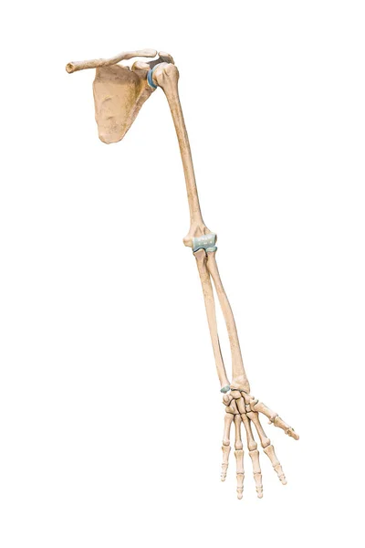
Accurate anterior or front view of the arm or upper limb bones of the human skeletal system isolated on white background 3D rendering illustration. Anatomy, medical, osteology concept.
Accurate Anterior Front View Arm Upper Limb Bones Human Skeletal — Stock Photo, Image
Spooky 3d human skeleton screaming
Spooky Human Skeleton Screaming — Stock Photo, Image
The arteries of the upper limb in palmar view.
Arteries Upper Limb Palmar View — Stock Photo, Image
Human skeleton anatomy Radius Bone 3D Rendering For Medical Concept
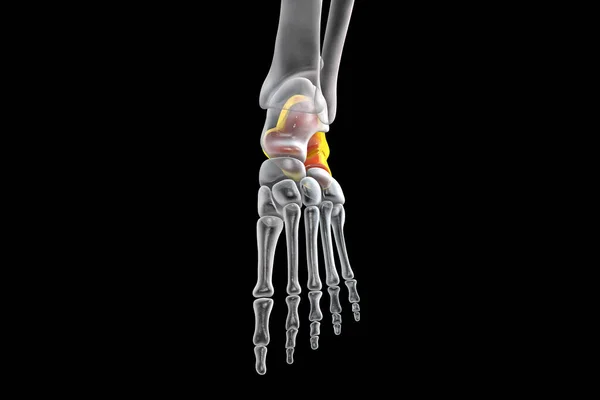
Human Skeleton Anatomy Radius Bone Rendering Medical Concept — Stock Photo, Image
Human Skeleton System Bones (Hands) Anatomy. 3D
Human Skeleton System Bones Hands Anatomy — Stock Photo, Image
3D rendering of a human skeleton isolated on white background
Rendering Human Skeleton Isolated White Background — Stock Photo, Image
Spinal cord
Spinal cord — Stock Photo, Image
Human skeleton anatomy Tibia Bone 3D Rendering For Medical Concept
Human Skeleton Anatomy Tibia Bone Rendering Medical Concept — Stock Photo, Image
Human Skeleton Tibia, Femur and Fibula Bones
Human Skeleton Tibia, Femur and Fibula Bones — Stock Photo, Image
2d cartoon illustration of arm bones
2d cartoon illustration of arm bones — Stock Photo, Image
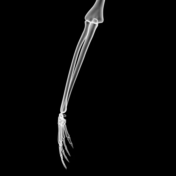
Human arm with elbow joint and bones x-ray on black background
Human Arm Elbow Joint Bones Ray Black Background — Stock Photo, Image
Fibula bone in skeleton of human body, computer illustration.
Fibula Bone Skeleton Human Body Computer Illustration — Stock Photo, Image
Human skeleton anatomy Tibia Bone 3D Rendering For Medical Concept
Human Skeleton Anatomy Tibia Bone Rendering Medical Concept — Stock Photo, Image
Right human femur bone, white background, 3d rendering
Right Human Femur Bone White Background Rendering — Stock Photo, Image
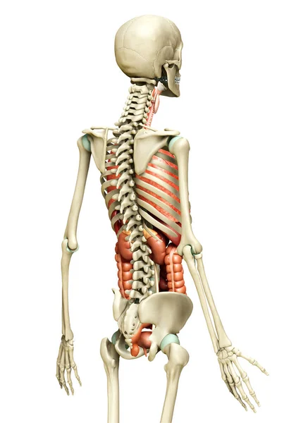
3d rendered medically accurate illustration of Internal organs and skeleton system
Rendered Medically Accurate Illustration Internal Organs Skeleton System — Stock Photo, Image
Abstract ink background. Marble style. Black paint stroke texture on white paper. Wallpaper for web and game design. Grunge mud art. Macro image of pen juice. Dark Smear
Abstract Ink Background Marble Style Black Paint Stroke Texture White — Stock Photo, Image
Tibia and Fibula Joints of Human Skeleton System Anatomy 3D Rendering
Tibia Fibula Joints Human Skeleton System Anatomy Rendering — Stock Photo, Image
Human skeleton anatomy Femur Bone 3D Rendering For Medical Concept
Human Skeleton Anatomy Femur Bone Rendering Medical Concept — Stock Photo, Image
Abstract black and white background template art, 3d render.
Abstract Black White Background Template Art Render — Stock Photo, Image
Computer illustration of a femur (thigh bone), the largest bone in the human body, showing its shape and structure in back view.
Computer Illustration Femur Thigh Bone Largest Bone Human Body Showing — Stock Photo, Image
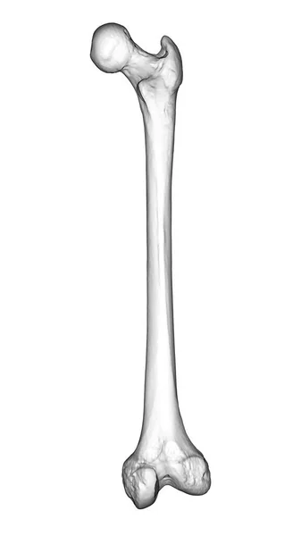
A femur bone, the largest bone in the human body located in the thigh, 3D illustration showcasing its shape and structure in back view
Femur Bone Largest Bone Human Body Located Thigh Illustration Showcasing — Stock Photo, Image
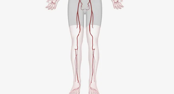
Lower extremity arterial interventions are procedures designed to restore blood flow to your legs and feet. 3D rendering
Lower Extremity Arterial Interventions Procedures Designed Restore Blood Flow Your — Stock Photo, Image
Human skeleton anatomy Fibula Bone 3D Rendering For Medical Concept
Human Skeleton Anatomy Fibula Bone Rendering Medical Concept — Stock Photo, Image
Skeleton
Skeleton — Stock Photo, Image
Human forearm skeleton anatomy bone
Human Forearm Skeleton Anatomy Bone — Stock Photo, Image
Human Body Bones Joint Pains (Leg Joints with Hip and Pelvis) - Illustration
Human Body Bones Joint Pains Leg Joints Hip Pelvis Illustration — Stock Photo, Image
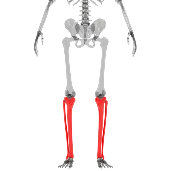
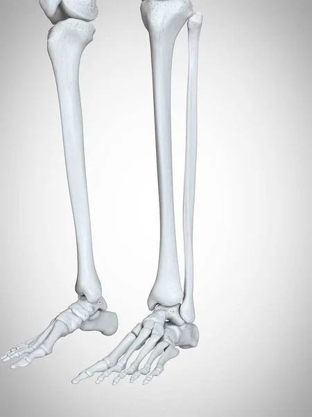
3d rendered illustration of lower legs and feet bones in human skeleton.
Rendered Illustration Lower Legs Feet Bones Human Skeleton — Stock Photo, Image
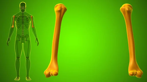
Human skeleton anatomy Humerus Bone 3D Rendering For Medical Concept
Human Skeleton Anatomy Humerus Bone Rendering Medical Concept — Stock Photo, Image
Human Skeleton
Human Skeleton — Stock Photo, Image
X-ray of a person's hand (3d rendering)
Ray Person Hand Rendering — Stock Photo, Image
3d rendered, medically accurate illustration of the skeleton system
Rendered Medically Accurate Illustration Skeleton System — Stock Photo, Image
Human Skeleton System Palm Hand Bone Joints Anatomy. 3D
Human Skeleton System Palm Hand Bone Joints Anatomy — Stock Photo, Image
Illustration of the calcaneus, or heel, bone of the foot.
Illustration Calcaneus Heel Bone Foot — Stock Photo, Image

Related image searches

Find the Perfect Ulna Images for Your Project

Ulna images are essential in the medical and anatomy fields for visualizing the forearm's inner structures and bones. If you're working on a project related to this field, you need high-quality images that can explain the intricate details of the ulna. In this article, we'll provide you with an overview of the different types of ulna images available, their usage, and where you can find them.

Types of Ulna Images

There are several types of ulna images that you can leverage to create an effective visual representation of the forearm's structure. These include:

  • Real-life photographs of the ulna bone
  • Illustrations of the ulna bone
  • 3D-rendered images of the ulna bone
  • X-ray images of the ulna bone

Each type of image has its unique advantages, and the type you choose largely depends on the purpose of your project and the level of detail you want to show. For instance, real-life photographs of the ulna bone are ideal for educational purposes and can be used in textbooks, while 3D-rendered images are perfect for medical presentations.

Where to Find Ulna Images

You can find ulna images from various sources such as stock image websites, academic resources, or even hospitals. However, interested parties should also seek access to publicly available bone scan databases which can help them find and publish images of ulnas.

Stock image websites are an effective way to access a diverse range of ulna images. Many of these sites offer a vast collection of ulna images in different formats, sizes, and resolutions to suit your project's needs. Additionally, university or hospital websites may provide 3D modelling services which can help in the creation of 3D-rendered images of the ulna bone.

Using Ulna Images Correctly

Choosing the right type of ulna image is just the starting point; using it correctly is essential in creating an impact. Here are some tips on how to use ulna images correctly:

  • Choose the right type of image that suits your project's purpose and level of detail.
  • Ensure the image's resolution is high enough for your project. Low-resolution images can appear pixelated when zoomed in, reducing their quality.
  • Use images consistently throughout your project for emphasis on the ulna's structure.
  • Use images to supplement or reinforce your text and not as a replacement for it.
  • Give credit if the image is not your original creation, and read the licensing agreement before using any image.

In Conclusion

Ulna images play an essential role in illustrating the intricate structures and details of the forearm bone. You can find them in different formats, shapes, and sizes, depending on your project's needs. Remember to use them correctly by including them as a supplement to your text and giving credit where needed. With these tips in mind, you can create effective, impactful projects with high-quality ulna images.