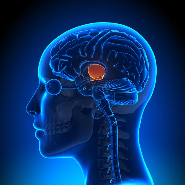
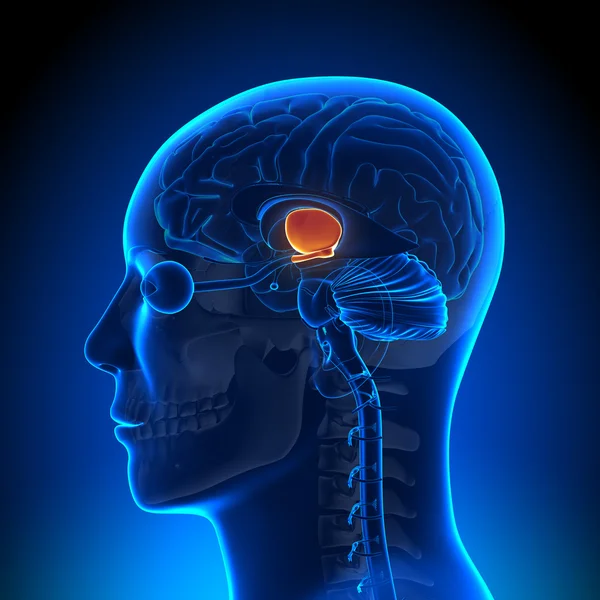
Hypothalamus - Female Brain Anatomy
Hypothalamus - Female Brain Anatomy — Stock Photo, Image
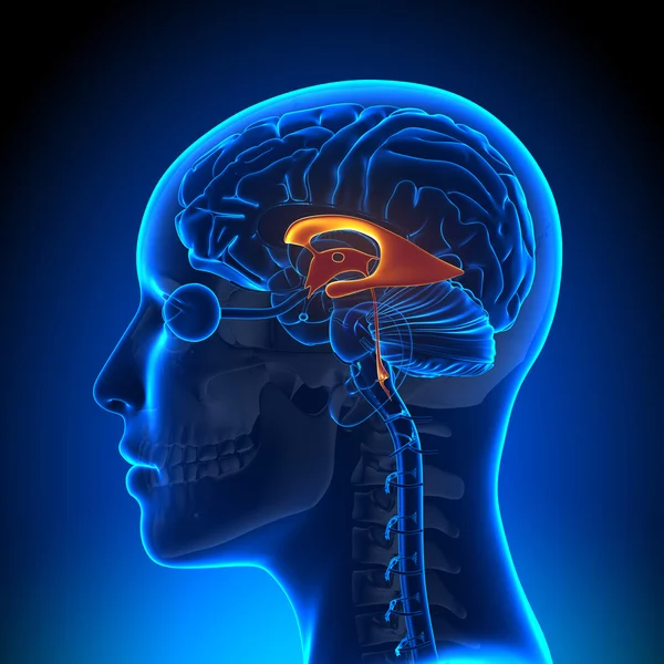
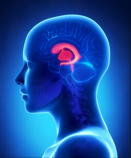
Female Ventricles - Anatomy Brain
Female Ventricles - Anatomy Brain — Stock Photo, Image
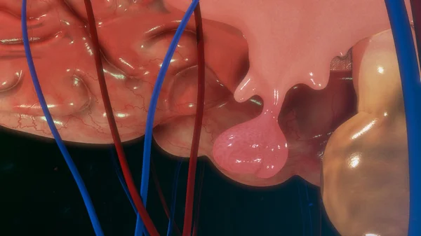
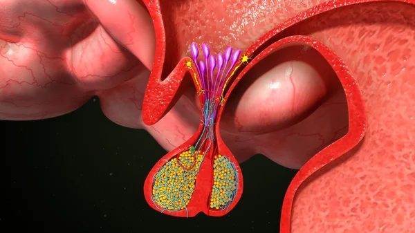
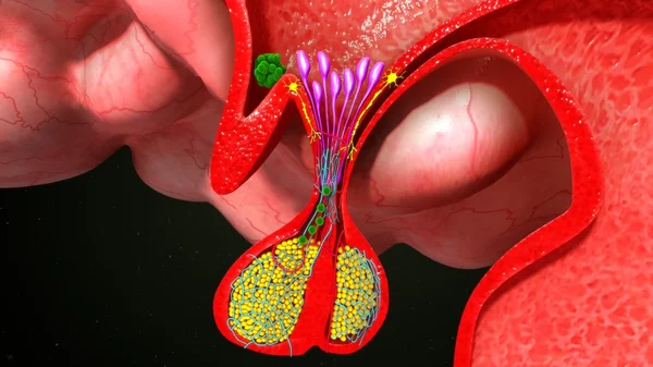
Pituitary gland anatomy
Pituitary gland anatomy — Stock Photo, Image
Human body of brain
Human body of brain — Stock Photo, Image
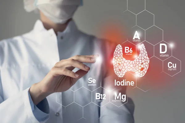
Essential nutrients for Thyroid Gland health including Iodine, Selenuim, Vitamin B6, Cuprum. Blurred portrait of doctor holding highlighted red Thyroid Gland.
Essential Nutrients Thyroid Gland Health Including Iodine Selenuim Vitamin Cuprum — Stock Photo, Image
Pituitary gland anatomy
Pituitary gland anatomy — Stock Photo, Image
Human brain antomy
Human brain antomy — Stock Photo, Image
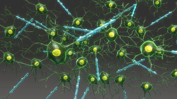
Structure of Neuron aerial
Structure of Neuron aerial — Stock Photo, Image
Human body of brain
Human body of brain — Stock Photo, Image
Pituitary gland anatomy
Pituitary gland anatomy — Stock Photo, Image
Brain Anatomy - Hippotalamus
Brain Anatomy - Hippotalamus — Stock Photo, Image
Woman eats night stole the refrigerator
Woman eats night stole the refrigerator — Stock Photo, Image
Pituitary gland anatomy
Pituitary gland anatomy — Stock Photo, Image
Front ventricles - female brain anatomy lateral view
Front ventricles - female brain anatomy lateral view — Stock Photo, Image
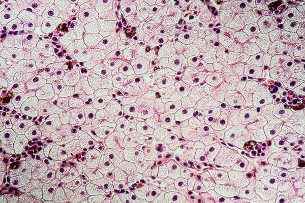
Liver cells of a rabit with Coccidia parasites.
Liver cells of a rabit with Coccidia parasites. — Stock Photo, Image
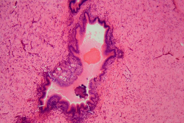
Neurofibromatosis, light micrograph, photo under microscope. A group of genetic disorders with formation of tumors in the nervous system
Neurofibromatosis Light Micrograph Photo Microscope Group Genetic Disorders Formation Tumors — Stock Photo, Image
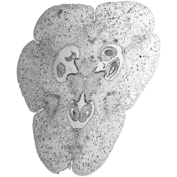
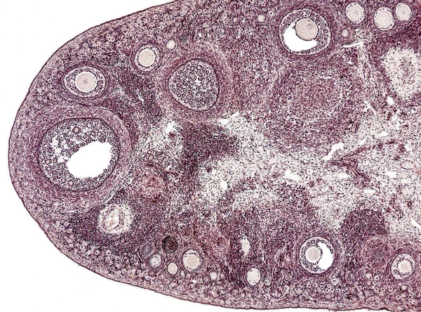
Black and white Lily ovary micrograph
Black and white Lily ovary micrograph — Stock Photo, Image
Mitosis cell in the Root tip of Onion under a microscope.
Mitosis Cell Root Tip Onion Microscope — Stock Photo, Image
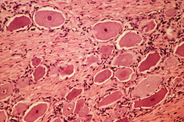
Spinal ganglion view from microscope
Spinal Ganglion View Microscope — Stock Photo, Image
Purulent meningitis, light micrograph, photo under microscope
Purulent Meningitis Light Micrograph Photo Microscope — Stock Photo, Image
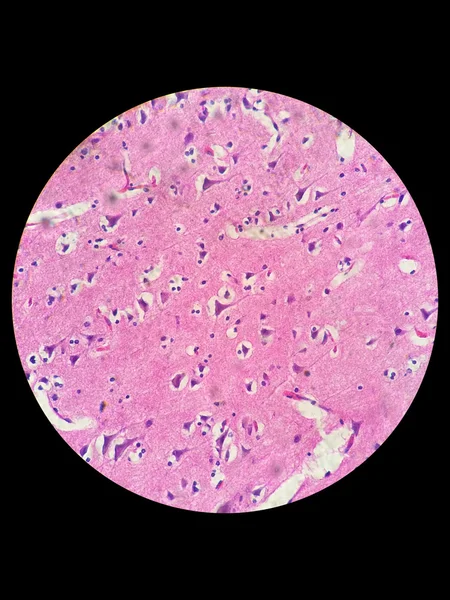
Nerve cells under light microscope
Nerve cells under light microscope — Stock Photo, Image
Microbiology, microscopic cells surface
Microbiology Microscopic Cells Surface — Stock Photo, Image
Abstract wallpaper, cells tissue microscope shot
Abstract Wallpaper Cells Tissue Microscope Shot — Stock Photo, Image
Adrenal Gland anatomy illustration. Gland and Kidney illustration
Adrenal Gland Anatomy Illustration Gland Kidney Illustration — Stock Photo, Image
Tissue with schistosomiasis sick tissue 100x
Tissue Schistosomiasis Sick Tissue 100X — Stock Photo, Image
Education Spinal cord and Motor Neuron under the microscope in Lab.
Education Spinal Cord Motor Neuron Microscope Lab — Stock Photo, Image
Illustration of a skeleton of an old human skull
Illustration Skeleton Old Human Skull — Stock Photo, Image
Cells of human uterus tissue with inoffensive tumor cells
Cells of human uterus tissue with inoffensive tumor cells — Stock Photo, Image
Axolot amphibians with liver cells 100x
Axolot Amphibians Liver Cells 100X — Stock Photo, Image
Microscope photo of a section through dog smooth muscle cells
Microscope Photo Section Dog Smooth Muscle Cells — Stock Photo, Image
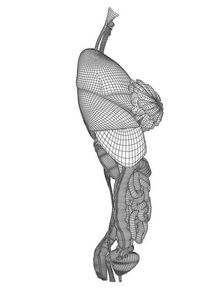
Wireframe digestive system
Wireframe digestive system — Stock Photo, Image
Red blood cells medical science background.
Red blood cells medical science background. — Stock Photo, Image
Thyroid follicular carcinoma, light micrograph, photo under microscope
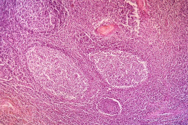
Thyroid Follicular Carcinoma Light Micrograph Photo Microscope — Stock Photo, Image
Thorax organs for anatomy designs
Thorax organs for anatomy designs — Stock Photo, Image
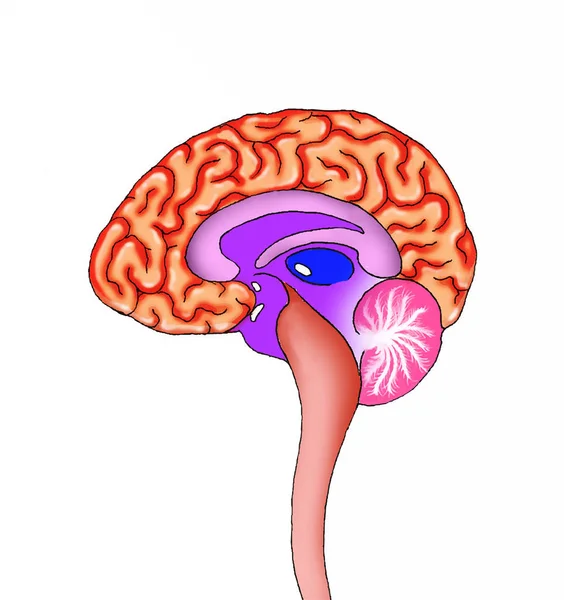
Human brain anatomy on white background
Human Brain Anatomy White Background — Stock Photo, Image
Microscopic cells texture, abstract background
Microscopic Cells Texture Abstract Background — Stock Photo, Image
Purple color weathered cement wall pattern.
Purple color weathered cement wall pattern. — Stock Photo, Image
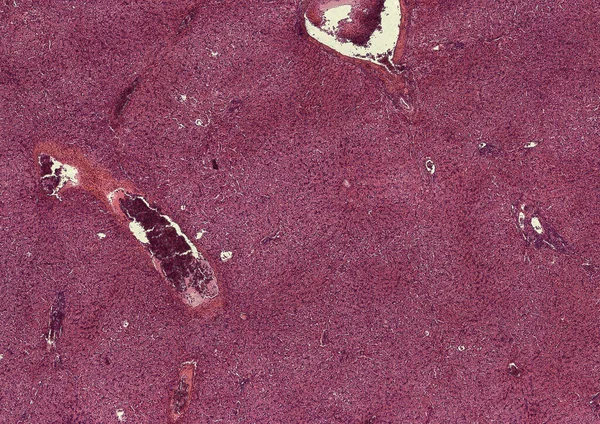
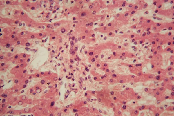
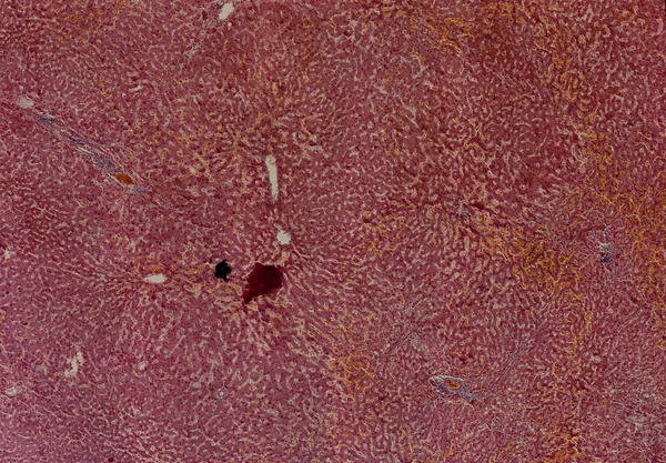
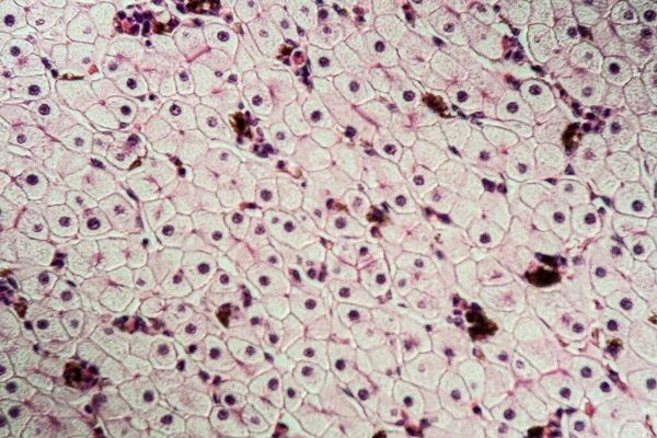
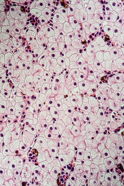
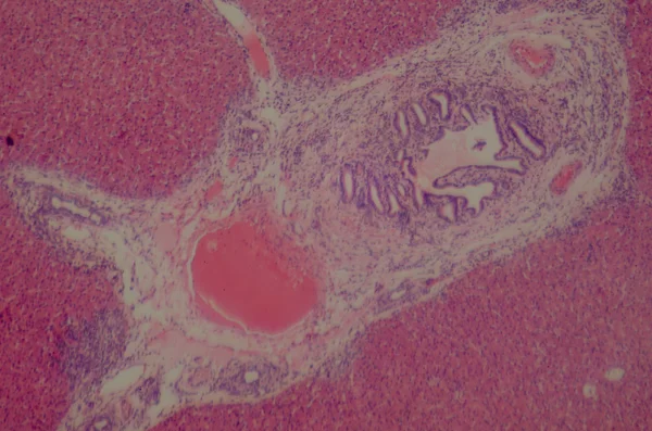
Microscopic section of human liver
Microscopic section of human liver — Stock Photo, Image
Light micrograph (LM) of a section through the fingertip.
Light Micrograph Section Fingertip — Stock Photo, Image
Micrograph of small intestinum tenue tissue
Micrograph of small intestinum tenue tissue — Stock Photo, Image
Biology cells, microscopic texture
Biology Cells Microscopic Texture — Stock Photo, Image
Cliated epithelium of human under the microscope in Lab.
Cliated Epithelium Human Microscope Lab — Stock Photo, Image
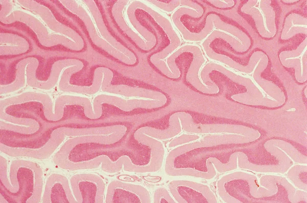
Cerebellum view from microscope
Cerebellum View Microscope — Stock Photo, Image
Abstract ink background. Marble style. Black paint stroke texture on white paper. Wallpaper for web and game design. Grunge mud art. Macro image of pen juice. Dark Smear
Abstract Ink Background Marble Style Black Paint Stroke Texture White — Stock Photo, Image
Stains of paint on paper. The texture of the splashes and spots of paint. EBRU- Ancient oriental drawing technique. Chaotic abstract organic design. Marbleized.
Stains Paint Paper Texture Splashes Spots Paint Ebru Ancient Oriental — Stock Photo, Image
Abstract wallpaper, cells tissue microscope shot
Abstract Wallpaper Cells Tissue Microscope Shot — Stock Photo, Image
Pork liver cut 100x
Pork Liver Cut 100X — Stock Photo, Image
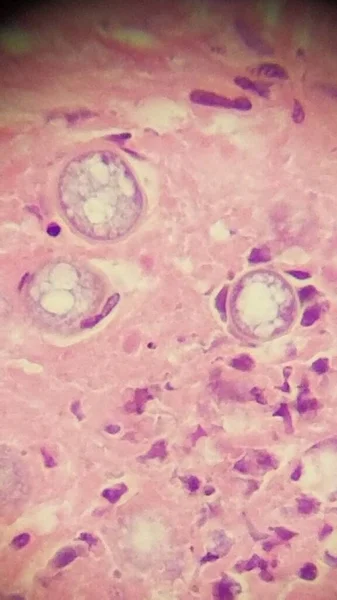
Coccidioides imitis spherules in skin biopsy specimen
Coccidioides imitis spherules in skin biopsy specimen — Stock Photo, Image
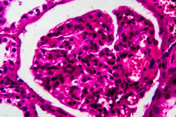
Cross section of the kidney 100x
Cross Section Kidney 100X — Stock Photo, Image
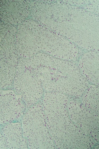
Testicle cross-section under the microscope 100x
Testicle Cross Section Microscope 100X — Stock Photo, Image
Human liver cells with tuberculosis under the microscope.
Human liver cells with tuberculosis under the microscope. — Stock Photo, Image
Abstract wallpaper, cells tissue microscope shot
Abstract Wallpaper Cells Tissue Microscope Shot — Stock Photo, Image
Hold the brain in hand black and white graphics, developmental psychology, icon, logo, vector,
Hold Brain Hand Black White Graphics Developmental Psychology Icon Logo — Stock Photo, Image
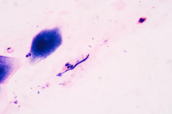
Budding yeast cells with pseudohyphae from sputum gram stain tes
Budding yeast cells with pseudohyphae from sputum gram stain tes — Stock Photo, Image
Mesh digestive system
Mesh digestive system — Stock Photo, Image
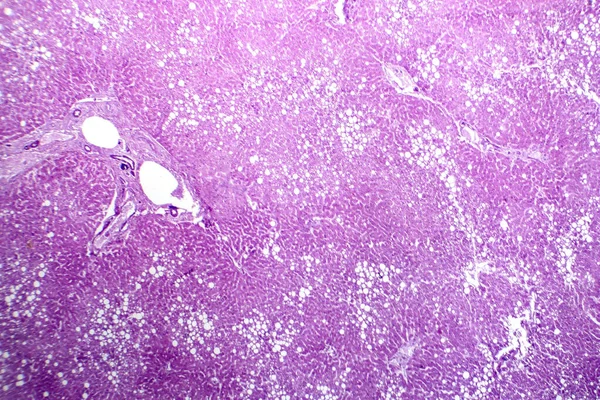
Photomicrograph of hepatic steatosis, revealing fat accumulation in liver cells, known as fatty liver disease.
Photomicrograph Hepatic Steatosis Revealing Fat Accumulation Liver Cells Known Fatty — Stock Photo, Image
Brain on blue background
Brain on blue background — Stock Photo, Image
Anatomy and Histological Ovary and Testis human cells under microscope.
Anatomy Histological Ovary Testis Human Cells Microscope — Stock Photo, Image
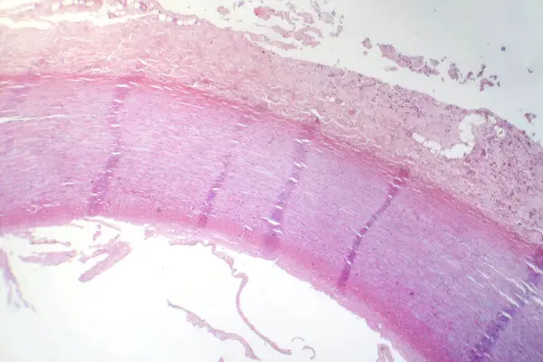
Photomicrograph of fibrinoid necrosis in a vessel wall, displaying damaged vessel integrity and immune response-related changes.
Photomicrograph Fibrinoid Necrosis Vessel Wall Displaying Damaged Vessel Integrity Immune — Stock Photo, Image
Abstract wallpaper, cells tissue microscope shot
Abstract Wallpaper Cells Tissue Microscope Shot — Stock Photo, Image
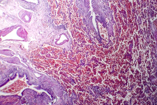
Photomicrograph of acute pulmonary hemorrhage, showing blood-filled alveoli and lung tissue damage due to bleeding.
Photomicrograph Acute Pulmonary Hemorrhage Showing Blood Filled Alveoli Lung Tissue — Stock Photo, Image
Original artwork photo of marble ink abstract art. High resolution photograph from exemplary original painting. Abstract painting was painted on HQ paper texture to create smooth marbling pattern.
Original Artwork Photo Marble Ink Abstract Art High Resolution Photograph — Stock Photo, Image
Red Bacteria cells in
Red Bacteria cells in — Stock Photo, Image
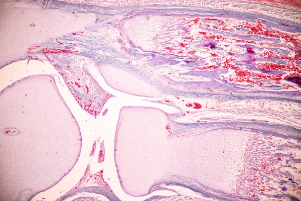
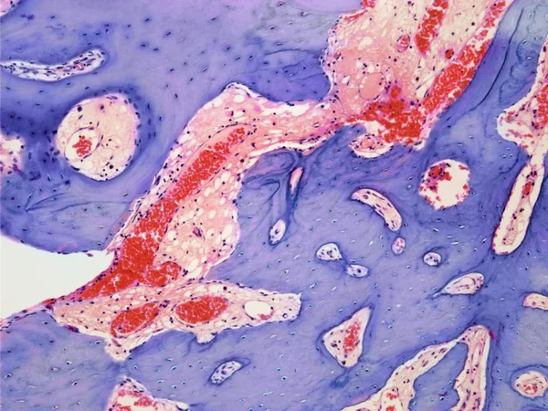
Anatomy and Histological Bone, Elastic cartilage human and Joint of human foetus under the microscope for education.
Anatomy Histological Bone Elastic Cartilage Human Joint Human Foetus Microscope — Stock Photo, Image
Abstract grunge background. Monochrome texture. Black and white textured background
Abstract Grunge Background Monochrome Texture Black White Textured Background — Stock Photo, Image
Abstract ink background. Marble style. Black paint stroke texture on white paper. Wallpaper for web and game design. Grunge mud art. Macro image of pen juice. Dark Smear
Abstract Ink Background Marble Style Black Paint Stroke Texture White — Stock Photo, Image
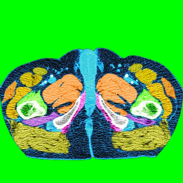
Man pelvis and perineum
Man pelvis and perineum — Stock Photo, Image
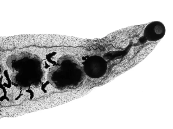
Liver fluke(Parasitic flatworm) of cattle and other grazing animals.
Liver Fluke Parasitic Flatworm Cattle Other Grazing Animals — Stock Photo, Image
Colon organ and digestive system
Colon organ and digestive system — Stock Photo, Image
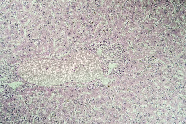
Liver mouse,schistosomiasis diseased tissue 100x
Liver Mouse Schistosomiasis Diseased Tissue 100X — Stock Photo, Image
Dramatic black, monochrome abstraction, reminiscent of dragon scales or caked lava, shimmering. Fragment of a modern artwork. Macro photography of the smallest details of a panting. Contemporary art. Artistic background image.
Dramatic Black Monochrome Abstraction Reminiscent Dragon Scales Caked Lava Shimmering — Stock Photo, Image
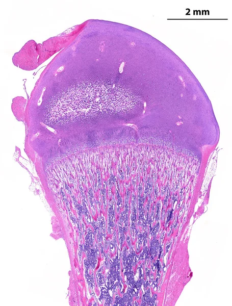
Low power light microscope micrograph showing a developing long bone (femur). On top, the epiphysis is made by hyaline cartilage that shows an initial secondary ossification centre. On bottom, the secondary diaphyseal ossification centre. The round l
Low Power Light Microscope Micrograph Showing Developing Long Bone Femur — Stock Photo, Image
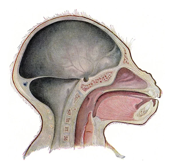
Median section through the head of a young gorilla, vintage engraving
Median Section Head Young Gorilla Vintage Engraving — Stock Photo, Image
Muscle cells
Muscle cells — Stock Photo, Image
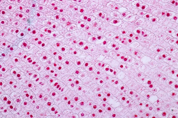
Root tip of Onion and Mitosis cell in the Root tip of Onion under a microscope.
Root Tip Onion Mitosis Cell Root Tip Onion Microscope — Stock Photo, Image
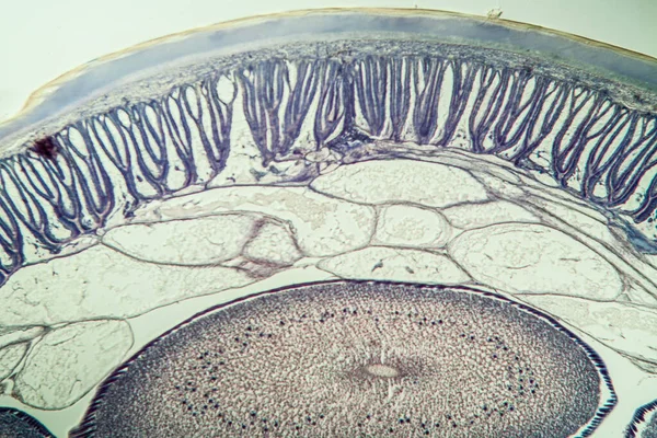
Horse roundworm in cross section with eggs 100x
Horse Roundworm Cross Section Eggs 100X — Stock Photo, Image
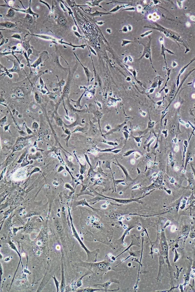
Cells of human find with microscope.
Cells of human find with microscope. — Stock Photo, Image
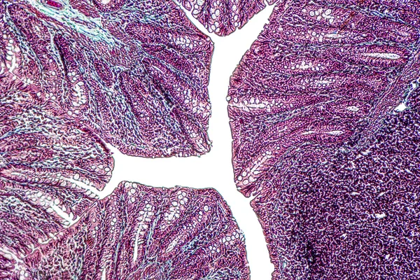
Photomicrograph of lung adenocarcinoma, illustrating malignant glandular cells characteristic of the most common type of lung cancer.
Photomicrograph Lung Adenocarcinoma Illustrating Malignant Glandular Cells Characteristic Most Common — Stock Photo, Image
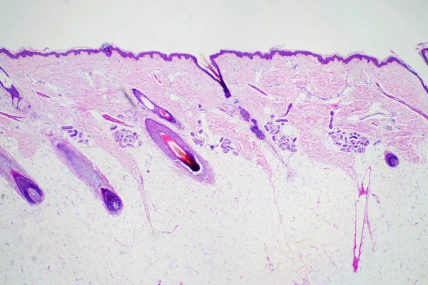
Cross section human skin head under microscope view for education histology. Histological for human physiology.
Cross Section Human Skin Head Microscope View Education Histology Histological — Stock Photo, Image
Root tip of Onion and Mitosis cell in the Root tip of Onion under a microscope.
Root Tip Onion Mitosis Cell Root Tip Onion Microscope — Stock Photo, Image
Reflex action system
Reflex action system — Stock Photo, Image
Cross section of a cerebellum. Light micrograph.
Cross Section Cerebellum Light Micrograph — Stock Photo, Image
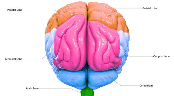
Central organ of Human Nervous System Brain Lobes with detailed Labels Anatomy.
Central Organ Human Nervous System Brain Lobes Detailed Labels Anatomy — Stock Photo, Image
Adrenocortical adenoma, light micrograph, photo under microscope. Benign tumor that emerges from the cells of the adrenal cortex
Adrenocortical Adenoma Light Micrograph Photo Microscope Benign Tumor Emerges Cells — Stock Photo, Image
Axolot amphibians with liver cells 100x
Axolot Amphibians Liver Cells 100X — Stock Photo, Image
Under high magnification microscopic view of labium minus
High Magnification Microscopic View Labium — Stock Photo, Image
Abstract ink background. Marble style. Black paint stroke texture on white paper. Wallpaper for web and game design. Grunge mud art. Macro image of pen juice. Dark Smear
Abstract Ink Background Marble Style Black Paint Stroke Texture White — Stock Photo, Image
Section through an osteoid osteoma
Section through an osteoid osteoma — Stock Photo, Image
The arterial supply to the nose with detailed information on a white background
Arterial Supply Nose Detailed Information White Background — Stock Photo, Image
Refuge in the Kottenforst-Ville Nature Park,Brhl,North Rhine-Westphalia,Germany
Refuge Kottenforst Ville Nature Park Brhl North Rhine Westphalia Germany — Stock Photo, Image
Marble patterned texture background
Marble Patterned Texture Background — Stock Photo, Image
Cross section of a human brain with brain, spinal bulb and cerebellum
Cross Section Human Brain Brain Spinal Bulb Cerebellum — Stock Photo, Image
Tubular atrophy, light micrograph, photo under microscope. High magnification
Tubular Atrophy Light Micrograph Photo Microscope High Magnification — Stock Photo, Image
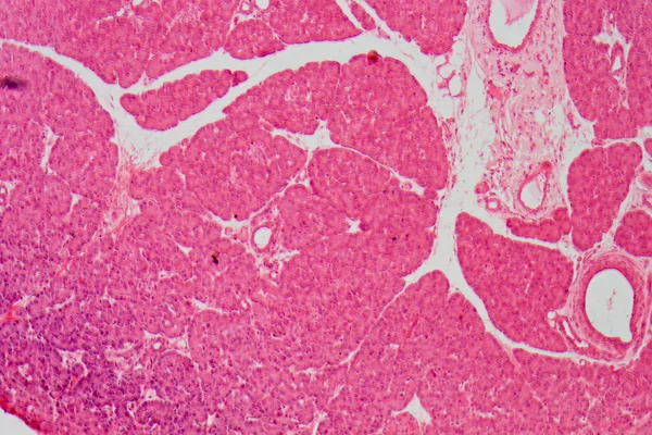
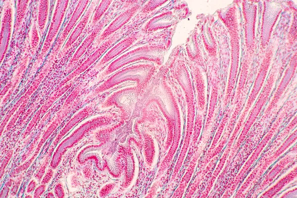
Backgrounds of Characteristics Tissue of Stomach Human, Small intestine Human, Pancreas Human and Large intestine Human under the microscope in Lab.
Backgrounds Characteristics Tissue Stomach Human Small Intestine Human Pancreas Human — Stock Photo, Image
Cross section of a cerebellum. Light micrograph.
Cross Section Cerebellum Light Micrograph — Stock Photo, Image
Large intestine of a rat
Large intestine of a rat — Stock Photo, Image
Endometrial adenocarcinoma, light micrograph, photo under microscope
Endometrial Adenocarcinoma Light Micrograph Photo Microscope — Stock Photo, Image
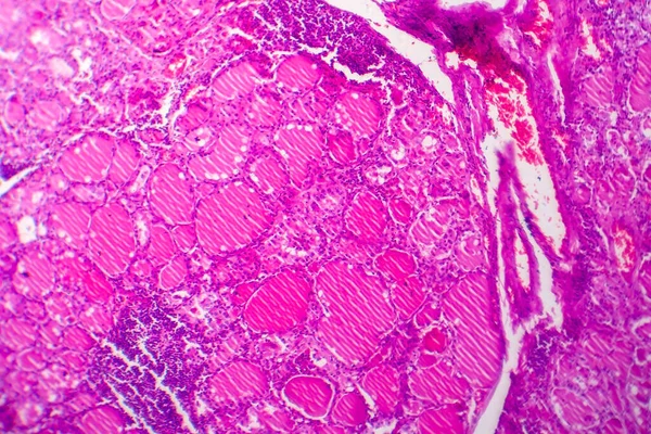
Simple goiter, light micrograph, photo under microscope. Enlargement of the thyroid gland. The most common cause of the simple goiter is iodine deficiency
Simple Goiter Light Micrograph Photo Microscope Enlargement Thyroid Gland Most — Stock Photo, Image

Related image searches

Hypothalamus Images: A Comprehensive Collection of High-Quality Stock Images

Are you in need of high-quality images of the hypothalamus for your medical research or educational project? Look no further because we have everything you need. Our collection of hypothalamus images features a wide range of visuals, including illustrations, diagrams, and real-life images in various file formats (JPG, AI, and EPS).

Why Choose Our Hypothalamus Images?

Our collection is packed with images that are perfect for your medical or educational projects. Our visuals will help you convey complex ideas and information in a clear and concise manner. Moreover, all our images are in high resolution, ensuring that they look great even when printed.

To ensure that our collection is up to date, we work with medical experts and professionals to curate and add new images regularly. From detailed illustrations to 3D models, we have everything you need to create engaging presentations and materials.

Our collection of hypothalamus images is perfect for medical researchers, educators, and students who need visuals to support their studies. With our images, you can create presentations, posters, brochures, and educational materials that are visually appealing and easy to understand.

Tips for Using Hypothalamus Images in Your Projects

Using images in your projects is a good way to make your ideas and information more engaging and easy to understand. However, you need to use them correctly to avoid confusion or misinterpretation. Here are some tips for using hypothalamus images effectively:

  • Choose the right image: Choose an image that best supports your topic and purpose. Look for images that are relevant and accurate.
  • Credit your sources: If you’re using images from other sources, make sure to credit them properly. This applies to both academic and commercial projects.
  • Use high-quality images: Use images that are high resolution so that they look great even when printed or displayed in large formats.
  • Use images sparingly: Don’t overwhelm your audience with too many images. Use them sparingly to support key points and ideas.
  • Use captions: Use captions to describe the images and explain their relevance to your topic.
  • Consider the audience: Consider your audience when choosing images. Use visuals that are appropriate for your audience's age and educational level.

Pricing and Licensing

Our collection of hypothalamus images is available for purchase on a per-image or subscription basis. All our images come with a standard license that allows you to use them for personal or commercial purposes with proper attribution. If you need to use our images for more specific purposes, such as in advertising or merchandise, we offer extended licenses to fit your needs.

In conclusion, our comprehensive collection of hypothalamus images is perfect for medical researchers, educators, and students who need high-quality visuals to support their projects and studies. With our images, you can create engaging and informative presentations, posters, and educational materials that will help you convey complex ideas and information in a clear and concise manner.