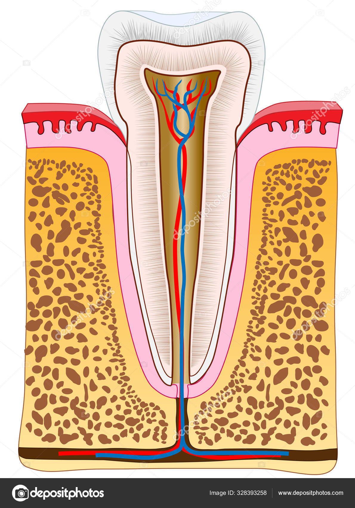
Anatomy Healthy Premolar Tooth Cross Section Single Tooth Gums Vessels — Stock Vector

Anatomy of the healthy premolar tooth. Cross section of single tooth with gums, vessels and bone. Inside the healthy human tooth. Vector dental 2D illustration. — Vector

Anatomy of the healthy premolar tooth. Cross section of single tooth with gums, vessels and bone. Inside the healthy human tooth. Vector dental 2D illustration.

 — Vector by AMoukhin

Same Series:

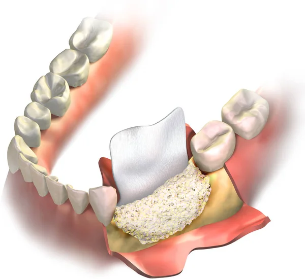
Dental barrier regenerative collagen membrane putting between cutted gums and bone graft, that part of vertical bone regeneration procedure. 3D illustration of Augmentation Surgery.
Dental Barrier Regenerative Collagen Membrane Putting Cutted Gums Bone Graft — Stock Photo, Image
Vertical Bone regeneration procedure of Augmentation Surgery. Close up, frontal view bone grafting and regenerative collagen dental membrane before warping above a graft. 3D illustration.
Vertical Bone Regeneration Procedure Augmentation Surgery Close Frontal View Bone — Stock Photo, Image
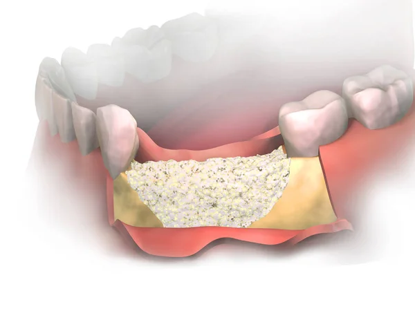
Adding new bone to Bone Loss on lower jaw after tooth removal. Bone grafting placement in front view. Before putting Membrane on bone graft. 3D illustration. Dental Augmentation Surgery.
Adding New Bone Bone Loss Lower Jaw Tooth Removal Bone — Stock Photo, Image
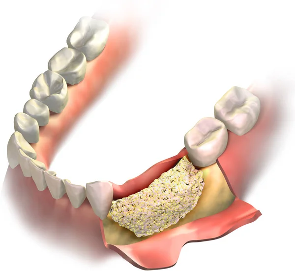
Bone graft placed on Loss bone in lower jaw before close it with gums. Vertical Bone regeneration procedure. 3D illustration.
Bone Graft Placed Loss Bone Lower Jaw Close Gums Vertical — Stock Photo, Image
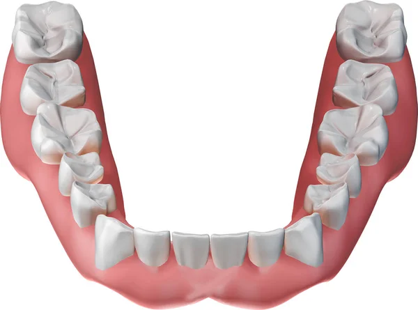
Removable lower dentures isolated on white background. 3D illustration.
Removable Lower Dentures Isolated White Background Illustration — Stock Photo, Image
Removable upper dentures coming to upper gums with no teeth. 3D illustration.
Removable Upper Dentures Coming Upper Gums Teeth Illustration — Stock Photo, Image
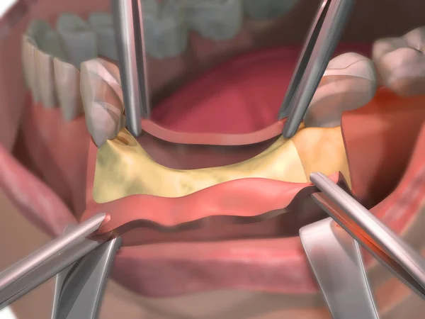
Bone Loss on lower jaw with opened gums before surgery using bone graft. Bone regeneration. 3D illustration.
Bone Loss Lower Jaw Opened Gums Surgery Using Bone Graft — Stock Photo, Image
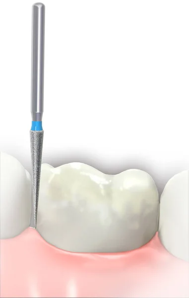
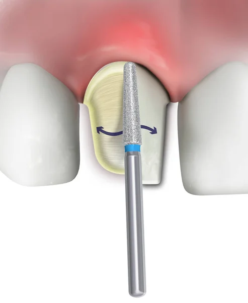
Preparing (shaping) the incisor tooth with diamond Dental Burs on white background. 3D illustration.
Preparing Shaping Incisor Tooth Diamond Dental Burs White Background Illustration — Stock Photo, Image
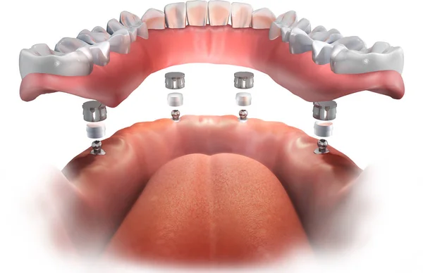
Ball Abutment installation. Lower Implant supported Complete Removable Denture. 3D illustration.
Ball Abutment Installation Lower Implant Supported Complete Removable Denture Illustration — Stock Photo, Image
Bone Loss After Tooth Removal. Opened gums before surgery using bone graft. Bone regeneration. Step of opening gums. 3D illustration.
Bone Loss Tooth Removal Opened Gums Surgery Using Bone Graft — Stock Photo, Image
Preparing (shaping) the incisor tooth with diamond Dental Burs on white background. 3D illustration.
Preparing Shaping Incisor Tooth Diamond Dental Burs White Background Illustration — Stock Photo, Image
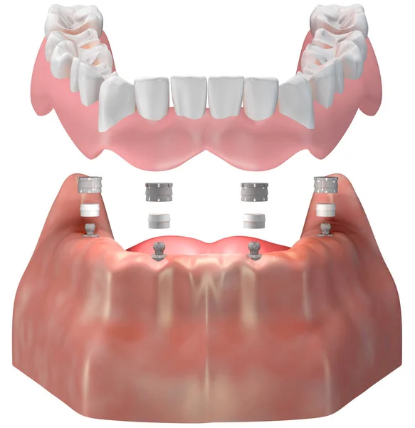
Lower Implant supported Complete Denture. All on 4 implants. Ball attachment superstructure. 3D illustration.
Lower Implant Supported Complete Denture All Implants Ball Attachment Superstructure — Stock Photo, Image
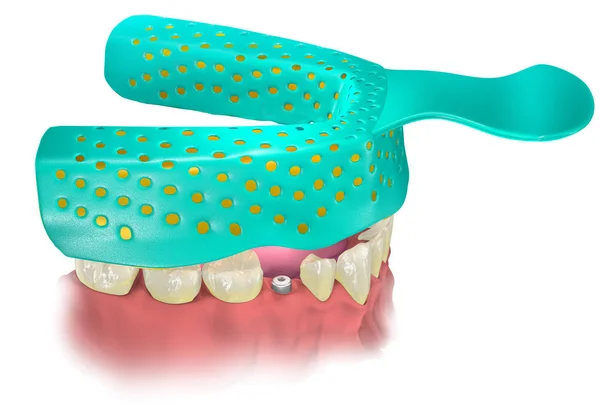
Impression Tray with Impression material putting on lower jaw with healing cap on the place of missing tooth. Close tray procedure. 3D illustration.
Impression Tray Impression Material Putting Lower Jaw Healing Cap Place — Stock Photo, Image
Bone Loss After Tooth Removal or Missing. Preparing a bone before insert the implants. Step 1 - cutting gums. Medical 3D illustration.
Bone Loss Tooth Removal Missing Preparing Bone Insert Implants Step — Stock Photo, Image
Complete Removable Denture putting on lower jaw via ball attachment. Implant supported. Transparent gums. 3D illustration.
Complete Removable Denture Putting Lower Jaw Ball Attachment Implant Supported — Stock Photo, Image
The ball attachment superstructure is intended to secure a removable prosthesis. The attachment is used in conjunction with a stainless steel cap and an intermediate silicone insert. Medical 3D illustration.
Ball Attachment Superstructure Intended Secure Removable Prosthesis Attachment Used Conjunction — Stock Photo, Image

Usage Information

You can use this royalty-free vector image "Anatomy of the healthy premolar tooth. Cross section of single tooth with gums, vessels and bone. Inside the healthy human tooth. Vector dental 2D illustration." for personal and commercial purposes according to the Standard or Extended License. The Standard License covers most use cases, including advertising, UI designs, and product packaging, and allows up to 500,000 print copies. The Extended License permits all use cases under the Standard License with unlimited print rights and allows you to use the downloaded vector files for merchandise, product resale, or free distribution.

This stock vector image is scalable to any size. You can buy and download it in high resolution up to 8641x11617. Upload Date: Dec 27, 2019