Chloroplast structure — Stock Vector

Chloroplast structure — Vector

Chloroplast structure. Plant cell, chloroplast, granum and thylakoid.

 — Vector by edesignua

Same Series:

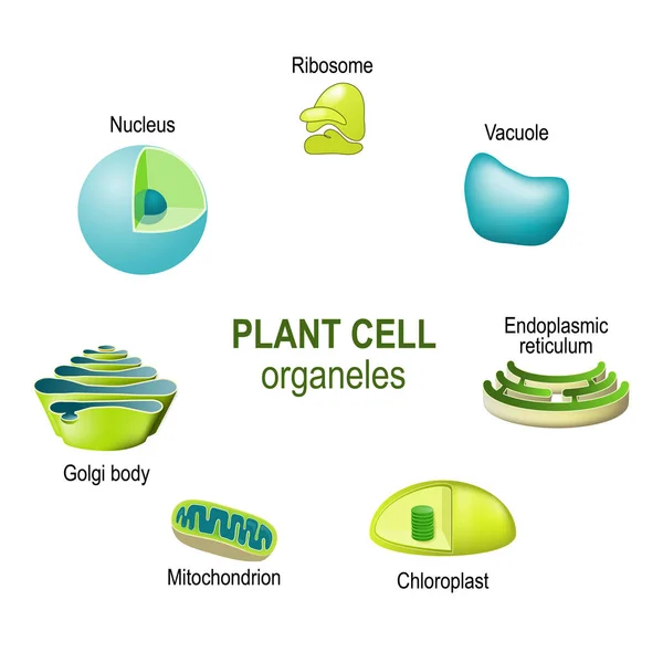
Organelles of plant cells. Vector illustration for biological, science and educational use
Organelles Plant Cells Vector Illustration Biological Science Educational Use — Stock Vector
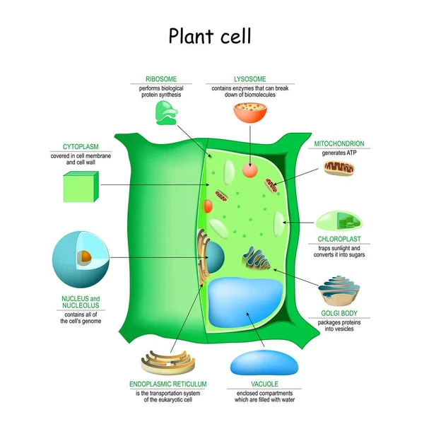
Plant cell anatomy. cross section. structure of a Eukaryotic cell. Vector diagram for your design, educational, medical, biological and science use
Plant Cell Anatomy Cross Section Structure Eukaryotic Cell Vector Diagram — Stock Vector
Plant cell anatomy structure. Close-up of Organelles of a plant cell. Educational infographic. Vector illustration
Plant Cell Anatomy Structure Close Organelles Plant Cell Educational Infographic — Stock Vector
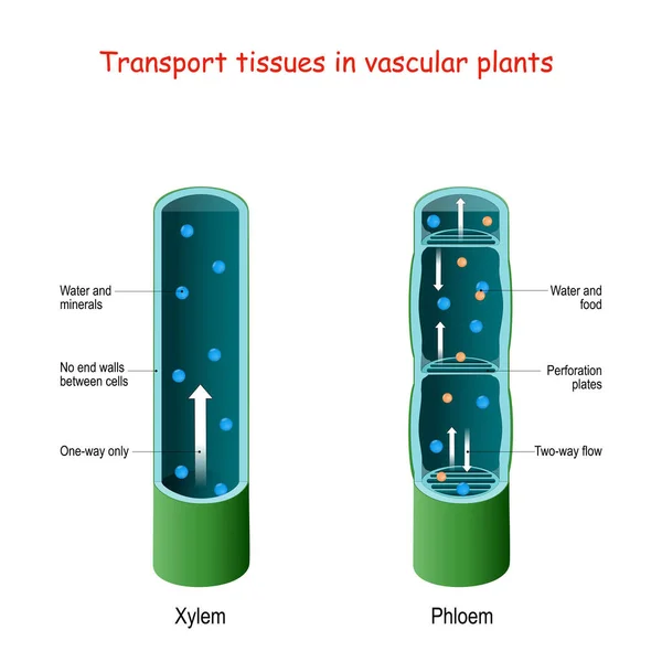
Xylem and phloem. biological structure scheme of inner vascular in Plant. cross section. function of xylem is to transport water and nutrients from roots to stems and leaves. phloem for transport organic compounds made during photosynthesis
Xylem Phloem Biological Structure Scheme Inner Vascular Plant Cross Section — Stock Vector
Structure of cell membrane. close-up of cell, phospholipid bilayer, and phospholipi
Structure of cell membrane — Stock Vector
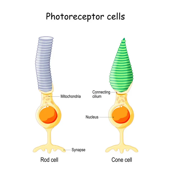
Anatomy of Photoreceptor. cell of a retina in the eye. Cone cells in respond to color vision and send signals to brain. Rod cells are used in peripheral vision
Anatomy Photoreceptor Cell Retina Eye Cone Cells Respond Color Vision — Stock Vector
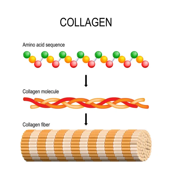
Collagen (fiber, molecule, and Amino acid sequence). Molecular structure. Three polypeptides coil to form tropocollagen. Tropocollagens bind together to form a fibril. Many fibrils bind together form a collagen fibre. Vector diagram for educational,
Collagen Fiber Molecule Amino Acid Sequence Molecular Structure Three Polypeptides — Stock Vector
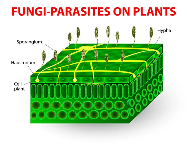
The parasitic fungi absorb their food material from the living tissues of the hosts on which they parasitize. Pathogens. education Vector diagram
Fungi-parasites on plants — Stock Vector
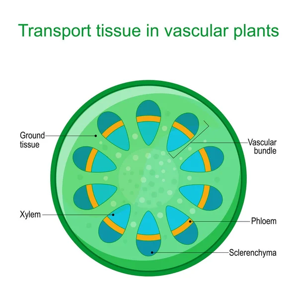
Cross section of vascular tissue system of a plant. Dicot vascular bundles of xylem and phloem are arranged in a ring. Vector diagram for educational use
Cross Section Vascular Tissue System Plant Dicot Vascular Bundles Xylem — Stock Vector
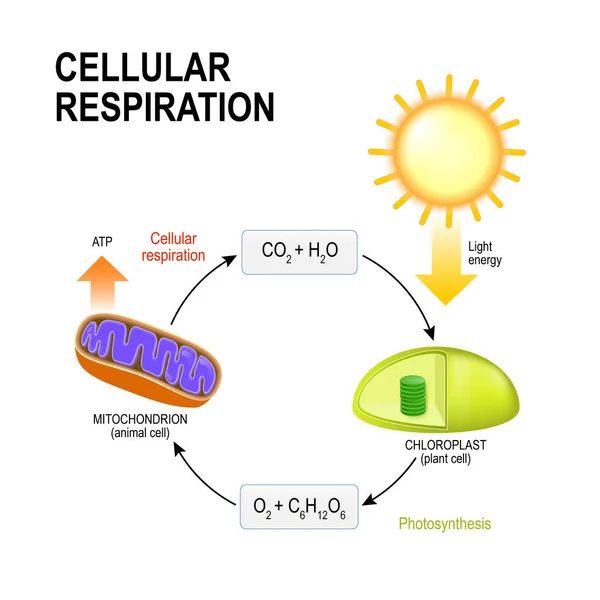
Cellular respiration. vector diagram presentation of the processes of aerobic cellular respiration. Connecting Cellular Respiration and Photosynthesis
Cellular respiration. Connecting Cellular Respiration and Photos — Stock Vector
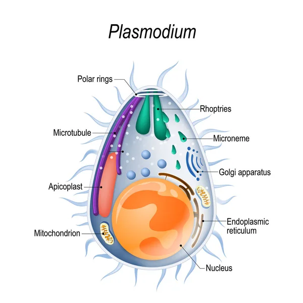
Plasmodium is the malaria parasite, is a large genus of parasitic protozoa. Infection with these protozoans is known as malaria, a deadly disease. Diagram of Plasmodium merozoites structure. vector illustration for medical, educational and science
Plasmodium Malaria Parasite Large Genus Parasitic Protozoa Infection Protozoans Known — Stock Vector
Antibiotics mechanisms of action. antibacterial drugs. From Disruption of cell membrane, and cell wall to Inhibition of RNA and DNA synthesis, protein synthesis, and folate synthesis. Vector illustration. Medical poster
Antibiotics Mechanisms Action Antibacterial Drugs Disruption Cell Membrane Cell Wall — Stock Vector
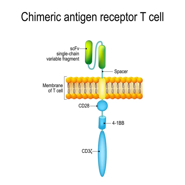
Chimeric antigen receptor T cell (CAR). Artificial T cell receptors are proteins that have been engineered for cancer therapy (killing of tumor cells). genetically engineered. Vector diagram for medical, educational and science use
Chimeric antigen receptor T cell (CAR) — Stock Vector
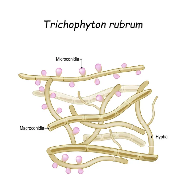
Trichophyton rubrum is a dermatophytic fungus that colonizes the upper layers of skin, and cause of athlete's foot, fungal infection of nail, and ringworm. Close-up of fungi with fertile hyphae, Macroconidia and Microconidia
Trichophyton rubrum. Close-up of fungi with fertile hyphae — Stock Vector
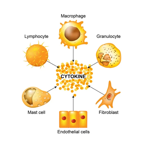
Cytokines are produced by macrophages, lymphocytes, mast cells, endothelial cells and fibroblasts. Cytokines include chemokines, interferons, interleukins, lymphokines, and tumour necrosis factors, but not hormones or growth factors
Cells that produce cytokines — Stock Vector
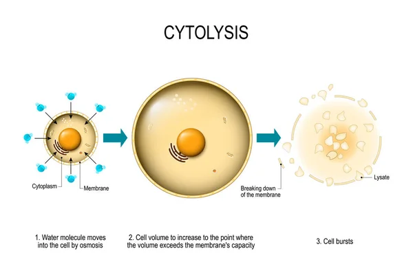
Cytolysis. Osmotic lysis. Water enter the cell and causes its volume to increase to the point where the volume exceeds the membrane's capacity and the cell bursts. Vector diagram for educational, medical, biological and science use
Cytolysis Osmotic Lysis Water Enter Cell Causes Its Volume Increase — Stock Vector

Similar Stock Videos:
Usage Information

You can use this royalty-free vector image "Chloroplast structure" for personal and commercial purposes according to the Standard or Extended License. The Standard License covers most use cases, including advertising, UI designs, and product packaging, and allows up to 500,000 print copies. The Extended License permits all use cases under the Standard License with unlimited print rights and allows you to use the downloaded vector files for merchandise, product resale, or free distribution.

This stock vector image is scalable to any size. You can buy and download it in high resolution up to 4921x3598. Upload Date: Jun 12, 2015