Peripheral Part Transverse Section Spinal Cord Showing Nerve Fibers Subdivided — Stock Vector

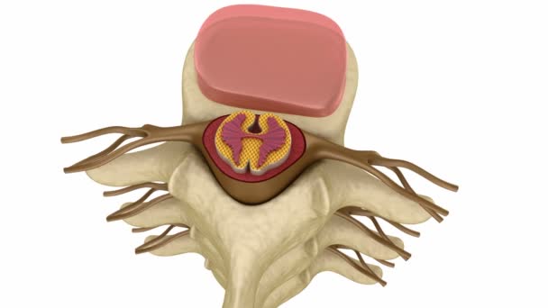
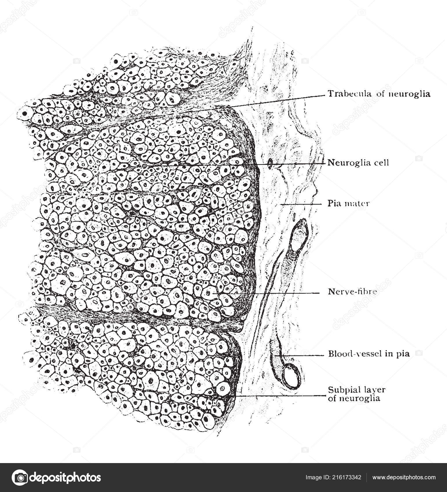
Peripheral part of transverse section of spinal cord showing nerve fibers subdivided into groups by ingrowth of subpial layer of neuroglia, vintage line drawing or engraving illustration. — Vector

Peripheral part of transverse section of spinal cord showing nerve fibers subdivided into groups by ingrowth of subpial layer of neuroglia, vintage line drawing or engraving illustration.

 — Vector by Morphart

Same Series:

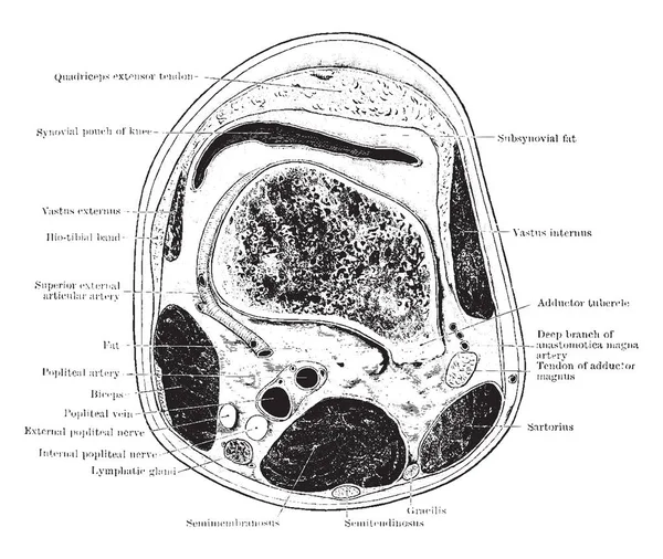
This illustration represents Section Through Thigh, vintage line drawing or engraving illustration.
Illustration Represents Section Thigh Vintage Line Drawing Engraving Illustration — Stock Vector
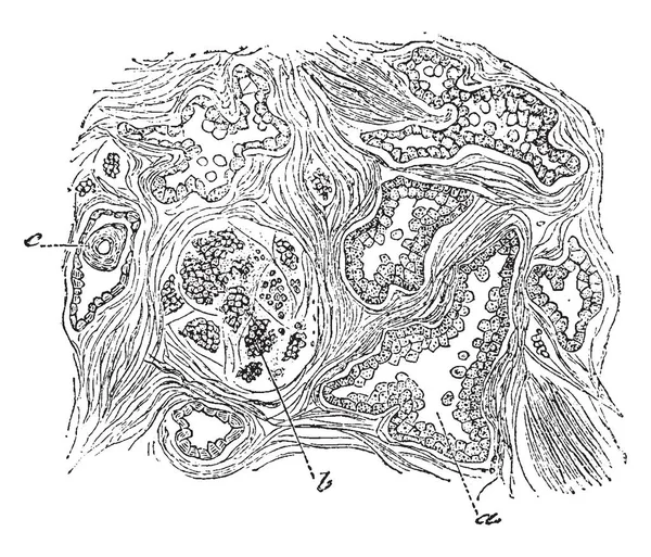
This illustration represents Section of the Prostate Gland, vintage line drawing or engraving illustration.
Illustration Represents Section Prostate Gland Vintage Line Drawing Engraving Illustration — Stock Vector
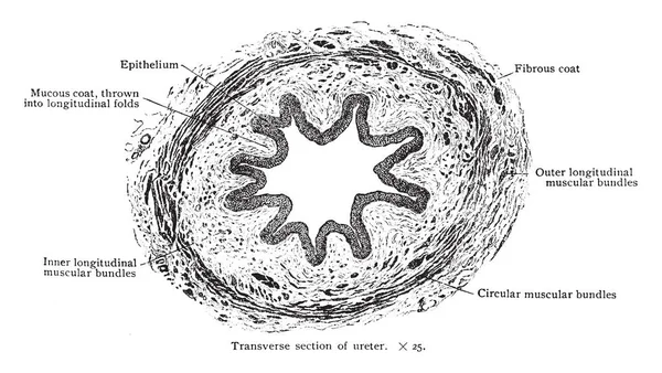
This illustration represents Transverse Section of Ureter, vintage line drawing or engraving illustration.
Illustration Represents Transverse Section Ureter Vintage Line Drawing Engraving Illustration — Stock Vector
Frontal section of the right ankle and foot and Viewed from in front, vintage line drawing or engraving illustration.
Frontal Section Right Ankle Foot Viewed Front Vintage Line Drawing — Stock Vector
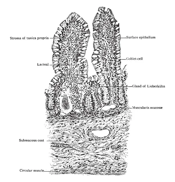
This illustration represents Transverse Section of Villi of Small Intestine, vintage line drawing or engraving illustration.
Illustration Represents Transverse Section Villi Small Intestine Vintage Line Drawing — Stock Vector
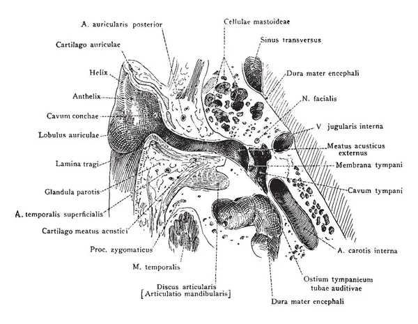
This illustration represents Auditory Apparatus of the Ear, vintage line drawing or engraving illustration.
Illustration Represents Auditory Apparatus Ear Vintage Line Drawing Engraving Illustration — Stock Vector
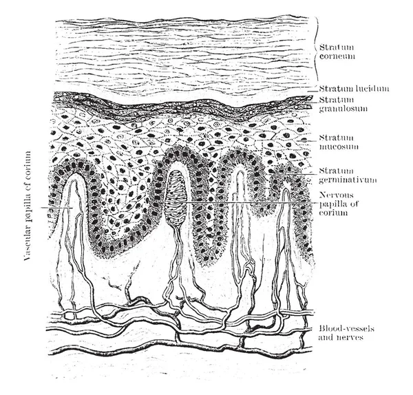
This illustration represents Vertical Section of Epidermis, vintage line drawing or engraving illustration.
Illustration Represents Vertical Section Epidermis Vintage Line Drawing Engraving Illustration — Stock Vector
The Vorticella is a protozoan and genus of bell-shaped ciliates, belongs to the Phyllum Ciliophora, vintage line drawing or engraving illustration.
Vorticella Protozoan Genus Bell Shaped Ciliates Belongs Phyllum Ciliophora Vintage — Stock Vector
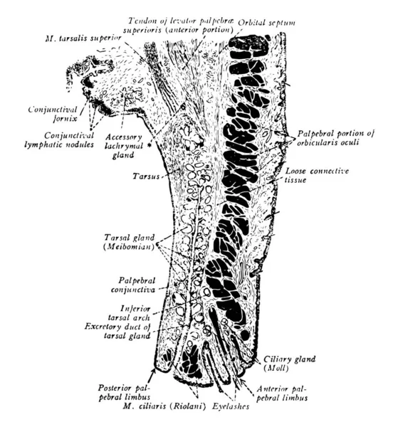
This illustration represents Eyelid, vintage line drawing or engraving illustration.
Illustration Represents Eyelid Vintage Line Drawing Engraving Illustration — Stock Vector
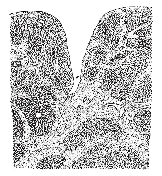
Syphilitic cirrhosis of the liver, vintage engraved illustration
Syphilitic cirrhosis of the liver, vintage engraving. — Stock Vector
Ramus Viseralis where median medullated fibers are present, vintage line drawing or engraving illustration.
Ramus Viseralis Median Medullated Fibers Present Vintage Line Drawing Engraving — Stock Vector
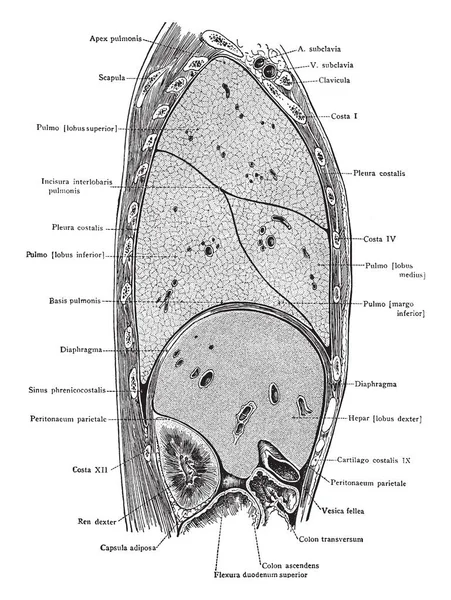
Sagittal section through the trunk 6 cm to the right of the median plane viewed from the right, vintage line drawing or engraving illustration.
Sagittal Section Trunk Right Median Plane Viewed Right Vintage Line — Stock Vector
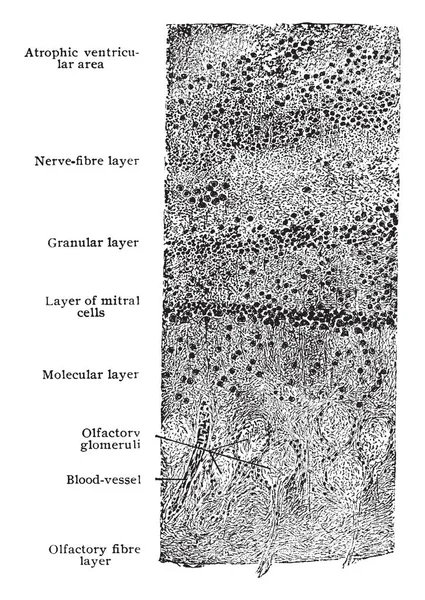
This illustration represents Transverse Section of Olfactory Bulb, vintage line drawing or engraving illustration.
Illustration Represents Transverse Section Olfactory Bulb Vintage Line Drawing Engraving — Stock Vector
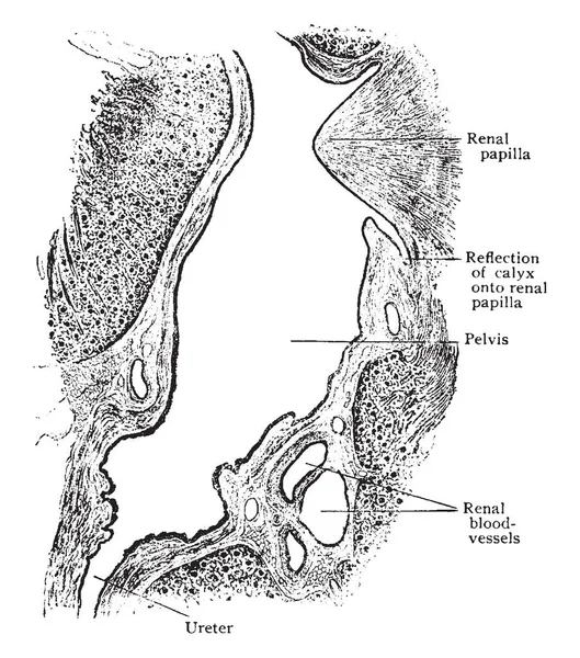
This illustration represents Sagittal Section Through Sinus of Kidney, vintage line drawing or engraving illustration.
Illustration Represents Sagittal Section Sinus Kidney Vintage Line Drawing Engraving — Stock Vector
This illustration represents Vertical Section of the Skin, vintage line drawing or engraving illustration.
Illustration Represents Vertical Section Skin Vintage Line Drawing Engraving Illustration — Stock Vector
A typical representation of the section of Stratified Thallus of Ricasolia herbacea, with the images, a, b and c, showing, cortical stratum, gonidial stratum and medullary stratum, respectively, vintage line drawing or engraving illustration.
Typical Representation Section Stratified Thallus Ricasolia Herbacea Images Showing Cortical — Stock Vector

Usage Information

You can use this royalty-free vector image "Peripheral part of transverse section of spinal cord showing nerve fibers subdivided into groups by ingrowth of subpial layer of neuroglia, vintage line drawing or engraving illustration." for personal and commercial purposes according to the Standard or Extended License. The Standard License covers most use cases, including advertising, UI designs, and product packaging, and allows up to 500,000 print copies. The Extended License permits all use cases under the Standard License with unlimited print rights and allows you to use the downloaded vector files for merchandise, product resale, or free distribution.

This stock vector image is scalable to any size. You can buy and download it in high resolution up to 7672x7954. Upload Date: Aug 19, 2018