Types Of Neurons — Stock Vector

Types Of Neurons — Vector

Basic neuron types. Unipolar, pseudo-unipolar neuron, bipolar, and multipolar Neurons. Neuron Cell Body. Different Types of Neurons

 — Vector by edesignua

Same Series:

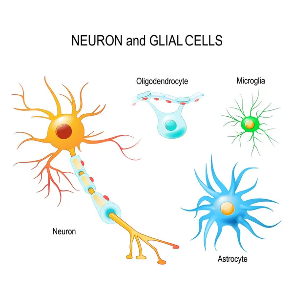
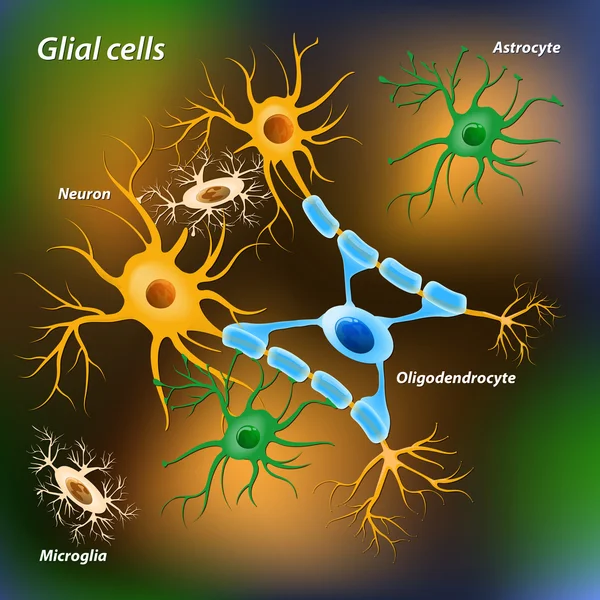
Cells of human's brain. Neuron and glial cells (Microglia, astrocyte and oligodendrocyte). Vector diagram for educational, medical, biological and science use
Cells Human Brain Neuron Glial Cells Microglia Astrocyte Oligodendrocyte Vector — Stock Vector
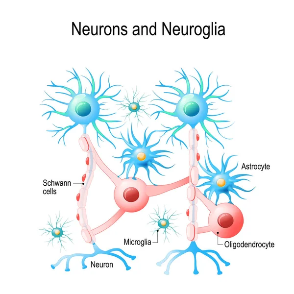
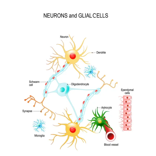
Neurons and neuroglial cells. Glial cells are non-neuronal cells in brain. There are different types of glial cells: oligodendrocyte, microglia, astrocytes and Schwann cells. Vector diagram for educational, medical, biological and science use
Neurons Neuroglial Cells Glial Cells Non Neuronal Cells Brain Different — Stock Vector
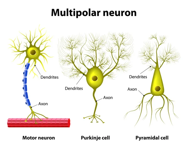
Different kinds of a multipolar neurons: pyramidal cell, Purkinje cell and motor neuron. Human anatomy
Types Of a multipolar Neurons — Stock Vector
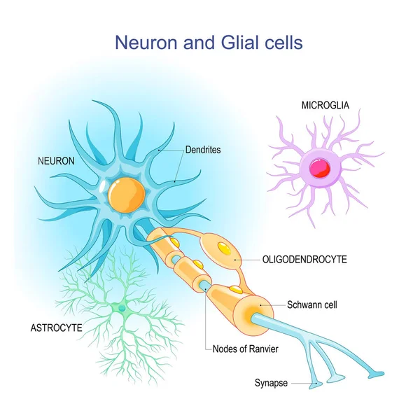
Neuron and Neuroglia. Structure and components of a neuron: dendrites, synapses, axon, myelin sheath, nodes of Ranvier, and Schwann cells. supportive glial cells: astrocytes, oligodendrocytes, and microglia. Vector infographic
Neuron Neuroglia Structure Components Neuron Dendrites Synapses Axon Myelin Sheath — Stock Vector
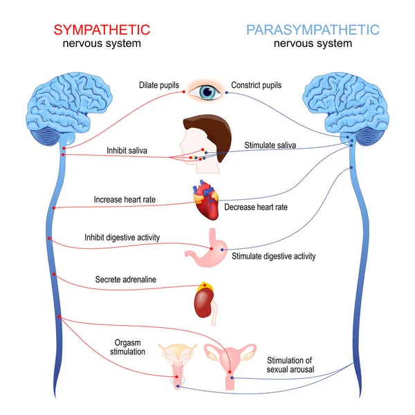
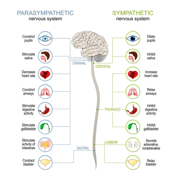
Sympathetic and parasympathetic nervous system. Difference and comparison. diagram with connected inner organs, brain and spinal cord. Educational guide of human anatomy. vector poster
Sympathetic Parasympathetic Nervous System Difference Comparison Diagram Connected Inner Organs — Stock Vector
Neurons and glial cells (Neuroglia) in brain (oligodendrocyte, microglia, astrocytes and Schwann cells), ependymal cells (ependymocytes). Vector diagram for educational, medical, biological and science use
Neurons Glial Cells Neuroglia Brain Oligodendrocyte Microglia Astrocytes Schwann Cells — Stock Vector
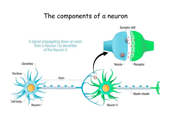
Neuron anatomy. Close-up of a Chemical synapse, Synaptic vesicle with neurotransmitter, and Receptors. Structure of Synaptic cleft. Vector poster
Neuron Anatomy Close Chemical Synapse Synaptic Vesicle Neurotransmitter Receptors Structure — Stock Vector
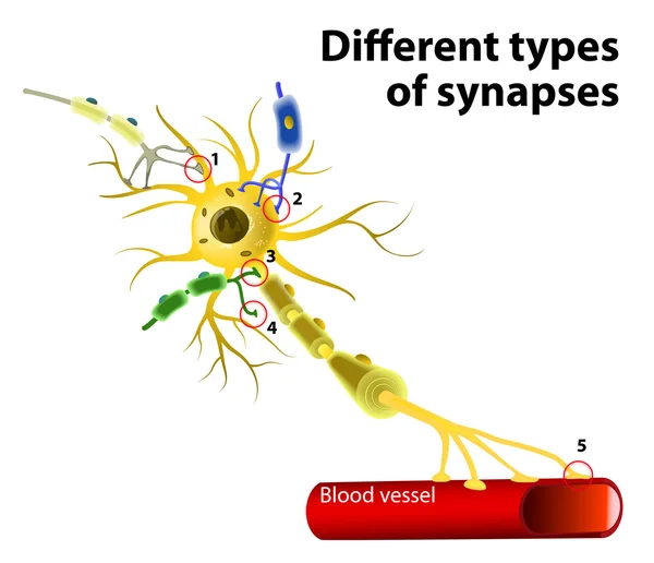
Different types of synapses. Neuron to Neuron Transmission
Different types of synapses — Stock Vector
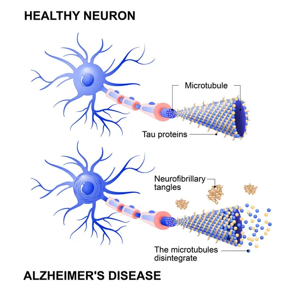
Alzheimer's disease is the change in tau protein that results in the breakdown of microtubules in brain cells. Mechanism of disease. Diagram shows two neurons: healthy cell and neuron with Alzheimer's disease. Tau hypothesis. Neurofibrillary tangles
Healthy cell and neurons with Alzheimer's disease. Tau hypothesi — Stock Vector
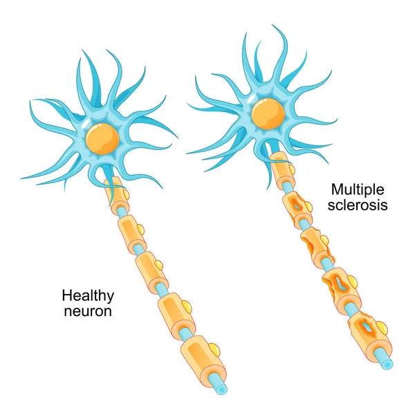
Multiple sclerosis. autoimmune disease. comparison of a healthy neuron and a neuron with a damaged myelin sheath. MS nerve damaged by immune system. Vector illustration
Multiple Sclerosis Autoimmune Disease Comparison Healthy Neuron Neuron Damaged Myelin — Stock Vector
Sympathetic And Parasympathetic Nervous System. Difference. diagram with connected inner organs, brain and spinal cord. vector illustration
Sympathetic Parasympathetic Nervous System Difference Diagram Connected Inner Organs Brain — Stock Vector
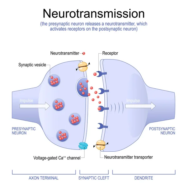
Neurotransmission. the presynaptic neuron releases a neurotransmitter, which activates receptors on the postsynaptic neuron. Synapse Structure. Neurotransmitter, synaptic vesicles and synaptic cleft. Anatomy of neurons. Vector Illustration.
Neurotransmission Presynaptic Neuron Releases Neurotransmitter Which Activates Receptors Postsynaptic Neuron — Stock Vector
Glial cells on the color background. Medical and science illustration
Glial cells — Stock Vector
Visual Pathways from Eyeball to Optic nerve, Lateral geniculate nucleus and Primary visual cortex. human head with a cross section of the brain.
Visual Pathways Eyeball Optic Nerve Lateral Geniculate Nucleus Primary Visual — Stock Vector
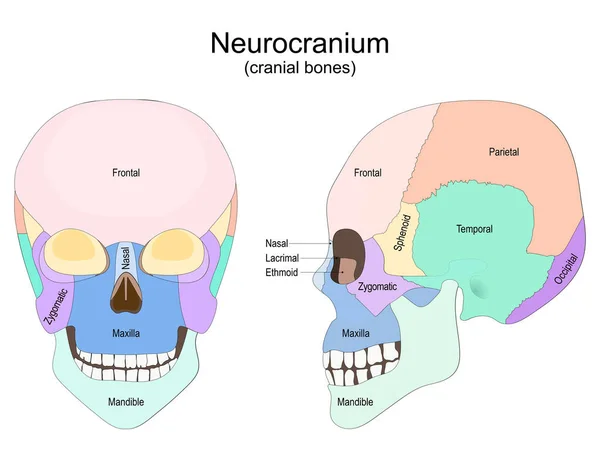
Human skull from the front and side view. Neurocranium. cranial bones. Vector poster for medical, and education use
Human Skull Front Side View Neurocranium Cranial Bones Vector Poster — Stock Vector

Similar Stock Videos:
Graphic animation showing the structure and composition of a coronavirus.
Graphic Animation Showing Structure Composition Coronavirus — Stock Video
Human Organs Animation on white background
Human Organs Animation — Stock Video
Human Organs Animation on white background
Human Organs Animation — Stock Video
Stomach upset animated line icon. stomach upset sign. isolated on white background
Stomach upset line icon animation — Stock Video
3d rendered medically illustration of male Internal organs
Rendered Medically Illustration Male Internal Organs — Stock Video
White Human stomach icon isolated on purple background. 4K Video motion graphic animation.
White Human stomach icon isolated on purple background. 4K Video motion graphic animation — Stock Video
Black line Virus cells in lung icon isolated on white background. Infected lungs. Coronavirus, COVID-19. 2019-nCoV. 4K Video motion graphic animation.
Black line Virus cells in lung icon isolated on white background. Infected lungs. Coronavirus, COVID-19. 2019-nCoV. 4K Video motion graphic animation — Stock Video
The stomach lies between the esophagus and the duodenum (the first part of the small intestine). It is on the left upper part of the abdominal cavity. The top of the stomach lies against the diaphragm.
Stomach Layers — Stock Video
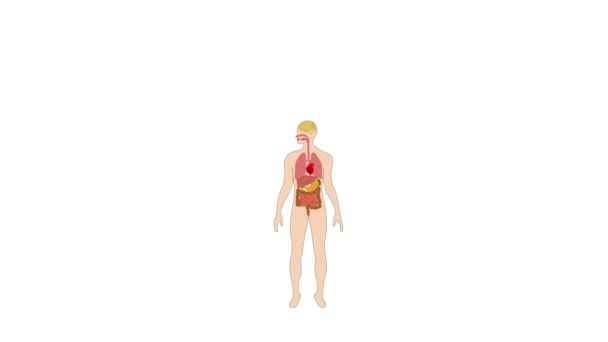
Animation of human organs internal diagram, Body of human internal organs, brain, heart, lungs, liver, stomach, intestine
Animation Human Organs Internal Diagram Body Human Internal Organs Brain — Stock Video
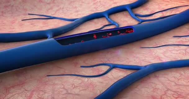
Red blood cells, blood vessel, inside the blood vessel, white blood cells, white blood cells inside the blood vessel, High quality 3d render of blood cells, pulsing down artery, Red and white blood cells in artery
Red Blood Cells Blood Vessel Blood Vessel White Blood Cells — Stock Video
Abstract futuristic motion background with graphic effects
Abstract Futuristic Motion Background Graphic Effects — Stock Video
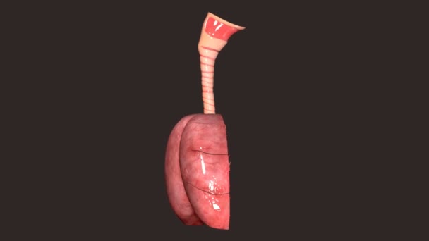
The lung is the essential respiration organ in many air-breathing animals, including most tetrapods, a few fish and a few snails. In mammals and the more complex life forms, the two lungs are located near the backbone on either side of the heart.
Lungs Anatomy — Stock Video
Pains ankle and wrist rheumatology disease animation ,4k video animated
Pains ankle and wrist rheumatology disease animation — Stock Video
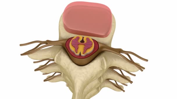
Prolapse of intervertebral disc
Prolapse of intervertebral disc — Stock Video
Heart, Lungs and Diaphragm in Skeleton Body on dark background
Heart, Lungs and Diaphragm — Stock Video
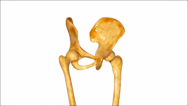
The hip bone (innominate bone, pelvic bone or coxal bone) is a large flat bone, constricted in the center and expanded above and below.
Pelvic Hip — Stock Video
Human Organs Animation on white background
Human Organs Animation — Stock Video
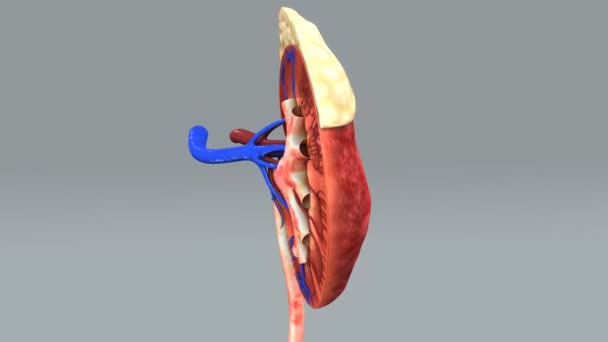
Human kidney half anatomy. 3d animation
Human kidney half anatomy — Stock Video

Usage Information

You can use this royalty-free vector image "Types Of Neurons" for personal and commercial purposes according to the Standard or Extended License. The Standard License covers most use cases, including advertising, UI designs, and product packaging, and allows up to 500,000 print copies. The Extended License permits all use cases under the Standard License with unlimited print rights and allows you to use the downloaded vector files for merchandise, product resale, or free distribution.

This stock vector image is scalable to any size. You can buy and download it in high resolution up to 5245x4110. Upload Date: Mar 30, 2015