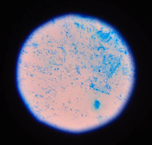
Candidiasis Stock Photos

100,000 Candidiasis pictures are available under a royalty-free license

  • Best Match
  • Fresh
  • Popular
1-100 of 100,000
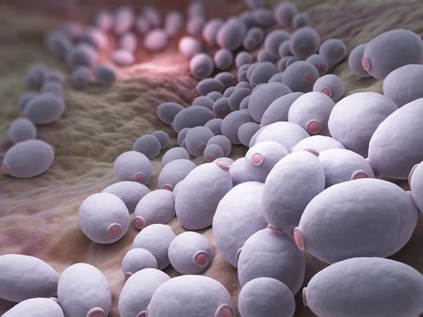
Candida albicans bacteria
Candida albicans bacteria — Stock Photo, Image
Vaginal mycosis of woman
Vaginal mycosis of woman — Stock Photo, Image
Vaginal Discomfort
Vaginal Discomfort — Stock Photo, Image
Women With Itching the Vagina
Women With Itching the Vagina — Stock Photo, Image
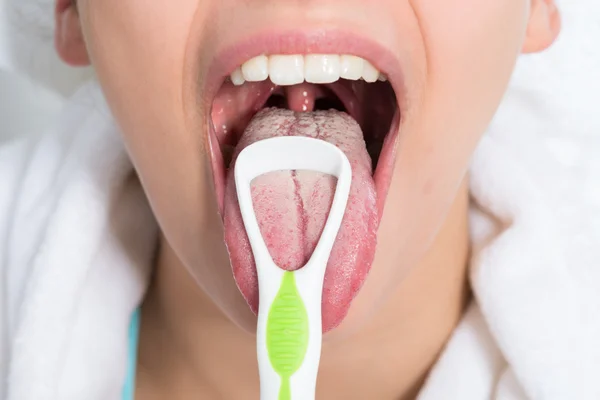
Woman Cleaning Tongue
Woman Cleaning Tongue — Stock Photo, Image
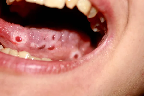
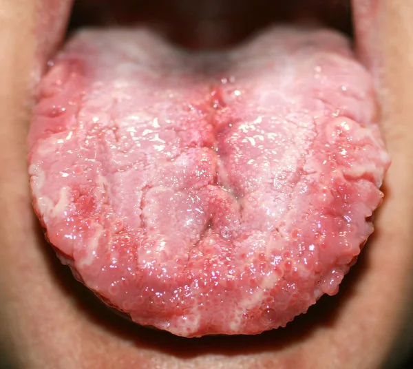
Amphotoid stomatitis. Candidiasis of the tongue. Ulcer on the tongue. Candida fungus.
Amphotoid stomatitis. Candidiasis of the tongue. Ulcer on the tongue. Candida fungus. — Stock Photo, Image
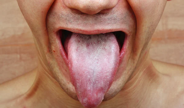
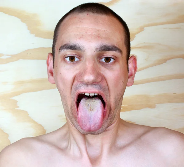
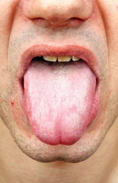
Candidiasis in the tongue. White coating. Fractured tongue. Thrush.
Candidiasis in the tongue. White coating. Fractured tongue. Thrush. — Stock Photo, Image
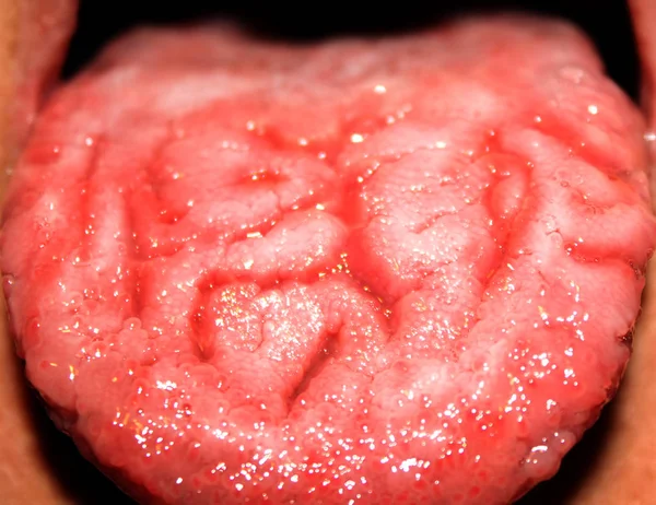
Geographic tongue, candidiasis, the language of the white coating candida
Geographic tongue, candidiasis, the language of the white coating candida — Stock Photo, Image
Candida albicans which cause candidiasis
Candida albicans which cause candidiasis — Stock Photo, Image
Word Candidiasis on a book and pills.
Word Candidiasis  on a book and pills. — Stock Photo, Image
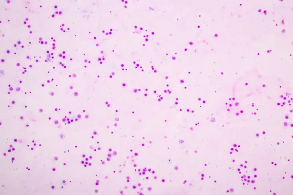
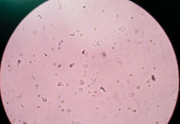
Yeast cells in urine sample.
Yeast cells in urine sample. — Stock Photo, Image
Infection tongue
Infection tongue — Stock Photo, Image
Doctor keeps a card with the name of the diagnosis - fungi
Doctor keeps a card with the name of the diagnosis - fungi — Stock Photo, Image
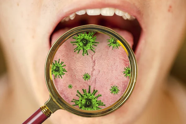
First sign of coronovirus, protruding white plaque on tongue, closeup
First Sign Coronovirus Protruding White Plaque Tongue Closeup — Stock Photo, Image
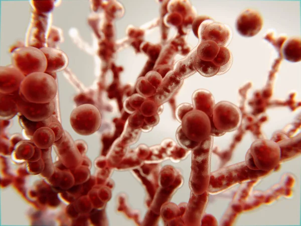
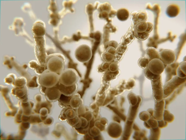
Candida fungi, Candida albicans, C. auris and other human pathogenic yeasts, 3D illustration
Candida Fungi Candida Albicans Auris Other Human Pathogenic Yeasts Illustration — Stock Photo, Image
A girl in a skirt sits on a chair and holds on to the groin area, pain in the lower abdomen, sexual infection
A girl in a skirt sits on a chair and holds on to the groin area, pain in the lower abdomen, sexual infection — Stock Photo, Image
Candida albicans, a pathogenic yeast
Candida albicans, a pathogenic yeast — Stock Photo, Image
Doctor holding yellow cubes with word Candida, closeup
Doctor Holding Yellow Cubes Word Candida Closeup — Stock Photo, Image
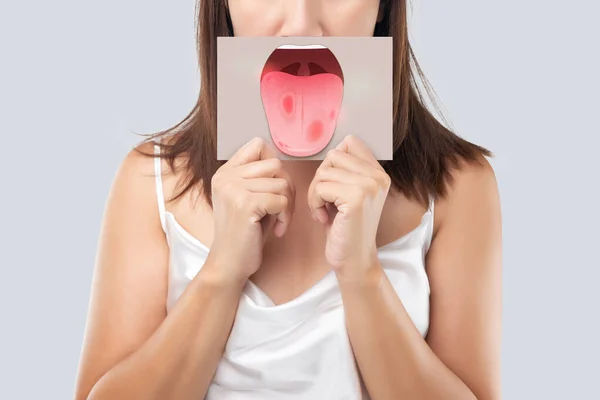
The woman show the picture of tongue problems, Illustration benign migratory glossitis on a brown paper, Behcet's Disease
Woman Show Picture Tongue Problems Illustration Benign Migratory Glossitis Brown — Stock Photo, Image
Candidiasis (Moniliasis, Thrush)
Candidiasis (Moniliasis, Thrush) — Stock Photo, Image
Candida yeast, a pathogen that causes candidiasis
Candida yeast, a pathogen that causes candidiasis — Stock Photo, Image
A man in a white shirt holds on to his groin, the concept of pain and inflammation in the groin, genital infections in men and prostatitis. White background, isolate
A man in a white shirt holds on to his groin, the concept of pain and inflammation in the groin, genital infections in men and prostatitis. White background, isolate — Stock Photo, Image
Infection tongue
Infection tongue — Stock Photo, Image
Itching Vagina
Itching Vagina — Stock Photo, Image
Newborn with candidiasis on tongue
Newborn with candidiasis on tongue — Stock Photo, Image
Chromosomes Human under the microscope for education.
Chromosomes Human Microscope Education — Stock Photo, Image
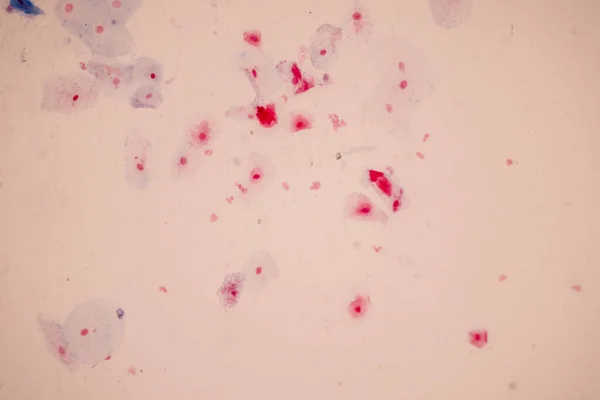
Histological sample Squamous epithelial cells under microscope,
Histological Sample Squamous Epithelial Cells Microscope — Stock Photo, Image
Chromosomes Human under the microscope for education.
Chromosomes Human Microscope Education — Stock Photo, Image
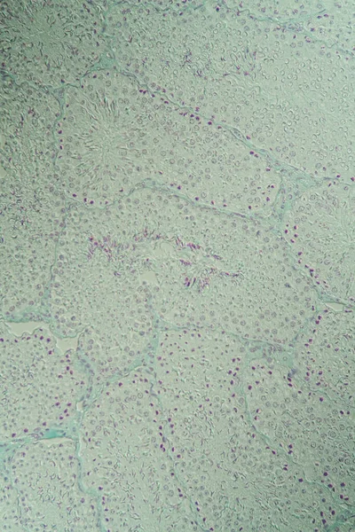
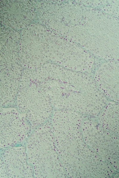
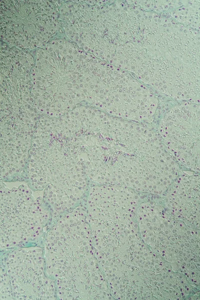
Testicle cross-section under the microscope 100x
Testicle Cross Section Microscope 100X — Stock Photo, Image
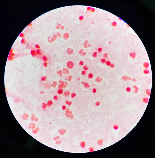
Fresh Branching budding yeast cells
Fresh Branching budding yeast cells — Stock Photo, Image
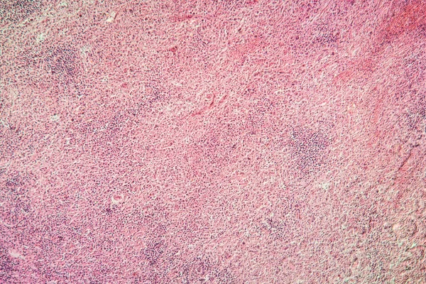
Lymphoid tissue myelin sarcoma diseased tissue 100x
Lymphoid Tissue Myelin Sarcoma Diseased Tissue 100X — Stock Photo, Image
Chromosomes in the nucleus of a cell
Chromosomes Nucleus Cell — Stock Photo, Image
Blue Alcohol Ink Marble. Gold Oriental Background. Golden Water Color Canvas. Foil Oil Paint. Gold Marble Watercolor. Bronze Water Color Watercolor. Geode Elegant Glitter. Luxury Abstract Template.
Blue Alcohol Ink Marble Gold Oriental Background Golden Water Color — Stock Photo, Image
Carcinoma in guinea pigs, tissue 100x
Carcinoma Guinea Pigs Tissue 100X — Stock Photo, Image
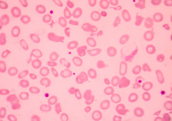
Thalassemia blood smear abnormal red blood cells morphology.Medical science concept.
Thalassemia Blood Smear Abnormal Red Blood Cells Morphology Medical Science — Stock Photo, Image
Big abstract shape with spheres
Big abstract shape with spheres — Stock Photo, Image
Gout crystals deposits in the kidney tissue under the microscope 100x
Gout Crystals Deposits Kidney Tissue Microscope 100X — Stock Photo, Image
Watercolor Multi Patterns. Purple Hippie Shirt.
Watercolor Multi Patterns. Purple Hippie Shirt. — Stock Photo, Image
Moderate gram nagative bacilli with white blood cells in urine sample.
Moderate Gram Nagative Bacilli White Blood Cells Urine Sample — Stock Photo, Image
Abstract background, texture of concrete wall
Abstract Background Texture Concrete Wall — Stock Photo, Image
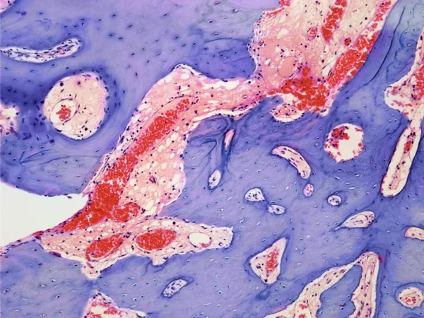
Section through an osteoid osteoma
Section through an osteoid osteoma — Stock Photo, Image
Bacteria or virus spheres in blood
Bacteria or virus spheres in blood — Stock Photo, Image
Texture of office linoleum close up. High-strength flooring for public spaces.
Texture Office Linoleum Close High Strength Flooring Public Spaces — Stock Photo, Image
Lymph glands cancer diseased tissue 100x
Lymph Glands Cancer Diseased Tissue 100X — Stock Photo, Image
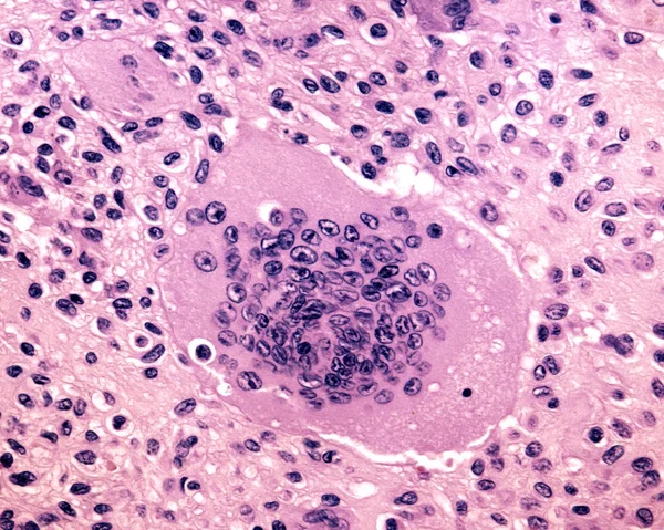
Mutinucleated giant cell
Mutinucleated giant cell — Stock Photo, Image
Histological sample Squamous epithelial cells under microscope,
Histological Sample Squamous Epithelial Cells Microscope — Stock Photo, Image
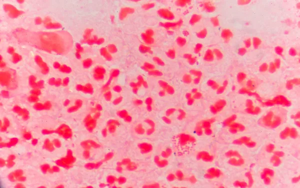
Moderate red white blood cells with gram negative diplococci.
Moderate red white blood cells with gram negative diplococci. — Stock Photo, Image
Hippie Pattern with Watercolor Circle. Color
Hippie Pattern with Watercolor Circle. Color — Stock Photo, Image
Abstract stones and water blue background
Abstract stones and water blue background — Stock Photo, Image
Abstract pink background, decorative texture
Abstract Pink Background Decorative Texture — Stock Photo, Image
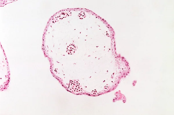
Under high magnification microscopic view of chorionic villus
High Magnification Microscopic View Chorionic Villus — Stock Photo, Image
Pink abstract background in watercolor style
Pink Abstract Background Watercolor Style — Stock Photo, Image
Illustration of Staphylococcus aureus (MRSA) coccoid bacteria. Staphylococcus aureus is a Gram-positive bacterium that causes food poisoning, toxic shock syndrome and skin and wound infections such as scalded skin syndrome
Illustration Staphylococcus Aureus Mrsa Coccoid Bacteria Staphylococcus Aureus Gram Positive — Stock Photo, Image
Gout crystals deposits in the kidney tissue under the microscope 100x
Gout Crystals Deposits Kidney Tissue Microscope 100X — Stock Photo, Image
Cross section under the microscope 200x
Cross Section Microscope 200X — Stock Photo, Image
Testicles guinea pig under a microscope
Testicles Guinea Pig Microscope — Stock Photo, Image
Abstract marbling pattern for fabric, design. Creative marbling background texture
Abstract Marbling Pattern Fabric Design Creative Marbling Background Texture — Stock Photo, Image
Pink stained paper
Pink stained paper — Stock Photo, Image
Photomicrograph of nasal polyps, displaying abnormal tissue growth in the nasal passages often causing congestion and discomfort.
Photomicrograph Nasal Polyps Displaying Abnormal Tissue Growth Nasal Passages Often — Stock Photo, Image
Triple phosphate crystals in human urine.
Triple phosphate crystals in human urine. — Stock Photo, Image
Inflamed tissue under the microscope 100x
Inflamed Tissue Microscope 100X — Stock Photo, Image
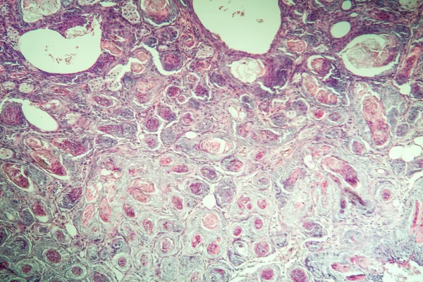
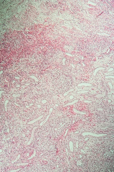
Poly nephritis,tissue under the microscope 100x
Poly Nephritis Tissue Microscope 100X — Stock Photo, Image
Close up of a sandstone brick - a textured background
Close Sandstone Brick Textured Background — Stock Photo, Image
Meiosis In Grasshopper Testis under the light microscope view.
Meiosis Grasshopper Testis Light Microscope View — Stock Photo, Image
Medical science background.
Medical science background. — Stock Photo, Image
Histopathology of interstitial nephritis, light micrograph, photo under microscope
Histopathology Interstitial Nephritis Light Micrograph Photo Microscope — Stock Photo, Image
Root tip of Onion and Mitosis cell in the Root tip of Onion under a microscope.
Root Tip Onion Mitosis Cell Root Tip Onion Microscope — Stock Photo, Image
Detailed pink grunge texture
Detailed pink grunge texture — Stock Photo, Image
Abstract colorful marbled patterned surface
Abstract Colorful Marbled Patterned Surface — Stock Photo, Image
Tissue with schistosomiasis sick tissue 100x
Tissue Schistosomiasis Sick Tissue 100X — Stock Photo, Image
Mycobacterium are to the bacteria that cause tuberculosis in fish or pet
Mycobacterium Bacteria Cause Tuberculosis Fish Pet — Stock Photo, Image
Breast cancer, light micrograph, photo under microscope
Breast Cancer Light Micrograph Photo Microscope — Stock Photo, Image
Nerve cells under light microscope
Nerve cells under light microscope — Stock Photo, Image
Anatomy and Histological Ovary, Testis and Sperm human cells under microscope.
Anatomy Histological Ovary Testis Sperm Human Cells Microscope — Stock Photo, Image
Orange liquid ink, digital wallpaper
Orange Liquid Ink Digital Wallpaper — Stock Photo, Image
Green Textured Paper Background With Marble Effects
Green Textured Paper Background Marble Effects — Stock Photo, Image
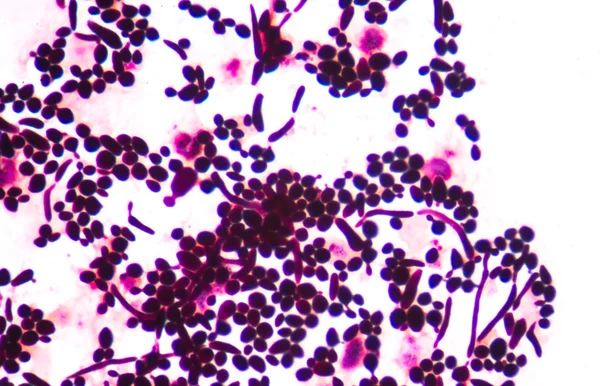
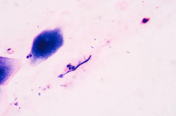
Budding yeast cells with pseudohyphae from sputum gram stain tes
Budding yeast cells with pseudohyphae from sputum gram stain tes — Stock Photo, Image
Red leukocyte in sputum gram stain with bacteria
Red leukocyte in sputum gram stain with bacteria — Stock Photo, Image
Abstract background. Monochrome texture. Black and white textured background.
Abstract Background Monochrome Texture Black White Textured Background — Stock Photo, Image
Abstract colorful marbled patterned surface
Abstract Colorful Marbled Patterned Surface — Stock Photo, Image
Moderate Neutrophil in synovial fluid inframatory cells.
Moderate Neutrophil Synovial Fluid Inframatory Cells — Stock Photo, Image
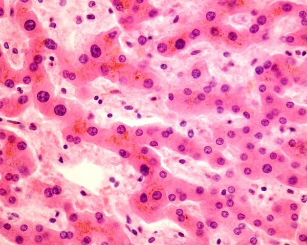
Hepatocytes
Hepatocytes — Stock Photo, Image
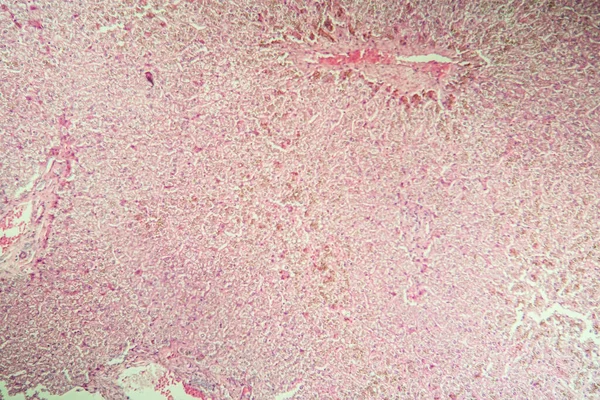
Billirubin in the liver disease in a microscope 100x
Billirubin Liver Disease Microscope 100X — Stock Photo, Image
Pink background with blue stains and drops
Pink background with blue stains and drops — Stock Photo, Image
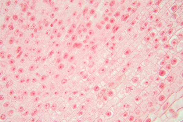
Root tip of Onion and Mitosis cell in the Root tip of Onion under a microscope.
Root Tip Onion Mitosis Cell Root Tip Onion Microscope — Stock Photo, Image
Abstract alcoholic ink texture. Abstraction from stains. Smoke and bubbles. Trendy background.
Abstract Alcoholic Ink Texture Abstraction Stains Smoke Bubbles Trendy Background — Stock Photo, Image
Bacterial infection disease tongue
Bacterial infection disease tongue — Stock Photo, Image
Creative color marble surface background
Creative Color Marble Surface Background — Stock Photo, Image
Microscope of Adenoid cystic carcinoma, rare type of cancer exist in many different body sites. This tumor occurs in the salivary glands, with close up macro 40x lens, microbiology laboratory concept.
Microscope Adenoid Cystic Carcinoma Rare Type Cancer Exist Many Different — Stock Photo, Image
Tissue of Blood human, Vein human, Artery human and Heart muscle human under the microscope in Lab.
Tissue Blood Human Vein Human Artery Human Heart Muscle Human — Stock Photo, Image
Pink abstract background in watercolor style
Pink Abstract Background Watercolor Style — Stock Photo, Image
Pathology study of human stem cell under the microscope.
Pathology Study Human Stem Cell Microscope — Stock Photo, Image
Abstract pink background, decorative texture
Abstract Pink Background Decorative Texture — Stock Photo, Image
Poly nephritis, tissue under the microscope 100x
Poly Nephritis Tissue Microscope 100X — Stock Photo, Image
Under high magnification microscopic view of lung
High Magnification Microscopic View Lung — Stock Photo, Image
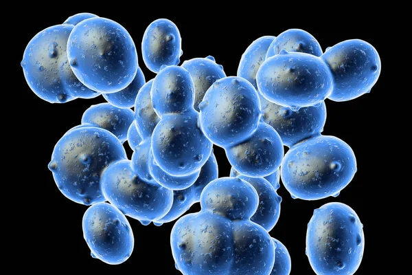
Bacteria blue cells
Bacteria blue cells — Stock Photo, Image
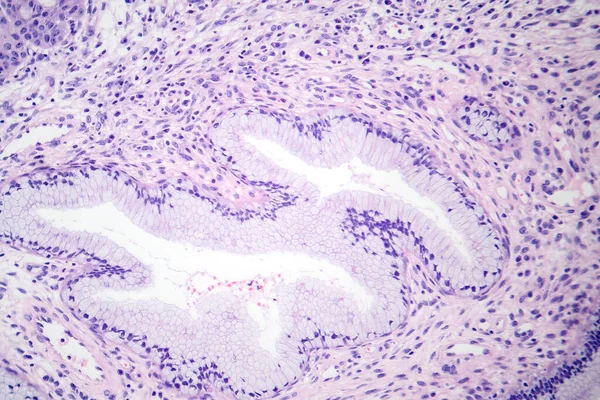
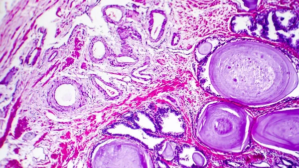
Histopathology of prostate gland hyperplasia
Histopathology of prostate gland hyperplasia — Stock Photo, Image
Moderate red white blood cells with gram negative diplococci.
Moderate red white blood cells with gram negative diplococci. — Stock Photo, Image
Poly nephritis, tissue under the microscope 100x
Poly Nephritis Tissue Microscope 100X — Stock Photo, Image
Gram staining, also called Gram's method, is a method of differe
Gram staining, also called Gram's method, is a method of differe — Stock Photo, Image

## Candidiasis Images: High-Quality Stock Photos for Medical Projects

Are you looking for candidiasis images to use in your medical projects? Look no further! We have a wide selection of high-quality stock photos available for download.

Our candidiasis images feature close-up shots of the affected areas, allowing for a clear and detailed view of the condition. The images are available in JPG, AI, and EPS formats, making them easy to use in a variety of projects.

Whether you are creating educational materials for patients, writing a medical thesis on candidiasis, or preparing a presentation for a medical conference, our candidiasis images are a valuable resource.
## Choosing the Right Candidiasis Images for Your Project

When choosing candidiasis images for your project, it's essential to consider your audience and the purpose of your project. Are you creating educational materials for patients, or are you presenting research findings to medical professionals?

For patient education materials, it's essential to choose images that are informative but not too graphic. Select images that clearly show the affected area but are not overly disturbing or sensationalized.

For presentations to medical professionals, you may want to choose images that are more detailed and technical, highlighting specific aspects of the condition and its treatment.

Remember, when using candidiasis images in medical projects, it's crucial to ensure that the images are accurate and appropriate for the intended audience. Always cite the source of the image and obtain permission to use it if necessary.

## Tips for Using Candidiasis Images Effectively

When using candidiasis images in your medical project, there are a few essential tips to keep in mind to ensure that the images are used effectively.

Firstly, ensure that the images are of high quality and resolution. Low-quality images can appear blurry and distorted, making it difficult to see the details of the condition.

Secondly, consider the placement of the images in your project. Ensure that the images are placed strategically to illustrate the points you are trying to make. For example, if you are discussing the symptoms of candidiasis, placing an image of the affected area next to the text will help to reinforce your message.

Lastly, ensure that the images are appropriately sized for the platform on which they will be displayed. Images that are too small will be difficult to see, while images that are too large can take up too much space and detract from the other content on the page.

## Where to Find Candidiasis Images

If you're looking for candidiasis images for your medical project, there are many places to find them. You can use stock image websites like Shutterstock or Getty Images, or you can search through medical image libraries like the National Library of Medicine's Visible Human Project.

When selecting candidiasis images, ensure that the images you choose are accurate and appropriate for your project. Consider the quality, resolution, and placement of the images to ensure that they are used effectively, and don't forget to cite the source of the image and obtain permission to use it if necessary.

By following these tips and selecting the right images, you can create a powerful and informative medical project that effectively communicates your message to your intended audience.