Epiglottis Stock Photos

100,000 Epiglottis pictures are available under a royalty-free license

  • Best Match
  • Fresh
  • Popular
1-100 of 100,000
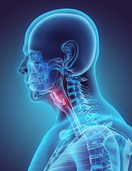
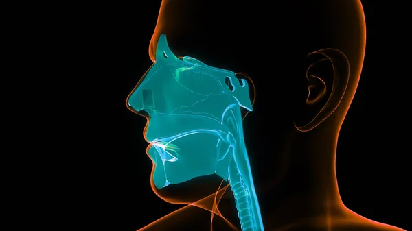
3D illustration of Epiglottis, medical concept.
3D illustration of Epiglottis, medical concept. — Stock Photo, Image
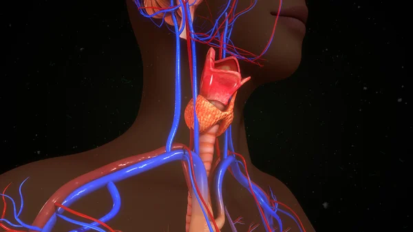
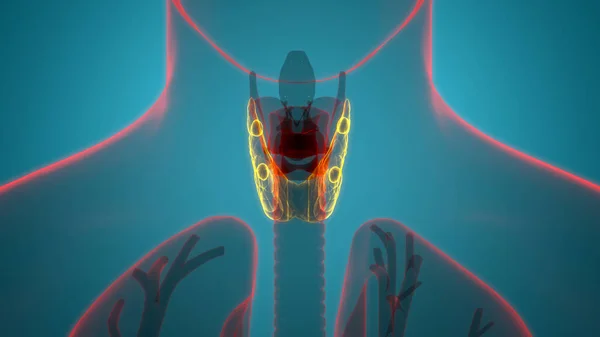
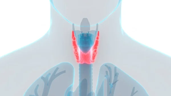
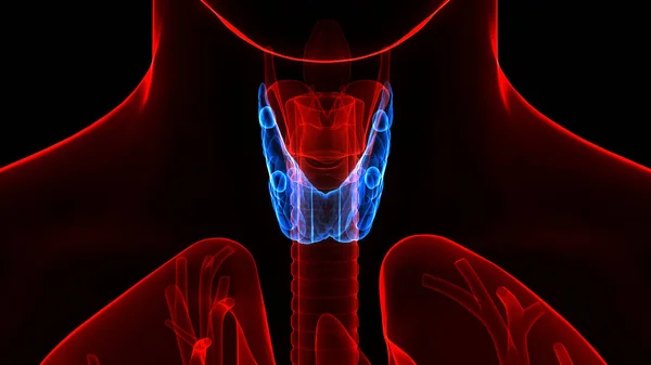
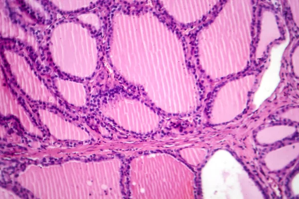
Human thyroid gland.
Human thyroid gland. — Stock Photo, Image
Endometrial adenocarcinoma, light micrograph, photo under microscope
Endometrial Adenocarcinoma Light Micrograph Photo Microscope — Stock Photo, Image
Man having sore throat and touching his neck isolated in clipping path coronavirus symptom
Man Having Sore Throat Touching His Neck Isolated Clipping Path — Stock Photo, Image
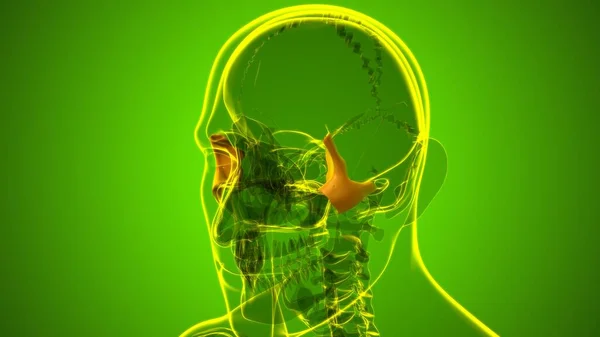
Human Skeleton Skull Zygomatic Bone Anatomy For Medical Concept 3D Illustration
Human Skeleton Skull Zygomatic Bone Anatomy Medical Concept Illustration — Stock Photo, Image
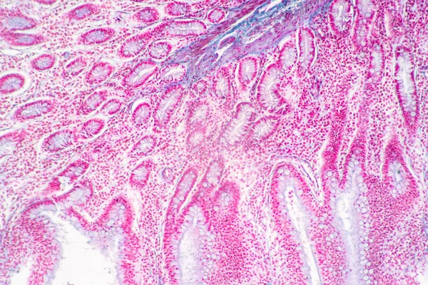
Backgrounds of Characteristics Tissue of Stomach Human, Small intestine Human, Pancreas Human and Large intestine Human under the microscope in Lab.
Backgrounds Characteristics Tissue Stomach Human Small Intestine Human Pancreas Human — Stock Photo, Image
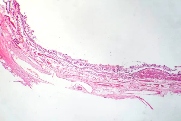
Photomicrograph of chronic bronchitis, showing inflamed bronchial lining with excess mucus production, characteristic of airway disease.
Photomicrograph Chronic Bronchitis Showing Inflamed Bronchial Lining Excess Mucus Production — Stock Photo, Image
Large intestine of a rat
Large intestine of a rat — Stock Photo, Image
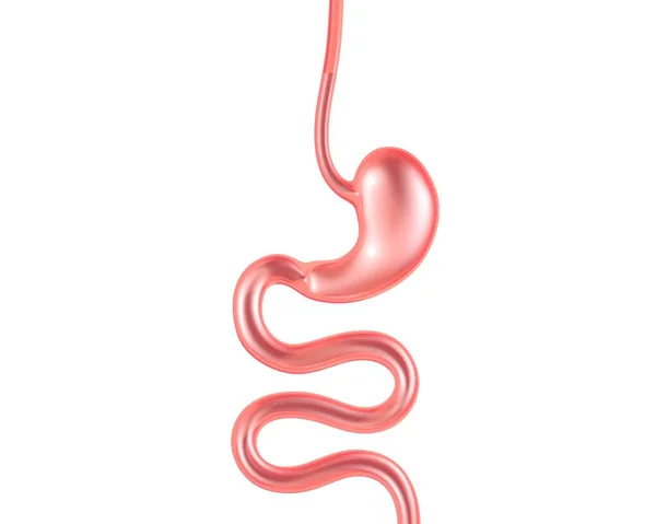
Transparent glass 3d illustration of the digestive system. Anatomy from the esophagus and stomach to the small intestine.
Transparent Glass Illustration Digestive System Anatomy Esophagus Stomach Small Intestine — Stock Photo, Image
Under high magnification microscopic view of labium minus
High Magnification Microscopic View Labium — Stock Photo, Image
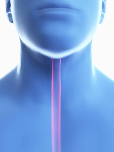
Illustration of oesophagus in male body silhouette, close-up.
Illustration Oesophagus Male Body Silhouette Close — Stock Photo, Image
View of fresh watermelon slice on black back
View of fresh watermelon slice on black back — Stock Photo, Image
Close up human cells with microscope.
Close up human cells with microscope. — Stock Photo, Image
3D Animation Concept of Human Glands Lobes of Thyroid Gland Anatomy
Animation Concept Human Glands Lobes Thyroid Gland Anatomy — Stock Photo, Image
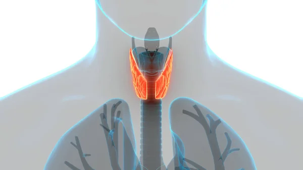
Human Body Glands Thyroid Gland Anatomy. 3D - Illustration
Human Body Glands Thyroid Gland Anatomy Illustration — Stock Photo, Image
Prostate cancer, light micrograph, photo under microscope
Prostate Cancer Light Micrograph Photo Microscope — Stock Photo, Image
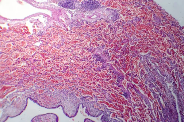
Photomicrograph of acute pulmonary hemorrhage, showing blood-filled alveoli and lung tissue damage due to bleeding.
Photomicrograph Acute Pulmonary Hemorrhage Showing Blood Filled Alveoli Lung Tissue — Stock Photo, Image
Pain
Pain — Stock Photo, Image
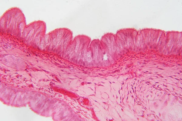
Cilliated epithelium tissue
Cilliated epithelium tissue — Stock Photo, Image
Microbiology, microscopic cells surface
Microbiology Microscopic Cells Surface — Stock Photo, Image
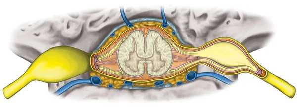
Nervous system, structure of spinal cord, lumbar spine, nerve root, lumbar vertebra, trunk wall, anatomy of human skeletal and nervous system, Intercostal veinus and venous plexuses of the vertebral canal superior view
Nervous System Structure Spinal Cord Lumbar Spine Nerve Root Lumbar — Stock Photo, Image
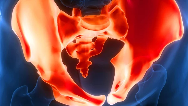
Human Skeleton System Pelvic Girdle Bone Joints Anatomy. 3D
Human Skeleton System Pelvic Girdle Bone Joints Anatomy — Stock Photo, Image
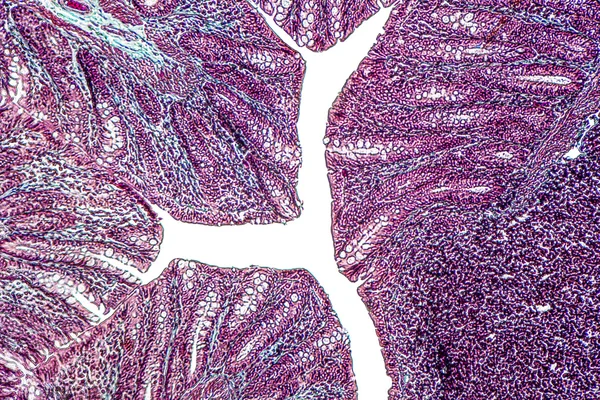
Microscope photo of a large intestine section with inflammation (Colitis).
Microscope photo of a large intestine section with inflammation (Colitis). — Stock Photo, Image
Condyloma acuminatum, also known as genital warts. Light micrograph, photo under microscope
Condyloma Acuminatum Also Known Genital Warts Light Micrograph Photo Microscope — Stock Photo, Image
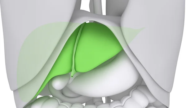
Liver with abdominal organs
Liver with abdominal organs — Stock Photo, Image
Human Skeleton System Skull Bone Parts Mandible Bone Anatomy. 3D
Human Skeleton System Skull Bone Parts Mandible Bone Anatomy — Stock Photo, Image
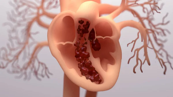
Blood pumping around a human heart, illustration.
Blood Pumping Human Heart Illustration — Stock Photo, Image
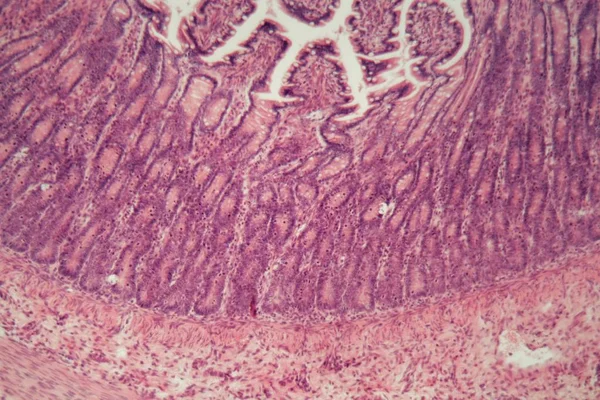
Microscopy photography. Small intestine transversal section.
Microscopy Photography Small Intestine Transversal Section — Stock Photo, Image
Human Body Glands Thyroid Gland Anatomy. 3D - Illustration
Human Body Glands Thyroid Gland Anatomy Illustration — Stock Photo, Image
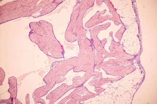
Tissue of Blood human, Vein human, Artery human and Heart muscle human under the microscope in Lab.
Tissue Blood Human Vein Human Artery Human Heart Muscle Human — Stock Photo, Image
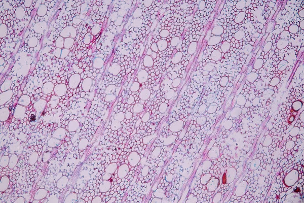
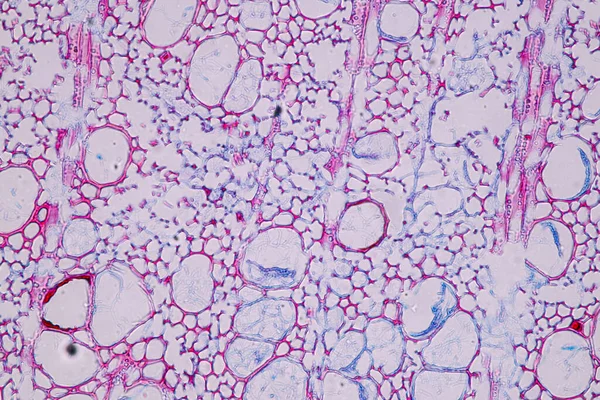
Characteristics of fungi living in wood as a group, are polyphyletic under the microscope for education.
Characteristics Fungi Living Wood Group Polyphyletic Microscope Education — Stock Photo, Image
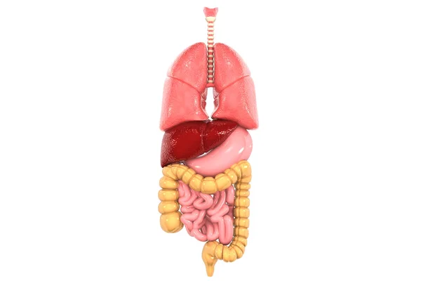
Human lungs and digestive system on white background. 3d illustration
Human Lungs Digestive System White Background Illustration — Stock Photo, Image
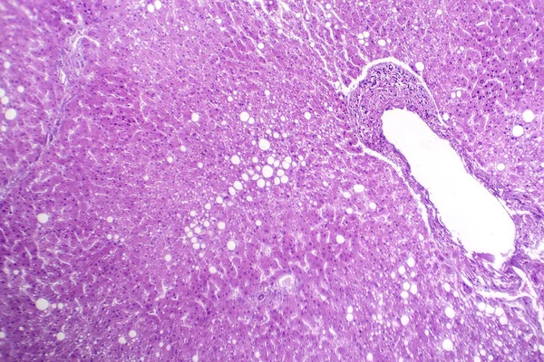
Photomicrograph of hepatic steatosis, revealing fat accumulation in liver cells, known as fatty liver disease.
Photomicrograph Hepatic Steatosis Revealing Fat Accumulation Liver Cells Known Fatty — Stock Photo, Image
Position of the mouth for the pronunciation of the 'I' in phonation.
Position Mouth Pronunciation Phonation — Stock Photo, Image
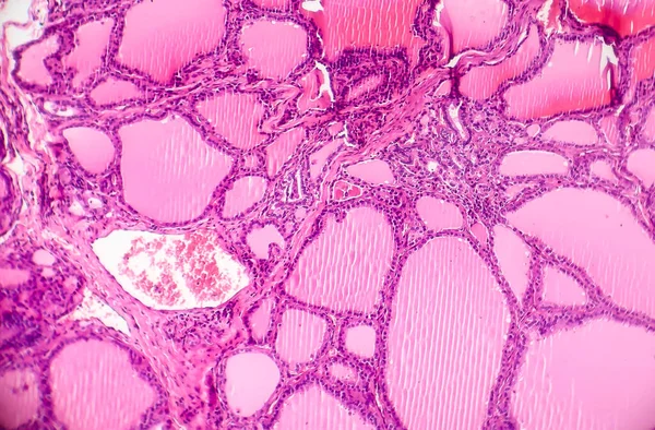
Toxic diffuse goiter, or Graves' disease, an autoimmune disease that affects the thyroid. Light micrograph shows hyperplastic thyroid follicles, decreased colloid
Toxic Diffuse Goiter Graves Disease Autoimmune Disease Affects Thyroid Light — Stock Photo, Image
Colon cancer, light micrograph
Colon cancer, light micrograph — Stock Photo, Image
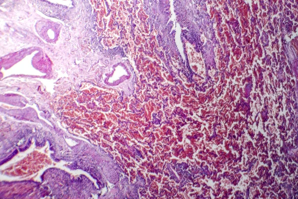
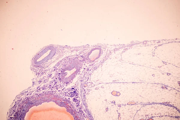
Ovarian cancer, light micrograph, photo under microscope. Photograph shows a fragment of a cancerous tumor in the female ovary. Selective focus
Ovarian Cancer Light Micrograph Photo Microscope Photograph Shows Fragment Cancerous — Stock Photo, Image
Artistic and imaginative digitally designed abstract 3D fractal background
Artistic Imaginative Digitally Designed Abstract Fractal Background — Stock Photo, Image
Close up of mouth for bad breath
Close up of mouth for bad breath — Stock Photo, Image
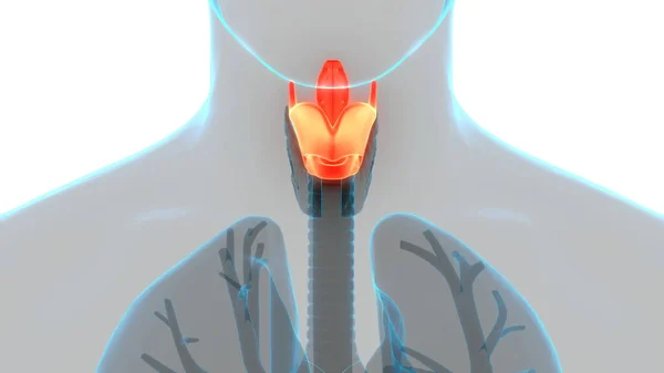
Human Respiratory System Larynx and Pharynx Anatomy. 3D
Human Respiratory System Larynx Pharynx Anatomy — Stock Photo, Image
Tissue of Blood human, Vein human, Artery human and Heart muscle human under the microscope in Lab.
Tissue Blood Human Vein Human Artery Human Heart Muscle Human — Stock Photo, Image
Microscopic cells texture, abstract background
Microscopic Cells Texture Abstract Background — Stock Photo, Image
Characteristics of fungi living in wood as a group, are polyphyletic under the microscope for education.
Characteristics Fungi Living Wood Group Polyphyletic Microscope Education — Stock Photo, Image
Tuberculous pleurisy, light micrograph
Tuberculous pleurisy, light micrograph — Stock Photo, Image
Human face cavity
Human face cavity — Stock Photo, Image
Human Body Glands Lobes of Thyroid Gland Anatomy. 3D
Human Body Glands Lobes Thyroid Gland Anatomy — Stock Photo, Image
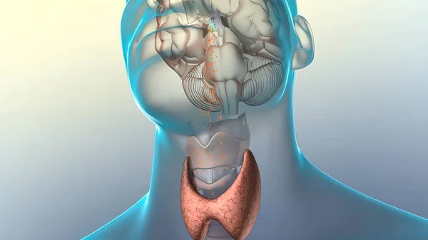
Hormone release in the brain 3d render
Hormone Release Brain Render — Stock Photo, Image
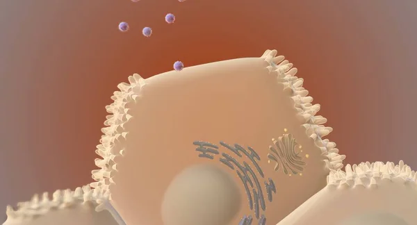
At the cellular level, the hepatitis C virus (HCV) invades liver cells (hepatocytes) and replicates. 3D rendering
Cellular Level Hepatitis Virus Hcv Invades Liver Cells Hepatocytes Replicates — Stock Photo, Image
Pseudostratified epithelium is a type of epithelium that, though comprising only a single layer of cells.
Pseudostratified Epithelium Type Epithelium Though Comprising Only Single Layer Cells — Stock Photo, Image
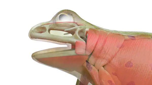
Frog's muscular system, illustration.

Frog Muscular System Illustration — Stock Photo, Image
Human sperm in the testis morphology under microscope. Micrograph showing spermatozoon for pathology education.
Human Sperm Testis Morphology Microscope Micrograph Showing Spermatozoon Pathology Education — Stock Photo, Image
Fantasy chaotic colorful fractal pattern. Abstract fractal shapes. 3D rendering illustration background or wallpaper
Fantasy chaotic colorful fractal pattern. Abstract fractal shapes. 3D rendering illustration background or wallpaper — Stock Photo, Image
Neurofibromatosis, light micrograph, photo under microscope. A group of genetic disorders with formation of tumors in the nervous system
Neurofibromatosis Light Micrograph Photo Microscope Group Genetic Disorders Formation Tumors — Stock Photo, Image
Larynx and trachea digital illustration on black background
Larynx Trachea Digital Illustration Black Background — Stock Photo, Image
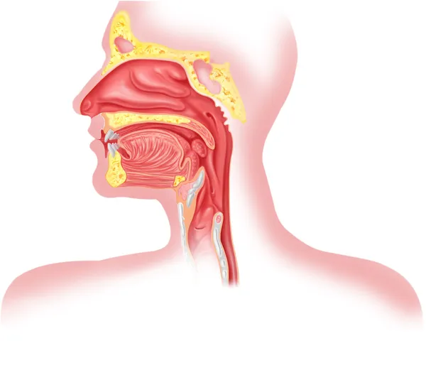
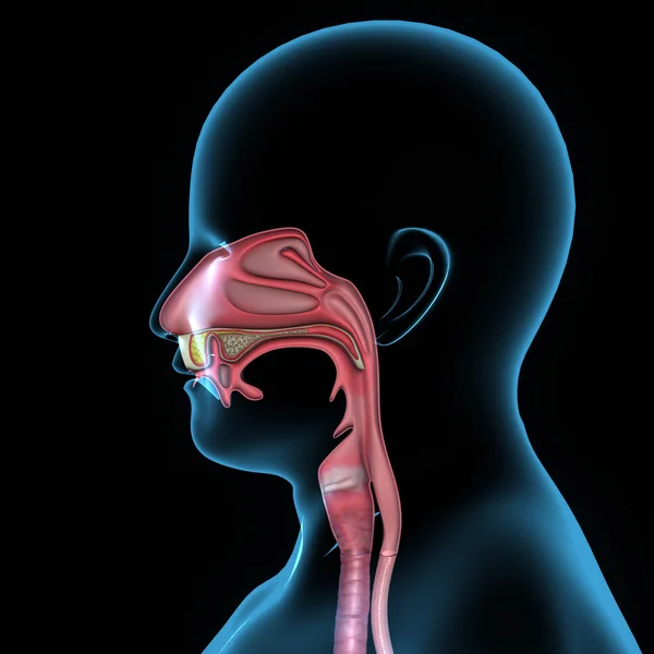
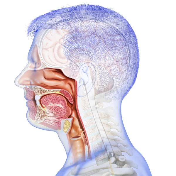
Human respiratory system cross section, head part.
Human respiratory system cross section, head part. — Stock Photo, Image
Root tip of Onion and Mitosis cell in the Root tip of Onion under a microscope.
Root Tip Onion Mitosis Cell Root Tip Onion Microscope — Stock Photo, Image
Human Body Glands Thyroid Gland Anatomy. 3D - Illustration
Human Body Glands Thyroid Gland Anatomy Illustration — Stock Photo, Image
Photomicrograph of acute pulmonary hemorrhage, showing blood-filled alveoli and lung tissue damage due to bleeding.
Photomicrograph Acute Pulmonary Hemorrhage Showing Blood Filled Alveoli Lung Tissue — Stock Photo, Image
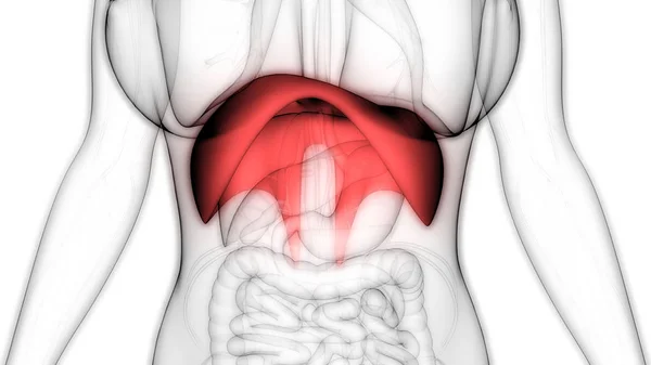
Human Diaphragm Anatomy. 3D illustration
Human Diaphragm Anatomy Illustration — Stock Photo, Image
Liver cirrhosis diseased tissue under the microscope 100x
Liver Cirrhosis Diseased Tissue Microscope 100X — Stock Photo, Image
Macro photography of a Schlumbergera flower from the inside, stamens close-up
Macro Photography Schlumbergera Flower Stamens Close — Stock Photo, Image
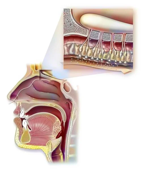
Smell: nose and nasopharynx and zoom on the organ of olfaction.
Smell Nose Nasopharynx Zoom Organ Olfaction — Stock Photo, Image
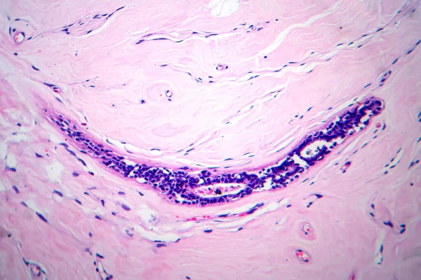
Trachea, light micrograph. Hyaline cartilage from trachea showing chondrocytes, collagen fibers and matrix.
Trachea Light Micrograph Hyaline Cartilage Trachea Showing Chondrocytes Collagen Fibers — Stock Photo, Image
Fractal pretty background. Cool design with colorful shapes and lines. Abstract artistic wallpaper. Hipster graphic image with interesting pattern.
Fractal Pretty Background Cool Design Colorful Shapes Lines Abstract Artistic — Stock Photo, Image
Pink abstract background in watercolor style
Pink Abstract Background Watercolor Style — Stock Photo, Image
Human Respiratory System Diaphragm Anatomy. 3D - Illustration
Human Respiratory System Diaphragm Anatomy Illustration — Stock Photo, Image
Pain in the neck
Pain in the neck — Stock Photo, Image
Dynamic trendy simple fluid color gradient abstract cool background with overlapping line effects. Illustration for wallpaper, banner, background, card, book, pamphlet,website. 2D illustration
Dynamic Trendy Simple Fluid Color Gradient Abstract Cool Background Overlapping — Stock Photo, Image
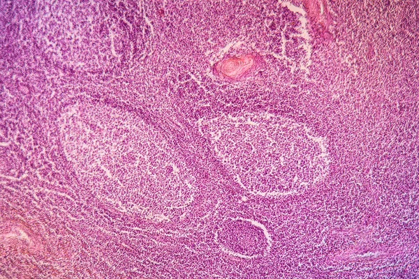
Uterus adenofibroma, light micrograph, photo under microscope. A rare benign tumor of the uterus composed of glandular and fibrous tissues
Uterus Adenofibroma Light Micrograph Photo Microscope Rare Benign Tumor Uterus — Stock Photo, Image
Characteristics of fungi living in wood as a group, are polyphyletic under the microscope for education.
Characteristics Fungi Living Wood Group Polyphyletic Microscope Education — Stock Photo, Image
Human Body Glands Thyroid Gland Anatomy. 3D
Human Body Glands Thyroid Gland Anatomy — Stock Photo, Image
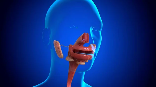
3D Illustration Human Mouth upper Esophageal For Medical Concept
Illustration Human Mouth Upper Esophageal Medical Concept — Stock Photo, Image
Human Body Glands Lobes of Thyroid Gland Anatomy. 3D
Human Body Glands Lobes Thyroid Gland Anatomy — Stock Photo, Image
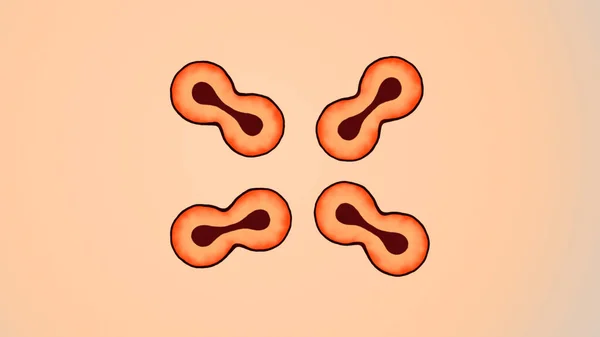
Illustration of Multiple Red Color Biology Cells Dividing
Illustration Multiple Red Color Biology Cells Dividing — Stock Photo, Image
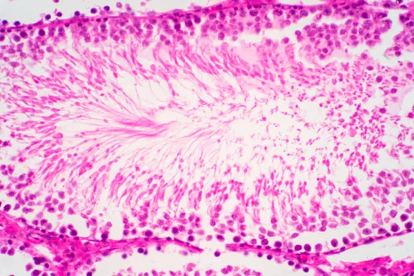
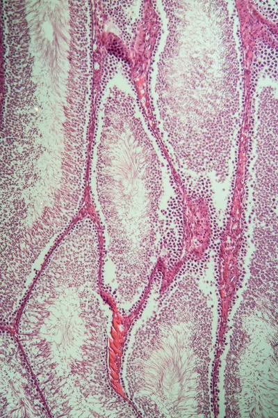
Testicles of the rat under the microscope
Testicles Rat Microscope — Stock Photo, Image
Mouth anatomy
Mouth anatomy — Stock Photo, Image
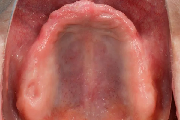
Edentulous upper jaw with perfect gingiva before implantation, seen through a mirror
Edentulous Upper Jaw Perfect Gingiva Implantation Seen Mirror — Stock Photo, Image
Artery section under the microscope
Artery section under the microscope — Stock Photo, Image
Swirls of alcohol ink in lilac, lavender, violet are reminiscent of marble and ripples of agate. Fluid modern Art banners, ethereal graphic design.
Swirls of alcohol ink in lilac, lavender, violet are reminiscent of marble and ripples of agate. Fluid modern Art banners, ethereal graphic design. — Stock Photo, Image
Lungworm under the microscope 100x
Lungworm Microscope 100X — Stock Photo, Image
Fractal pretty background. Cool design with colorful shapes and lines. Abstract artistic wallpaper. Hipster graphic image with interesting pattern.
Fractal Pretty Background Cool Design Colorful Shapes Lines Abstract Artistic — Stock Photo, Image
Human Body Glands Lobes of Thyroid Gland Anatomy. 3D
Human Body Glands Lobes Thyroid Gland Anatomy — Stock Photo, Image
Sore throat
Sore throat — Stock Photo, Image
Histopathology of portal cirrhosis
Histopathology of portal cirrhosis — Stock Photo, Image
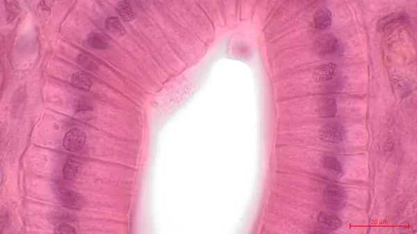
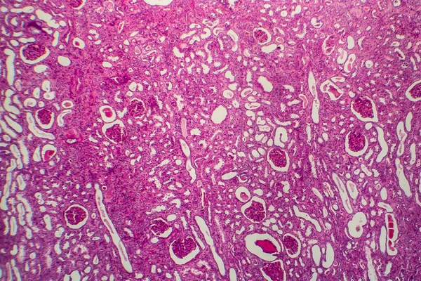
Simple columnar epithelium. Light micrograph of a renal medulla showing several collecting ducts lined by a simple columnar epithelium. Magnification: 1000
Simple Columnar Epithelium Light Micrograph Renal Medulla Showing Several Collecting — Stock Photo, Image
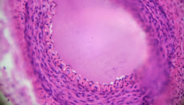
Ovarian mucinous cystadenoma (15-20% of ovarian tumours) is a usually large and benign tumour showing glands and cysts, with filiform papillae with fibrovascular cores, lined by benign columnar epithelium.
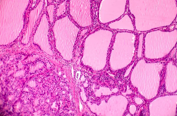
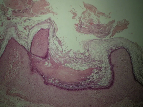
Ovarian Mucinous Cystadenoma Ovarian Tumours Usually Large Benign Tumour Showing — Stock Photo, Image
Human Respiratory System Larynx and Pharynx Anatomy. 3D
Human Respiratory System Larynx Pharynx Anatomy — Stock Photo, Image
Asian woman having throat pain
Asian woman having throat pain — Stock Photo, Image
Photomicrograph of a toxic goiter tissue sample under a microscope, revealing hypertrophy of thyroid follicular cells, increased vascularity, and colloid depletion.
Photomicrograph Toxic Goiter Tissue Sample Microscope Revealing Hypertrophy Thyroid Follicular — Stock Photo, Image
Cancer of cervix. Light micrograph of cervical biopsy. Photo under microscope. Selective focus
Cancer Cervix Light Micrograph Cervical Biopsy Photo Microscope Selective Focus — Stock Photo, Image
Cancer of cervix. Light micrograph of cervical biopsy. Photo under microscope. Selective focus
Cancer Cervix Light Micrograph Cervical Biopsy Photo Microscope Selective Focus — Stock Photo, Image
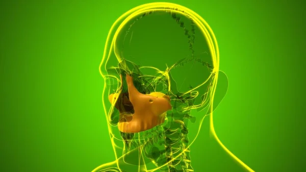
Human Skeleton Skull Maxillal Bone Anatomy For Medical Concept 3D Illustration
Human Skeleton Skull Maxillal Bone Anatomy Medical Concept Illustration — Stock Photo, Image
Human Skeleton Skull Zygomatic Bone Anatomy For Medical Concept 3D Illustration
Human Skeleton Skull Zygomatic Bone Anatomy Medical Concept Illustration — Stock Photo, Image
Chronic pyelonephritis, light micrograph, photo under microscope
Chronic Pyelonephritis Light Micrograph Photo Microscope — Stock Photo, Image
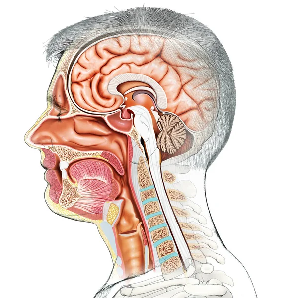
3d rendered, medically accurate illustration of Cross section of male head
Rendered Medically Accurate Illustration Cross Section Male Head — Stock Photo, Image
3d rendered, medically accurate illustration of male Trachea and esophagus anatom
Rendered Medically Accurate Illustration Male Trachea Esophagus Anatom — Stock Photo, Image
Toxic diffuse goiter, or Graves' disease, an autoimmune disease that affects the thyroid. Light micrograph shows hyperplastic thyroid follicles, decreased colloid
Toxic Diffuse Goiter Graves Disease Autoimmune Disease Affects Thyroid Light — Stock Photo, Image
Bazex syndrome or paraneoplastic acrokeratosis is a psoriasiform eruption that is seen more frequently in adult Caucasian males and is characterized by a predominantly acral distribution with nail dystrophy.
Bazex Syndrome Paraneoplastic Acrokeratosis Psoriasiform Eruption Seen More Frequently Adult — Stock Photo, Image
Cliated epithelium of human under the microscope in Lab.
Cliated Epithelium Human Microscope Lab — Stock Photo, Image

Find the Best Epiglottis Images for Your Project

If you are searching for epiglottis images, you have come to the right place. We offer a wide range of stock images that are perfect for medical publications, presentations, and educational materials. Our images are available in different formats, including JPG, AI, and EPS, so you can choose the one that works best for your project.

If you need to illustrate the epiglottis, an essential part of your respiratory system, you can find the perfect image on our website. Our images depict this small flap of tissue at the base of the tongue in high resolution, allowing you to showcase its structure, function, and importance.

Whether you need an image for a medical textbook, a journal article, or an online course, we have everything you need. Our collection includes images of the epiglottis in various angles, sizes, and colors, so you can pick the one that fits your design concept.

Tips for Using Epiglottis Images

When using epiglottis images, always keep your audience in mind. If your project is for medical professionals, you can use detailed, scientific images that show the epiglottis in detail. However, if your project targets a non-medical audience, it's best to use simpler, more straightforward images that focus on the epiglottis' function in the body.

Always choose images that are of high quality and resolution to ensure they look sharp and clear, even when printed at a larger size. If possible, choose images with a transparent background, so you can easily incorporate them into your design.

When designing your project, consider the placement of the image carefully. You want to make sure that the image adds value to your text and draws the reader's eye to the most important information. Additionally, you should consider the color scheme and font choice to complement the image and make your project cohesive.

Discover Our Extended Collection

We offer a comprehensive collection of stock images that are ideal for medical and scientific projects. Our images cover various topics, including anatomy, physiology, pathology, and more. With our extensive collection, you can find the perfect images to complement your project and make it stand out.

In addition to our epiglottis images, you can also discover our other related images, such as larynx, trachea, vocal cords, and more. Our images come with different licensing options, allowing you to use them for personal or commercial projects.

Choose the Perfect Epiglottis Images Today

Whether you are a medical professional or a designer, using the right epiglottis images can make or break your project. At our website, you can find high-quality images that are perfect for your needs. Browse our collection today and select the perfect images to illustrate your project beautifully.

By choosing our top-quality images, you can ensure that your project is informative, engaging, and visually appealing. Invest in great images today and bring your project to the next level.