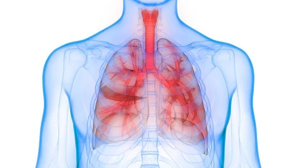
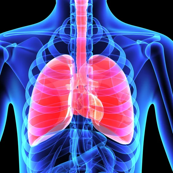
Human Respiratory System Lungs Anatomy. 3D
Human Respiratory System Lungs Anatomy — Stock Photo, Image
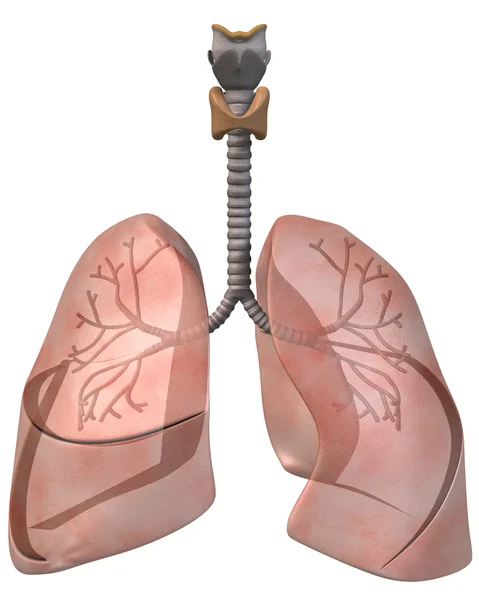
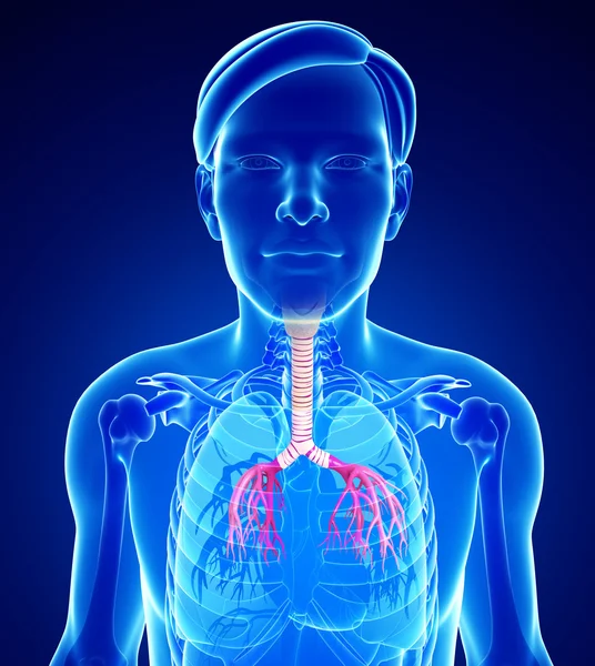
Lungs and Bronchi, Front View
Lungs and Bronchi, Front View — Stock Photo, Image
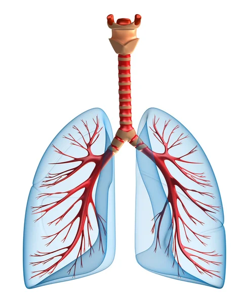
Lungs - pulmonary system. Front view
Lungs - pulmonary system. Front view — Stock Photo, Image
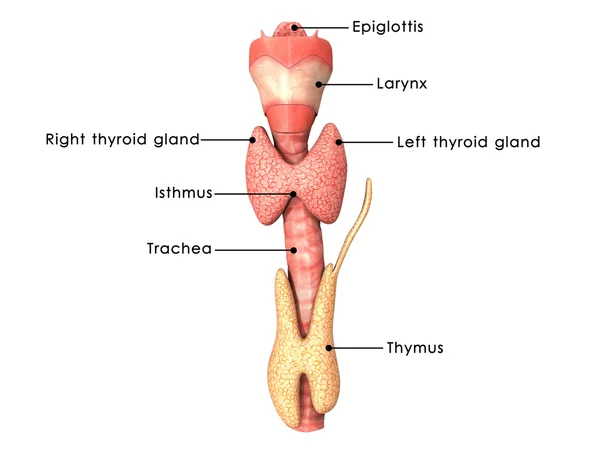
Thyroid gland
Thyroid gland — Stock Photo, Image
Male throat anatomy
Male throat anatomy — Stock Photo, Image
Bronchi
Bronchi — Stock Photo, Image
Human lungs anatomy
Human lungs anatomy — Stock Photo, Image
Thyroid glands anatomy
Thyroid glands anatomy — Stock Photo, Image
3D Concept of Human Respiratory System Lungs Anatomy
Concept Human Respiratory System Lungs Anatomy — Stock Photo, Image
Human lungs anatomy
Human lungs anatomy — Stock Photo, Image
Human Lungs
Human Lungs — Stock Photo, Image
Lungs Foreign Object - inhaled or swallowed
Lungs Foreign Object - inhaled or swallowed — Stock Photo, Image
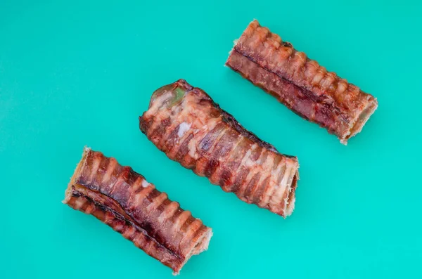
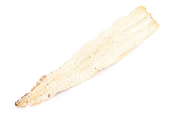
Three Dried Beef Tracheas. Natural chewing treats for dogs. Love for domestic animals. Pet supplies. Side view. Selective focus. Close-up.
Three Dried Beef Tracheas Natural Chewing Treats Dogs Love Domestic — Stock Photo, Image
Male throat anatomy
Male throat anatomy — Stock Photo, Image
Lungs
Lungs — Stock Photo, Image
Male urinary system
Male urinary system — Stock Photo, Image
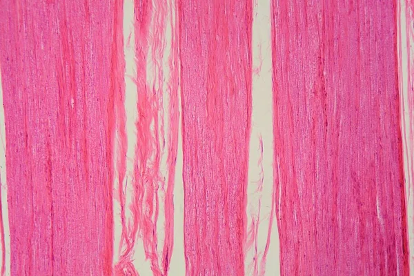
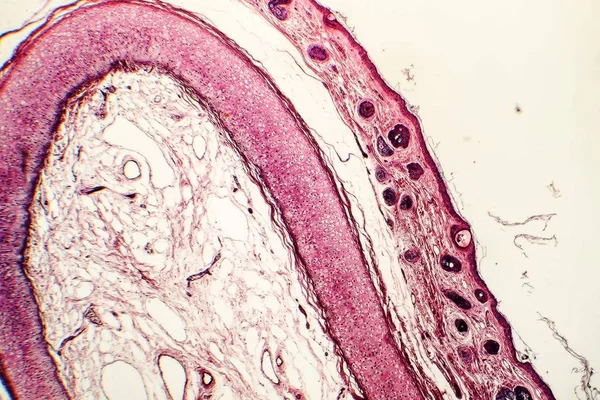
Trachea, light micrograph. Hyaline cartilage from trachea showing chondrocytes, collagen fibers and matrix.
Trachea Light Micrograph Hyaline Cartilage Trachea Showing Chondrocytes Collagen Fibers — Stock Photo, Image
A longitudinal section of human nerve cells under the microscope.
Longitudinal Section Human Nerve Cells Microscope — Stock Photo, Image
Manchurian Walnut (Juglans mandshurica). Chambered Pith Closeup
Manchurian Walnut Juglans Mandshurica Chambered Pith Closeup — Stock Photo, Image
Background of the bark of a palm tree. Close-up of fragments of the bark of a tree in chaotic abstract design. In category of creative background of exotic summer relaxation, screen saver, wallpaper.
Background Bark Palm Tree Close Fragments Bark Tree Chaotic Abstract — Stock Photo, Image
Background of the bark of a palm tree. Close-up of fragments of
Background of the bark of a palm tree. Close-up of fragments of — Stock Photo, Image
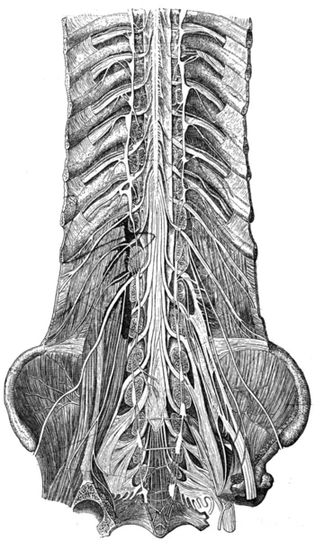
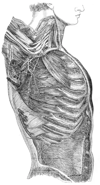
Nerves of the lower trunk, the spinal canal is open, front view
Nerves of the lower trunk, the spinal canal is open, front view — Stock Photo, Image
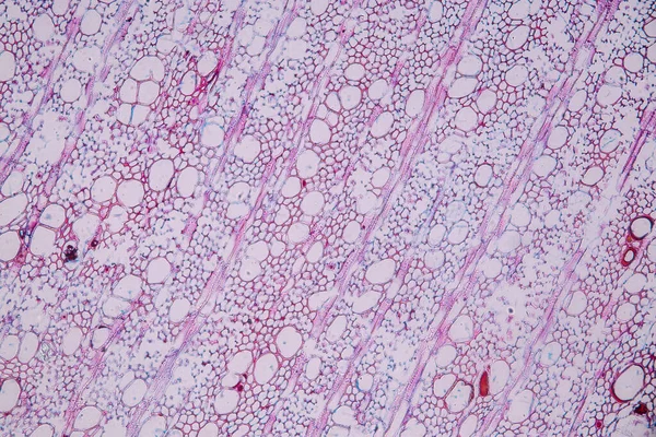
Plant vascular tissue under the light microscope view for education.
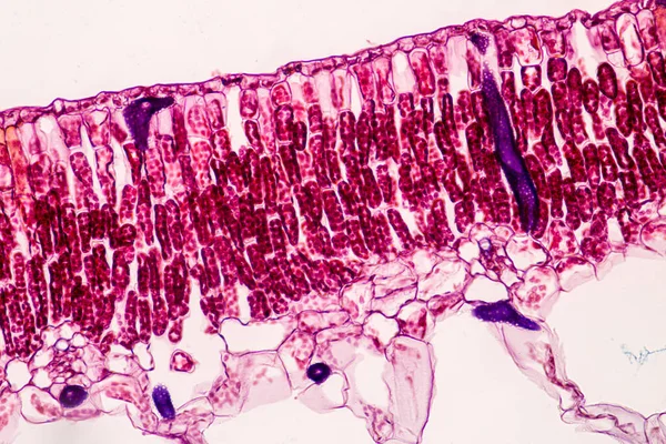
Plant Vascular Tissue Light Microscope View Education — Stock Photo, Image
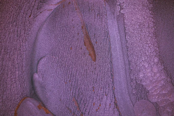
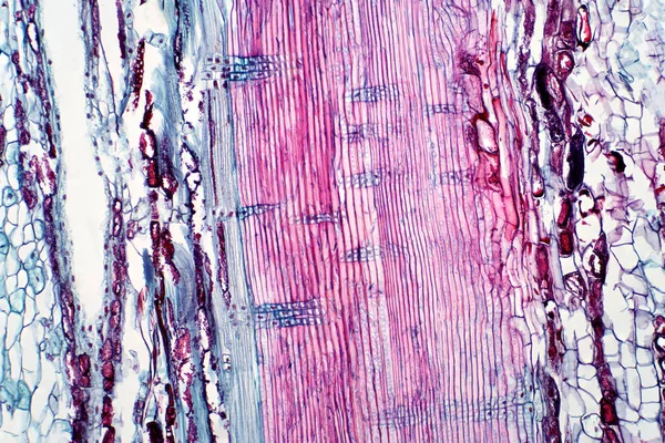
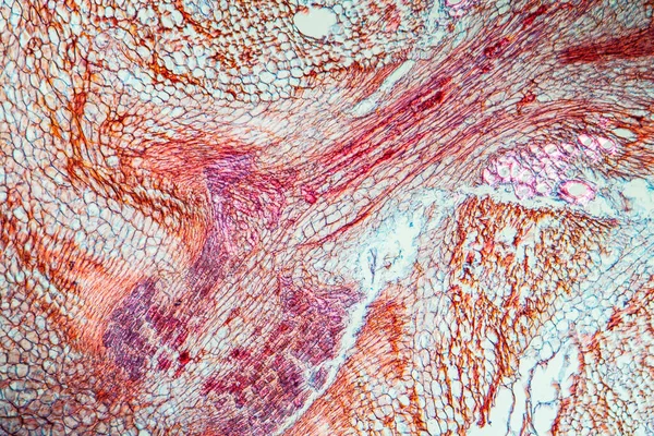
Plant vascular tissue, light micrograph. Haematoxylin and eosin stain.
Plant Vascular Tissue Light Micrograph Haematoxylin Eosin Stain — Stock Photo, Image
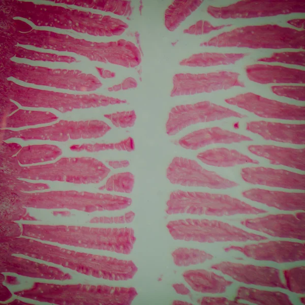
Close up human cells with microscope.
Close up human cells with microscope. — Stock Photo, Image
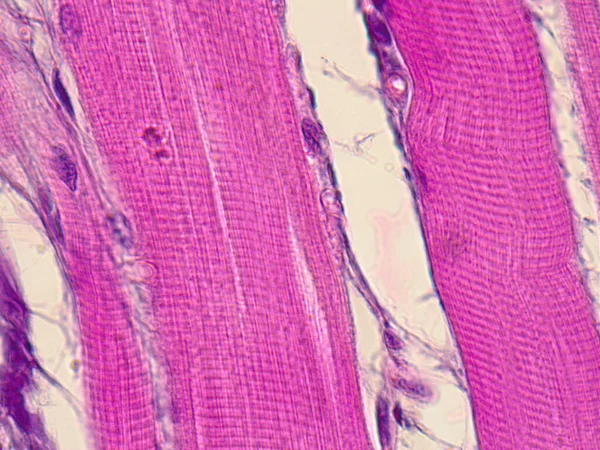
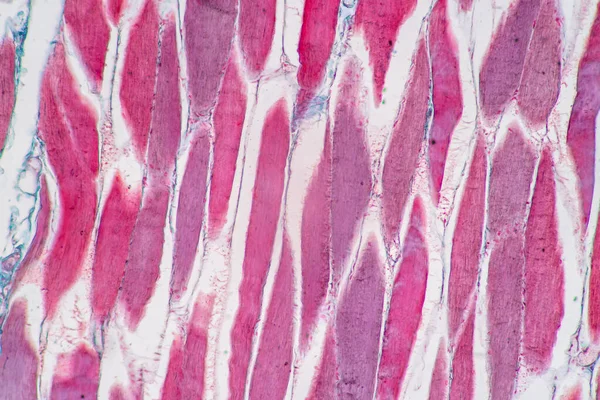
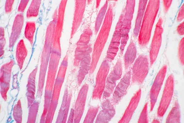
Striated muscle tissue under the microscope.
Striated muscle tissue under the microscope. — Stock Photo, Image
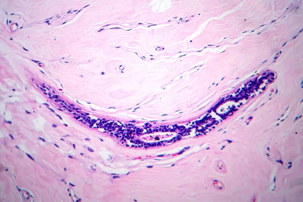
Photomicrograph of fibroadenoma, showcasing benign glandular and fibrous tissue growth within the breast, a common noncancerous tumor.
Photomicrograph Fibroadenoma Showcasing Benign Glandular Fibrous Tissue Growth Breast Common — Stock Photo, Image
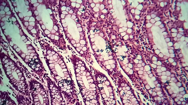
Bacillary dysentery, light micrograph, photo under microscope showing presence of bacteria and accumulation of inflammatory cells in intestinal epithelium
Bacillary Dysentery Light Micrograph Photo Microscope Showing Presence Bacteria Accumulation — Stock Photo, Image
Tree bark with moss
Tree Bark Moss — Stock Photo, Image
The modern art background of high-quality abstract Chinese landscape painting presented in alcohol ink.
Modern Art Background High Quality Abstract Chinese Landscape Painting Presented — Stock Photo, Image
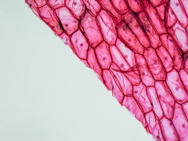
Onion epidermus micrograph
Onion epidermus micrograph — Stock Photo, Image
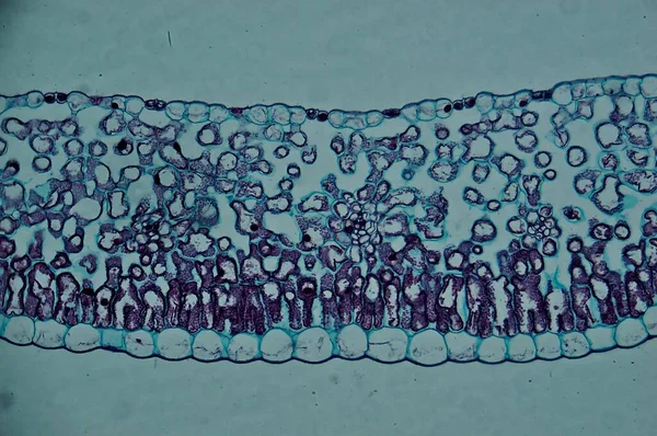
Lily leaf c.s. under microscope
Lily Leaf Microscope — Stock Photo, Image
Removed human tooth with caries and Tartar
Removed human tooth with caries and Tartar — Stock Photo, Image
Abstract black and white background template art, 3d render.
Abstract Black White Background Template Art Render — Stock Photo, Image
The study Plant tissue of under the microscope for classroom education.
Study Plant Tissue Microscope Classroom Education — Stock Photo, Image
The study Plant tissue of under the microscope for classroom education.
Study Plant Tissue Microscope Classroom Education — Stock Photo, Image
Dramatic black, monochrome abstraction, reminiscent of dragon scales or caked lava, shimmering. Fragment of a modern artwork. Macro photography of the smallest details of a panting. Contemporary art. Artistic background image.
Dramatic Black Monochrome Abstraction Reminiscent Dragon Scales Caked Lava Shimmering — Stock Photo, Image
Endometrial adenocarcinoma, light micrograph, photo under microscope
Endometrial Adenocarcinoma Light Micrograph Photo Microscope — Stock Photo, Image
Science medical anthropotomy physiology microscopic human small
Science medical anthropotomy physiology microscopic human small — Stock Photo, Image
Cilliated epithelium tissue
Cilliated epithelium tissue — Stock Photo, Image
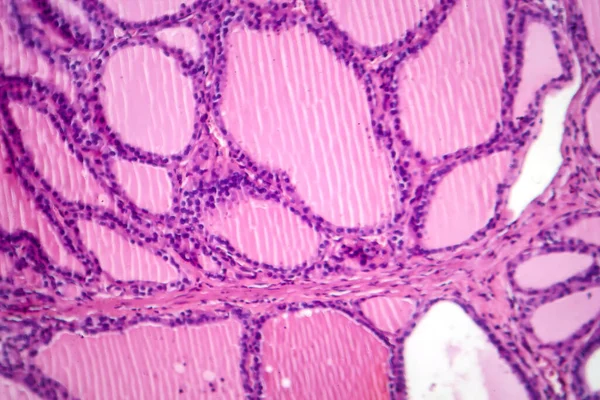
Photomicrograph of a toxic goiter tissue sample under a microscope, revealing hypertrophy of thyroid follicular cells, increased vascularity, and colloid depletion.
Photomicrograph Toxic Goiter Tissue Sample Microscope Revealing Hypertrophy Thyroid Follicular — Stock Photo, Image
Old wood cracked texture,Seamless tree bark texture,Endless wooden background for web page fill or graphic design. Selective focus.
Old Wood Cracked Texture Seamless Tree Bark Texture Endless Wooden — Stock Photo, Image
Relief bark
Relief bark — Stock Photo, Image
The study Plant tissue of under the microscope for classroom education.
Study Plant Tissue Microscope Classroom Education — Stock Photo, Image
Abstract ink background. Marble style. Black paint stroke texture on white paper. Wallpaper for web and game design. Grunge mud art. Macro image of pen juice. Dark Smear
Abstract Ink Background Marble Style Black Paint Stroke Texture White — Stock Photo, Image
Close up of bark texture on a sweet gum tree, a large, deciduous tree. commonly called sweetgum (sweet gum in the UK), gum, redgum, satin-walnut, or American storax. Common tree in Northern California
Close Bark Texture Sweet Gum Tree Large Deciduous Tree Commonly — Stock Photo, Image
Magenta Female Knitted Scarf
Magenta Female Knitted Scarf — Stock Photo, Image
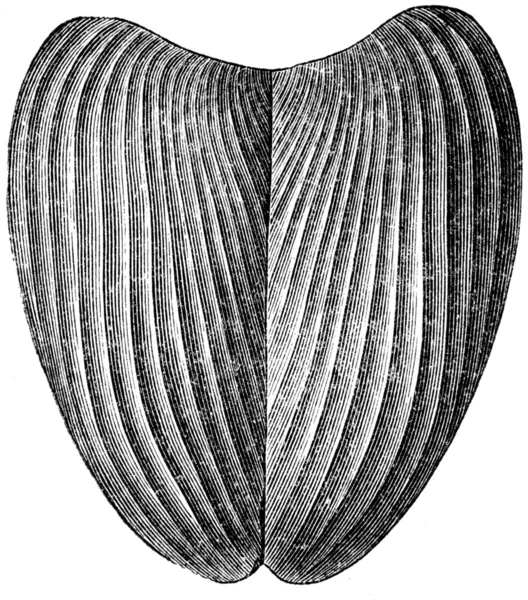
Aptyhus camellosis
Aptyhus camellosis — Stock Photo, Image
Illustration of a snake
Illustration Snake — Stock Photo, Image
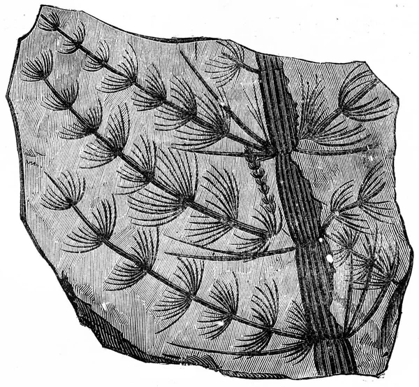
Ancient algae
Ancient algae — Stock Photo, Image
Education anatomy and Histological sample of Human under the microscope.
Education Anatomy Histological Sample Human Microscope — Stock Photo, Image
Uterus adenofibroma, light micrograph, photo under microscope. A rare benign tumor of the uterus composed of glandular and fibrous tissues
Uterus Adenofibroma Light Micrograph Photo Microscope Rare Benign Tumor Uterus — Stock Photo, Image
Carrot with root across 100x
Carrot Root 100X — Stock Photo, Image
Cicada larva
Cicada larva — Stock Photo, Image
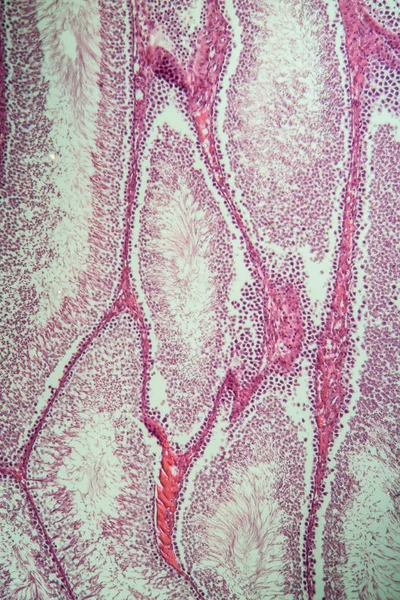
Testicles of the rat under the microscope
Testicles Rat Microscope — Stock Photo, Image
A closeup shot of a tree bark texture
Closeup Shot Tree Bark Texture — Stock Photo, Image
Nerve trunk and nerve plexus of arm
Nerve trunk and nerve plexus of arm — Stock Photo, Image
Bacillary dysentery, light micrograph, photo under microscope showing presence of bacteria and accumulation of inflammatory cells in intestinal epithelium
Bacillary Dysentery Light Micrograph Photo Microscope Showing Presence Bacteria Accumulation — Stock Photo, Image
Onion epidermus micrograph
Onion epidermus micrograph — Stock Photo, Image
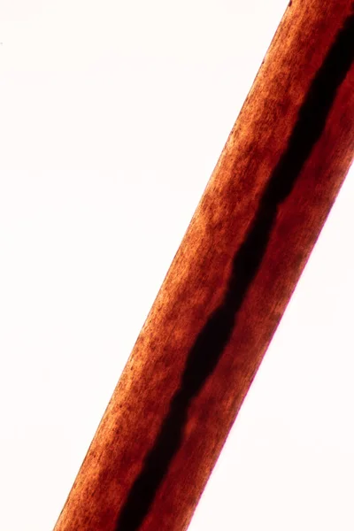
Characteristics of Hair cell of human under microscope view for education in laboratory.
Characteristics Hair Cell Human Microscope View Education Laboratory — Stock Photo, Image
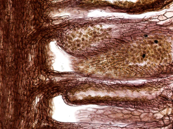
Characteristics of fungi living in wood as a group, are polyphyletic under the microscope for education.
Characteristics Fungi Living Wood Group Polyphyletic Microscope Education — Stock Photo, Image
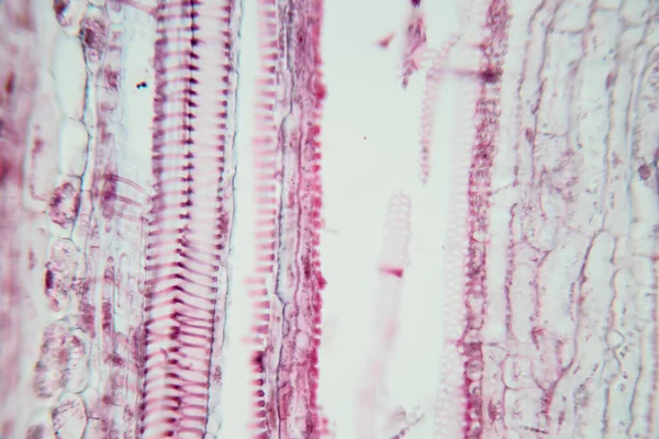
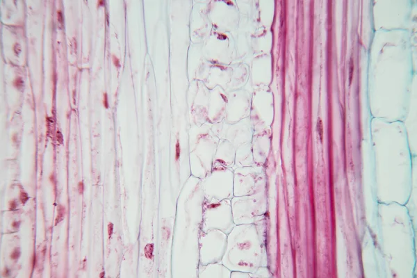
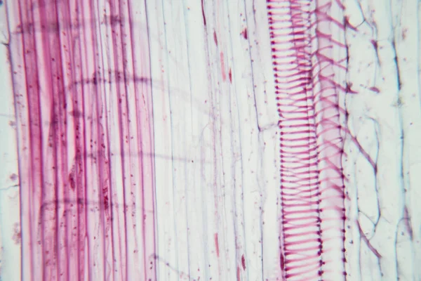
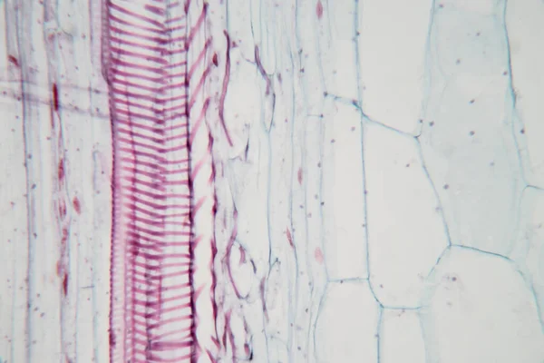
Long section plant vascular tissue under the light microscope view for education.
Long Section Plant Vascular Tissue Light Microscope View Education — Stock Photo, Image
Stem with sieve cells under the microscope
Stem with sieve cells under the microscope — Stock Photo, Image
Abstract background. Monochrome texture. Black and white textured background.
Abstract Background Monochrome Texture Black White Textured Background — Stock Photo, Image
Characteristics of fungi living in wood as a group, are polyphyletic under the microscope for education.
Characteristics Fungi Living Wood Group Polyphyletic Microscope Education — Stock Photo, Image
Black white abstract acrylic painting color texture on white paper background by using rorschach inkblot method
Black White Abstract Acrylic Painting Color Texture White Paper Background — Stock Photo, Image
Wood texture 151
Wood texture 151 — Stock Photo, Image
Background of mulberry paper, Thailand.
Background Mulberry Paper Thailand — Stock Photo, Image
Background of mulberry paper, Thailand.
Background Mulberry Paper Thailand — Stock Photo, Image
Bark background There are cracks in the bark.
Bark Background Cracks Bark — Stock Photo, Image
Large-Leaved Lime (Tilia platyphyllos). Twig Detail Closeup
Large Leaved Lime Tilia Platyphyllos Twig Detail Closeup — Stock Photo, Image
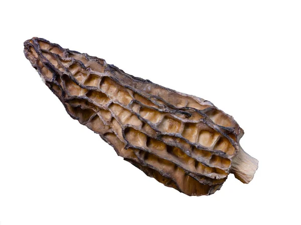
Super macro shoot of dried morel mushrooms very close in detail. Ideal food background
Super Macro Shoot Dried Morel Mushrooms Very Close Detail Ideal — Stock Photo, Image
The study Plant tissue of under the microscope for classroom education.
Study Plant Tissue Microscope Classroom Education — Stock Photo, Image
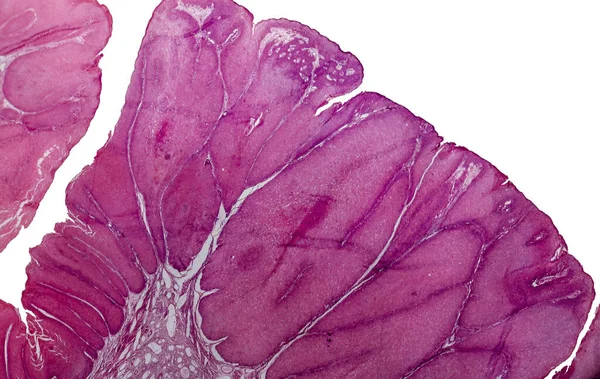
Condyloma acuminatum, also known as genital warts. Light micrograph, photo under microscope
Condyloma Acuminatum Also Known Genital Warts Light Micrograph Photo Microscope — Stock Photo, Image
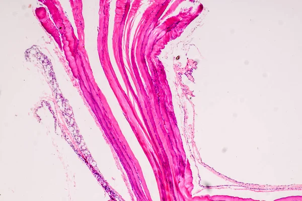
Fish's gills greatly enlarged, 100x
Fish Gills Greatly Enlarged 100X — Stock Photo, Image
Cross section of a cerebellum. Light micrograph.
Cross Section Cerebellum Light Micrograph — Stock Photo, Image
Background of the bark of a palm tree. Close-up of fragments of
Background of the bark of a palm tree. Close-up of fragments of — Stock Photo, Image
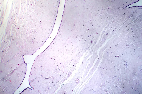
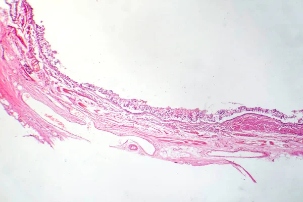
Photomicrograph of chronic bronchitis, showing inflamed bronchial lining with excess mucus production, characteristic of airway disease.
Photomicrograph Chronic Bronchitis Showing Inflamed Bronchial Lining Excess Mucus Production — Stock Photo, Image
Dramatic black, monochrome abstraction, reminiscent of dragon scales or caked lava, shimmering. Fragment of a modern artwork. Macro photography of the smallest details of a panting. Contemporary art. Artistic background image.
Dramatic Black Monochrome Abstraction Reminiscent Dragon Scales Caked Lava Shimmering — Stock Photo, Image
Characteristics of anatomy and Histological sample Striated (Skeletal) muscle of mammal Tissue under the microscope.
Characteristics Anatomy Histological Sample Striated Skeletal Muscle Mammal Tissue Microscope — Stock Photo, Image
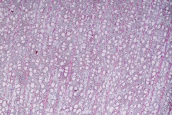
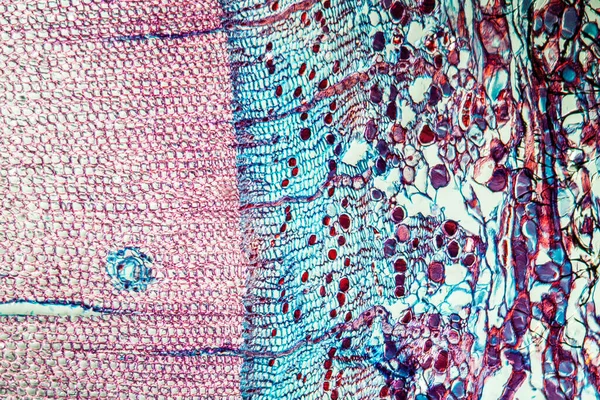
Pine tree trunk across 100x
Pine Tree Trunk 100X — Stock Photo, Image
Squid
Squid — Stock Photo, Image
Elastic cartilage of human outer ear
Elastic cartilage of human outer ear — Stock Photo, Image
Characteristics of Hair cell of human under microscope view for education in laboratory.
Characteristics Hair Cell Human Microscope View Education Laboratory — Stock Photo, Image
Black white background with watercolor texture
Black White Background Watercolor Texture — Stock Photo, Image
Bark of old linden tree. Relief bark is like view high mountains from above. old bark texture
Bark Old Linden Tree Relief Bark View High Mountains Old — Stock Photo, Image
Old wood background texture
Old Wood Background Texture — Stock Photo, Image
Chandelier raster drawing. Pendant light element. Modern chandelier on white background. Decor accessory design. Raster illustration sketch
Chandelier Raster Drawing Pendant Light Element Modern Chandelier White Background — Stock Photo, Image
Abstract ink background. Marble style. Black paint stroke texture on white paper. Wallpaper for web and game design. Grunge mud art. Macro image of pen juice. Dark Smear
Abstract Ink Background Marble Style Black Paint Stroke Texture White — Stock Photo, Image
Complex mucosal folding of seminal vesicle
Complex mucosal folding of seminal vesicle — Stock Photo, Image
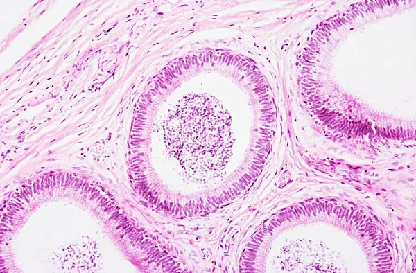
Under high magnification microscopic view of epididymis
High Magnification Microscopic View Epididymis — Stock Photo, Image
Bark palm
Bark palm — Stock Photo, Image
The study Plant tissue of under the microscope for classroom education.
Study Plant Tissue Microscope Classroom Education — Stock Photo, Image
Carrot root across 100x
Carrot Root 100X — Stock Photo, Image
Tree bark texture background. - Image a tree of Thailand.
Tree bark texture background. - Image a tree of Thailand. — Stock Photo, Image
Texture of tree
Texture of tree — Stock Photo, Image
I painted acrylic colors on white paper and I folded the paper together similar to the rorschach inkblot method that create the texture of mixed acrylic colors.
Painted Acrylic Colors White Paper Folded Paper Together Similar Rorschach — Stock Photo, Image
Characteristics of anatomy and Histological sample Striated (Skeletal) muscle of mammal Tissue under the microscope.
Characteristics Anatomy Histological Sample Striated Skeletal Muscle Mammal Tissue Microscope — Stock Photo, Image
Stalagmite closeup
Stalagmite closeup — Stock Photo, Image
Chandelier raster drawing. Pendant light element. Modern chandelier on white background. Decor accessory design. Raster illustration sketch
Chandelier Raster Drawing Pendant Light Element Modern Chandelier White Background — Stock Photo, Image

Related image searches

Trachea Images: High-Quality Visuals for Medical Professionals

Trachea images are an essential element for medical professionals looking to enhance their presentations, publications, and medical research. At our stock images platform, we offer a vast collection of trachea images in different file formats, including JPG, AI, and EPS. Our images are carefully designed with all the visual details required to accurately represent the trachea.

Types of Trachea Images Available

We understand that medical professionals require different types of trachea images for various projects. Therefore, we offer several types of trachea images, including:

  • Frontal view of the trachea
  • Lateral view of the trachea
  • Close-up view of the trachea
  • Trachea with surrounding structures
  • Tracheotomy procedure images

These trachea images are available in multiple angles, including 2D and 3D. They present a comprehensive and accurate representation of the trachea, making them useful for medical presentations, research, and training materials.

Best Use Cases for Trachea Images

Trachea images are vital for medical professionals in various fields, including pulmonologists, otolaryngologists, thoracic surgeons, and medical students. These images can be used in different projects, including:

  • Medical textbooks and educational materials
  • Research publications and presentations
  • Patient education materials
  • Medical illustrations and diagrams
  • Medical websites and blogs

With our collection of trachea images, medical professionals have access to visuals that accurately depict the trachea's anatomy, making it easy to educate and inform patients and colleagues.

Choosing the Right Trachea Image

When selecting trachea images, medical professionals are advised to consider the following:

  • The project's objective: Medical professionals should choose images that reflect the project's objective accurately. For instance, if the image's purpose is to educate patients on the tracheotomy procedure, the image chosen should represent the procedure clearly.
  • The image quality: High-quality trachea images with high resolution are essential for medical publications and presentations. High-quality images make the visuals more professional and accurate.
  • Permissions: It's important to ensure you have the necessary permissions to use the trachea images for your intended purpose. At our stock images platform, we provide safe and licensed images for all medical professionals.

With these factors in mind, medical professionals can make informed decisions when selecting trachea images that best represent their projects.

Conclusion

Trachea images are an essential element for medical professionals in various fields. They make it easier to educate and inform patients and colleagues on trachea anatomy, procedures, and medical research. Our platform offers a wide range of high-quality trachea images that medical professionals can select from, depending on their project's objective. Medical professionals can be sure of finding the best trachea images that accurately represent the trachea's anatomy and enhance their presentations, publications, and research with accurate, professional visuals.