Skin cross section Stock Photos

100,000 Skin cross section pictures are available under a royalty-free license

  • Best Match
  • Fresh
  • Popular
1-100 of 100,000
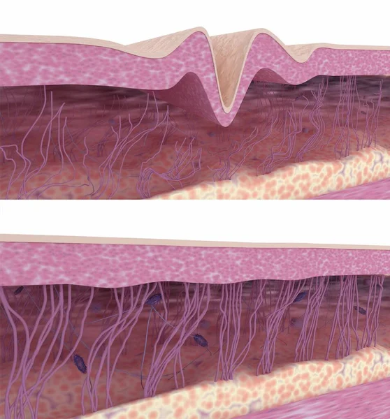
Skin rejuvenation. Collagen and elastin fibers rebuilding. 3D rendered illustration.
Skin Rejuvenation Collagen Elastin Fibers Rebuilding Rendered Illustration — Stock Photo, Image
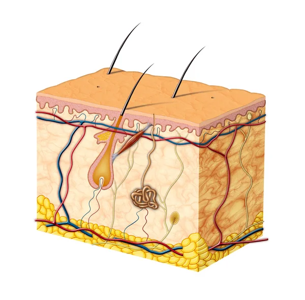
Skin anatomy
Skin anatomy — Stock Photo, Image
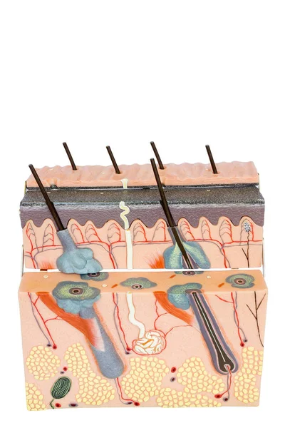
Model human skin tissue on white background
Model human skin tissue on white background — Stock Photo, Image
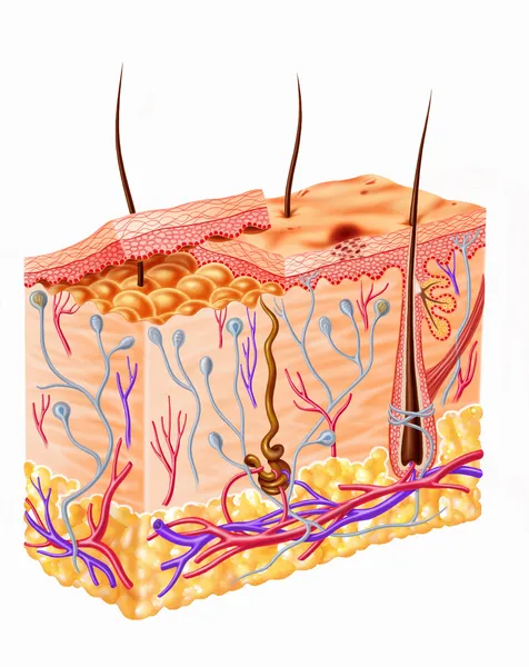
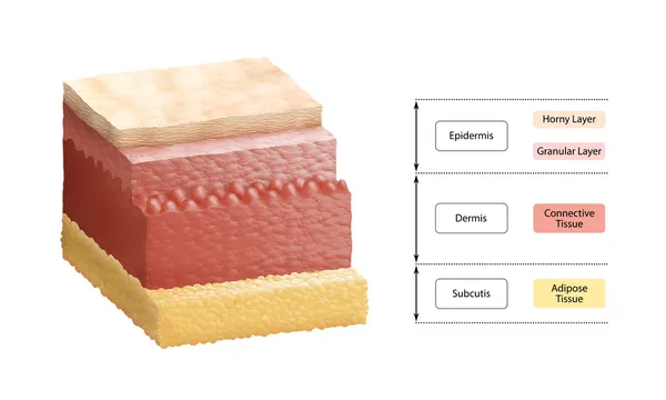
Human skin section diagram
Human skin section diagram — Stock Photo, Image
Layers Of Human Skin
Layers Of Human Skin — Stock Photo, Image
Structure of the human skin, cross section of hair follicle with descriptions - 3d illustration
Structure of the human skin, cross section of hair follicle with descriptions - 3d illustration — Stock Photo, Image
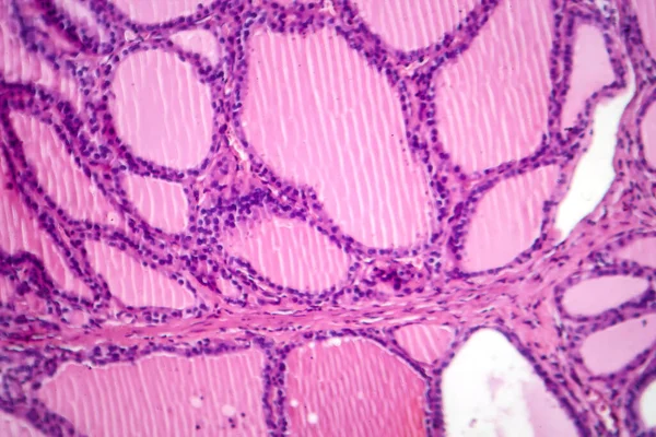
Photomicrograph of a toxic goiter tissue sample under a microscope, revealing hypertrophy of thyroid follicular cells, increased vascularity, and colloid depletion.
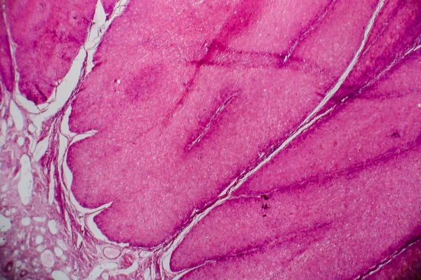
Photomicrograph Toxic Goiter Tissue Sample Microscope Revealing Hypertrophy Thyroid Follicular — Stock Photo, Image
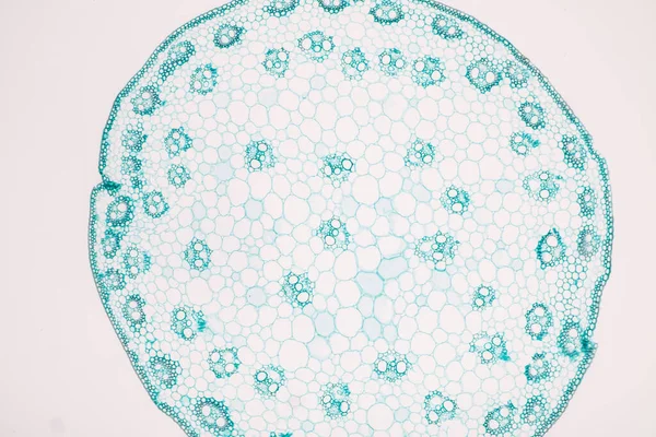
Characteristics of fungi living in wood as a group, are polyphyletic under the microscope for education.
Characteristics Fungi Living Wood Group Polyphyletic Microscope Education — Stock Photo, Image
Leiden leaf c.s. under microscope
Leiden Leaf Microscope — Stock Photo, Image
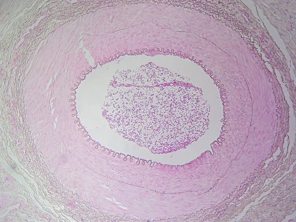
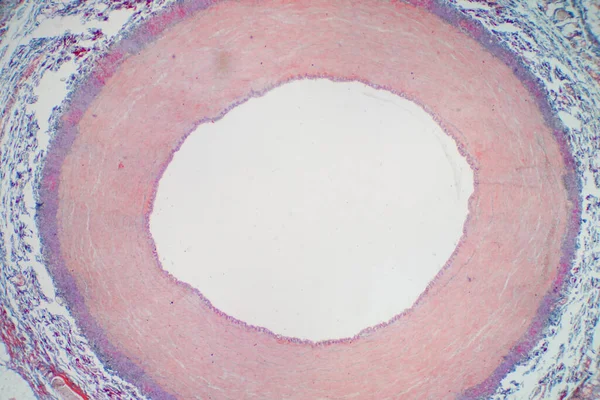
The Anatomy of Blood Vessels: Muscular Artery in High Detail
Anatomy Blood Vessels Muscular Artery High Detail — Stock Photo, Image
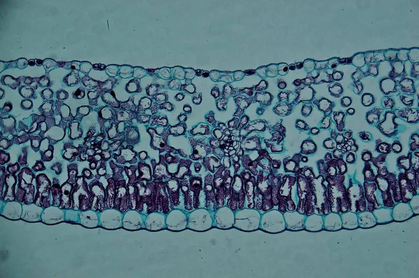
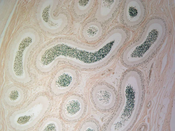
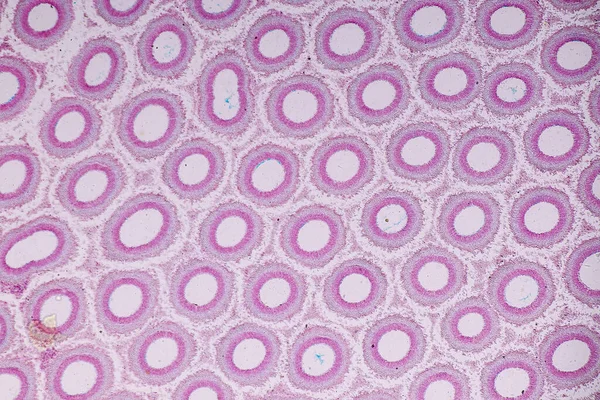
Sperm's Journey to Maturity: Epididymis in High Magnification
Sperm Journey Maturity Epididymis High Magnification — Stock Photo, Image
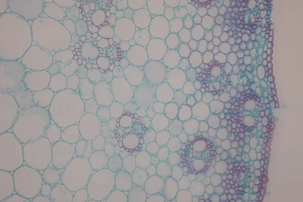
The study Plant tissue of under the microscope for classroom education.
Study Plant Tissue Microscope Classroom Education — Stock Photo, Image
Vicia faba root micrograph
Vicia faba root micrograph — Stock Photo, Image
Honeycomb close up - food
Honeycomb Close Food — Stock Photo, Image
Characteristics of fungi living in wood as a group, are polyphyletic under the microscope for education.
Characteristics Fungi Living Wood Group Polyphyletic Microscope Education — Stock Photo, Image
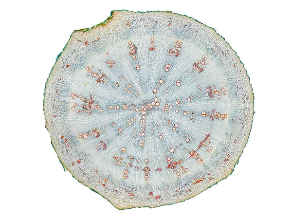
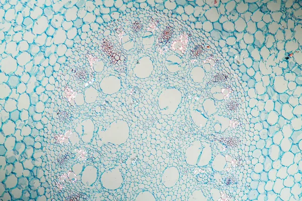
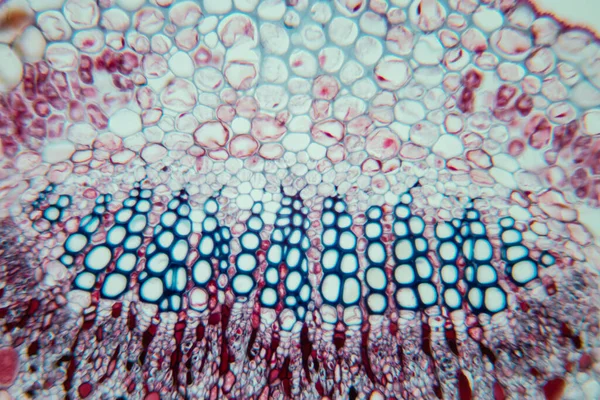
Cross section cut slice of plant stem under the microscope microscopic view of plant cells for botanic education high quality
Cross Section Cut Slice Plant Stem Microscope Microscopic View Plant — Stock Photo, Image
Stem with sieve cells under the microscope
Stem with sieve cells under the microscope — Stock Photo, Image
Pink abstract background in watercolor style
Pink Abstract Background Watercolor Style — Stock Photo, Image
Plant cell under the microscope view for education
Plant Cell Microscope View Education — Stock Photo, Image
Background or texture leather texture
Background or texture leather texture — Stock Photo, Image
Melon background texture bands
Melon background texture bands — Stock Photo, Image
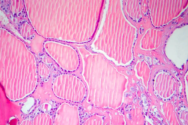
Follicles of colloid in the thyroid
Follicles Colloid Thyroid — Stock Photo, Image
Brown cow leather
Brown cow  leather — Stock Photo, Image
Background of mulberry paper, Thailand.
Background Mulberry Paper Thailand — Stock Photo, Image
Background of mulberry paper, Thailand.
Background Mulberry Paper Thailand — Stock Photo, Image
Blue spruce needle in cross section 100x
Blue Spruce Needle Cross Section 100X — Stock Photo, Image
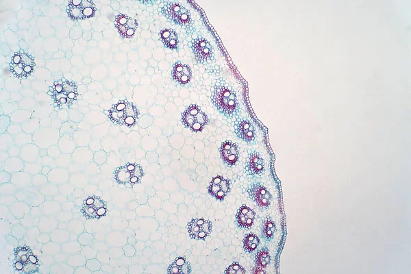
Cross sections of monocot stem under light microscope view for botany education.
Cross Sections Monocot Stem Light Microscope View Botany Education — Stock Photo, Image
Cross section of a cerebellum. Light micrograph.
Cross Section Cerebellum Light Micrograph — Stock Photo, Image
Trauerweide Blatt im Querschnitt 100x
Trauerweide Blatt Querschnitt 100X — Stock Photo, Image
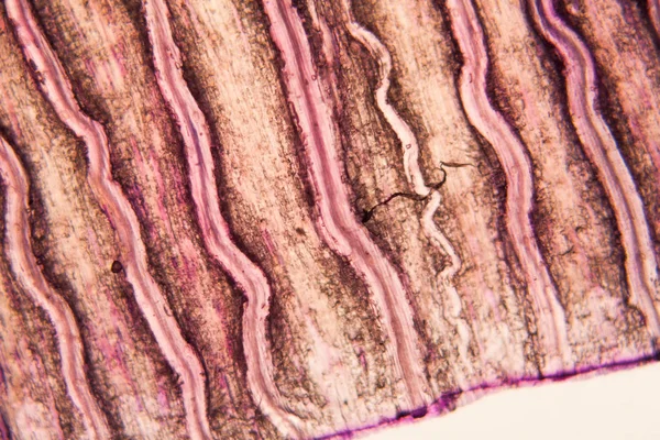
Parasitic nematode worm (Ascaris sp.) under the microscope
Parasitic nematode worm (Ascaris sp.) under the microscope — Stock Photo, Image
Microscopic photography of the biological objects
Microscopic Photography Biological Objects — Stock Photo, Image
The study Plant tissue of under the microscope for classroom education.
Study Plant Tissue Microscope Classroom Education — Stock Photo, Image
Characteristics of fungi living in wood as a group, are polyphyletic under the microscope for education.
Characteristics Fungi Living Wood Group Polyphyletic Microscope Education — Stock Photo, Image
Artery blood vessel under microscope view for education histology. Histological for human physiology.
Artery Blood Vessel Microscope View Education Histology Histological Human Physiology — Stock Photo, Image
The Beauty of Cartilage: Hyaline Cartilage in Detailed View Under the Microscope
Beauty Cartilage Hyaline Cartilage Detailed View Microscope — Stock Photo, Image
Condyloma acuminatum, also known as genital warts. Light micrograph, photo under microscope
Condyloma Acuminatum Also Known Genital Warts Light Micrograph Photo Microscope — Stock Photo, Image
The study of plant tissues under the microscope in the laboratory.
Study Plant Tissues Microscope Laboratory — Stock Photo, Image
The study Plant tissue of under the microscope for classroom education.
Study Plant Tissue Microscope Classroom Education — Stock Photo, Image
Lily leaf c.s. under microscope
Lily Leaf Microscope — Stock Photo, Image
Abstract black ink stain. Chinese ink background on watercolor paper.
Abstract Black Ink Stain Chinese Ink Background Watercolor Paper — Stock Photo, Image
Tulip petal at the microscope
Tulip Petal Microscope — Stock Photo, Image
Close up Core Tree of Cut Tree Trunk Background.
Close Core Tree Cut Tree Trunk Background — Stock Photo, Image
Blueberry leaf cross section under the microscope, 100x
Blueberry Leaf Cross Section Microscope 100X — Stock Photo, Image
Light micrograph of a cross sections of monocot stem.
Light Micrograph Cross Sections Monocot Stem — Stock Photo, Image
Granite surface texture for background, detail of the polished granite texture
Granite Surface Texture Background Detail Polished Granite Texture — Stock Photo, Image
Abstract ice texture background
Abstract ice texture background — Stock Photo, Image
Artery blood vessel under microscope view for education histology. Histological for human physiology.
Artery Blood Vessel Microscope View Education Histology Histological Human Physiology — Stock Photo, Image
Follicular adenoma of thyroid gland, light micrograph. Histopathology of thydoid adenoma. Photo under microscope
Follicular Adenoma Thyroid Gland Light Micrograph Histopathology Thydoid Adenoma Photo — Stock Photo, Image
Aerial root across 100x
Aerial Root 100X — Stock Photo, Image
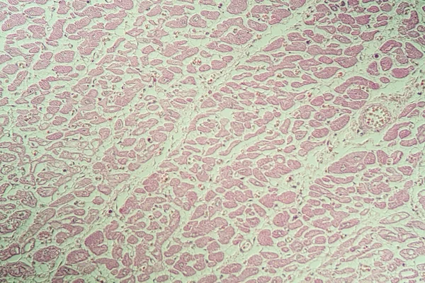
Inflamed heart muscle sick tissue under the microscope 200x
Inflamed Heart Muscle Sick Tissue Microscope 200X — Stock Photo, Image
Stem with sieve cells under the microscope
Stem with sieve cells under the microscope — Stock Photo, Image
The study Plant tissue of under the microscope for classroom education.
Study Plant Tissue Microscope Classroom Education — Stock Photo, Image
Microbiology, microscopic cells surface
Microbiology Microscopic Cells Surface — Stock Photo, Image
Fern stems in cross section 100x
Fern Stems Cross Section 100X — Stock Photo, Image
Cross sections of plants stem show plant vascular tissue under microscope view for education.
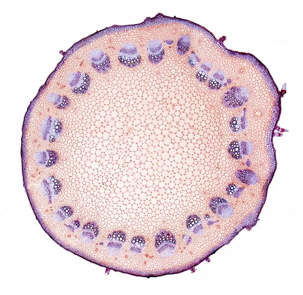
Cross Sections Plants Stem Show Plant Vascular Tissue Microscope View — Stock Photo, Image
Leopard skin texture for background
Leopard skin texture for background — Stock Photo, Image
Cardiac muscle after infarction, tissue section 100x
Cardiac Muscle Infarction Tissue Section 100X — Stock Photo, Image
Characteristics of anatomy and Histological sample Striated (Skeletal) muscle of mammal Tissue under the microscope.
Characteristics Anatomy Histological Sample Striated Skeletal Muscle Mammal Tissue Microscope — Stock Photo, Image
Brown textured leather background pattern
Brown textured leather background pattern — Stock Photo, Image
Light micrograph (LM) of a section through the fingertip.
Light Micrograph Section Fingertip — Stock Photo, Image
Dandelion taproot with milk tubes 100x along
Dandelion Taproot Milk Tubes 100X — Stock Photo, Image
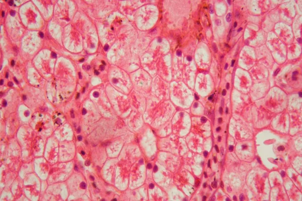
Microscope photo of a section through a frog liver.
Microscope photo of a section through a frog liver. — Stock Photo, Image
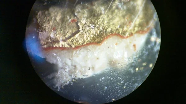
A macro shot of gold under the microscope
Macro Shot Gold Microscope — Stock Photo, Image
The study Plant tissue of under the microscope for classroom education.
Study Plant Tissue Microscope Classroom Education — Stock Photo, Image
Bird root root across 100x
Bird Root Root 100X — Stock Photo, Image
Tulip petal at the microscope
Tulip Petal Microscope — Stock Photo, Image
Sciatic nerve showing a bundle of nerve fibres
Sciatic nerve showing a bundle of nerve fibres — Stock Photo, Image
Vegetable marrow isolated on white background
Vegetable Marrow Isolated White Background — Stock Photo, Image
Fixed stained slide - mammal femur
Fixed Stained Slide Mammal Femur — Stock Photo, Image
Shiny pink leaf gold foil texture background
Shiny Pink Leaf Gold Foil Texture Background — Stock Photo, Image
Beautiful silver christmas beads
Beautiful silver christmas beads — Stock Photo, Image
Cells of a tobacco leaf (Nicotiana tabacum).
Cells of a tobacco leaf (Nicotiana tabacum). — Stock Photo, Image
Wood texture old
Wood texture old — Stock Photo, Image
The study Plant tissue of under the microscope for classroom education.
Study Plant Tissue Microscope Classroom Education — Stock Photo, Image
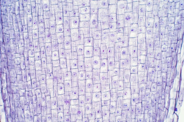
Root tip of Onion and Mitosis cell in the Root tip for education
Root Tip Onion Mitosis Cell Root Tip Education — Stock Photo, Image
Eucalyptus cross section through leaf 100x
Eucalyptus Cross Section Leaf 100X — Stock Photo, Image
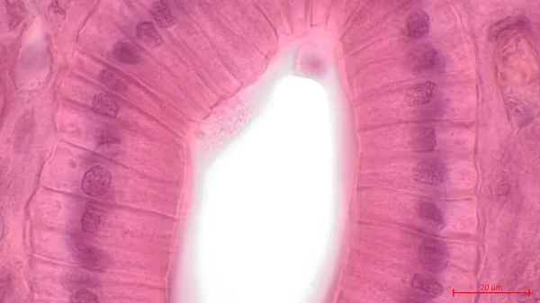
Simple columnar epithelium. Light micrograph of a renal medulla showing several collecting ducts lined by a simple columnar epithelium. Magnification: 1000
Simple Columnar Epithelium Light Micrograph Renal Medulla Showing Several Collecting — Stock Photo, Image
Melon skin texture or background
Melon Skin Texture Background — Stock Photo, Image
Fern with roots across 100x
Fern Roots 100X — Stock Photo, Image
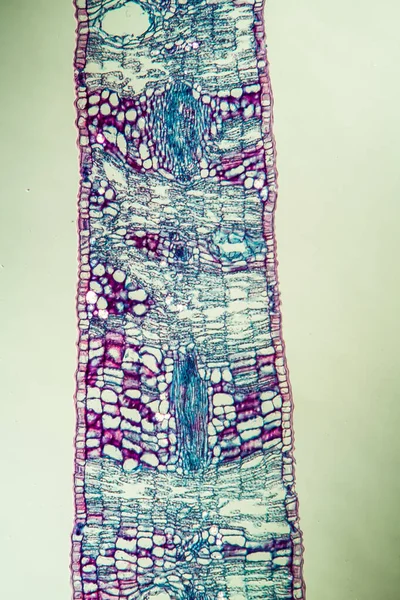
Sheet cross section 100x
Sheet Cross Section 100X — Stock Photo, Image
Aerial view of Dry cracked earth. Climate change and drought land. Global warming.
Aerial View Dry Cracked Earth Climate Change Drought Land Global — Stock Photo, Image
Rough weathered natural Wood Texture Background
Rough Weathered Natural Wood Texture Background — Stock Photo, Image
Microscopic photography. Young Root of plant, transversal section
Microscopic photography. Young Root of plant, transversal section — Stock Photo, Image
Silk bast with root, cross section 100x
Silk Bast Root Cross Section 100X — Stock Photo, Image
Gladiolus with stem 100x across
Gladiolus Stem 100X — Stock Photo, Image
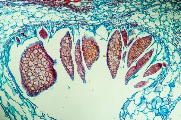
Capsella old embryo sec. under microscope
Capsella Old Embryo Sec Microscope — Stock Photo, Image
The study Plant tissue of under the microscope for classroom education.
Study Plant Tissue Microscope Classroom Education — Stock Photo, Image
Nestwurz orchid root cross 100x
Nestwurz Orchid Root Cross 100X — Stock Photo, Image
Section of a dog ciliated columnar epithelium
Section of a dog ciliated columnar epithelium — Stock Photo, Image
Horse chestnut with flower bud in cross section 100x
Horse Chestnut Flower Bud Cross Section 100X — Stock Photo, Image
Pine Wood micrograph
Pine Wood micrograph — Stock Photo, Image
Rough weathered natural Wood Texture Background
Rough Weathered Natural Wood Texture Background — Stock Photo, Image
Crocodile leather
Crocodile leather — Stock Photo, Image
Endemic goiter, light micrograph, abnormal enlargement of the thyroid gland due to dietary iodine deficiency. Photomicrograph shows follicles of varying size, abundant colloid, lymphocytic infiltrate
Endemic Goiter Light Micrograph Abnormal Enlargement Thyroid Gland Due Dietary — Stock Photo, Image
Pipe bush with trunk 100x across
Pipe Bush Trunk 100X — Stock Photo, Image
Closeup of grass roots close-up.
Closeup Grass Roots Close — Stock Photo, Image
Book spider of the garden spider under the microscope 100x
Book Spider Garden Spider Microscope 100X — Stock Photo, Image
Microscope shot of bacteria on white background
Microscope Shot Bacteria White Background — Stock Photo, Image
Helianthus stem. Light micrograph (LM) of stem of perennial sunflower (Helianthus sp.) in cross-section showing vascular bundles (green, yellow). Vascular tissues transport water and dissolved substances inside plant
Helianthus Stem Light Micrograph Stem Perennial Sunflower Helianthus Cross Section — Stock Photo, Image

Skin Cross Section Images - Stock Images

The Power of Visuals: Exploring Skin Cross Section Images

When it comes to visual storytelling, high-quality images can make a significant impact. With the rise of online platforms and digital marketing, the importance of captivating visuals cannot be overstated. In particular, skin cross section images have gained popularity due to their ability to educate and engage audiences in various industries. Whether you are a medical professional, a skincare enthusiast, or a graphic designer in need of visuals for a project, our collection of stock images provides a diverse range of options to suit your needs.

1. Understanding the Different Types of Skin Cross Section Images

Our collection offers an extensive array of skin cross section images captured in different techniques and formats. From histological slides to high-resolution photographs, each image is designed to provide a detailed glimpse into the intricate layers of human skin. Whether you require images showcasing the epidermis, dermis, or subcutaneous tissue, our collection will help you find the perfect visual for your specific purpose.

2. Versatile Usage Across Various Projects

Skin cross section images have a multitude of applications across diverse fields. In the medical industry, these visuals are invaluable for educational purposes, allowing researchers, doctors, and students to understand the cellular structure and composition of the skin. Additionally, skincare brands can utilize these images to showcase the effects of their products on different skin layers, enhancing their credibility and demonstrating their expertise.

Moreover, designers can seamlessly integrate skin cross section images into their projects, be it for website design, infographic creation, or even medical journal publications. The versatility of these visuals allows for endless possibilities, helping you convey your message effectively and professionally.

3. Choosing the Right Image for Your Project

When selecting skin cross section images, it is crucial to consider the intended purpose and target audience of your project. For medical professionals and scientific publications, histological slides captured at various magnifications offer a comprehensive view of the skin structure. On the other hand, graphic designers and web developers may prefer high-resolution photographs or vector illustrations that can be customized and integrated seamlessly into their designs.

Furthermore, it is essential to ensure the chosen image aligns with the tone and style of your project. For a modern and sleek aesthetic, high-contrast images with vibrant colors can be highly impactful. Conversely, if you are striving for a more technical or clinical feel, grayscale images with fine details can convey authenticity and precision.

4. Unleashing the Power of Skin Cross Section Images

When used strategically, skin cross section images can elevate your project to new heights. These visuals help simplify complex information, making it more accessible and engaging for your audience. By incorporating these images into your educational materials, marketing campaigns, or design projects, you can capture attention, increase understanding, and leave a lasting impression.