Spinal nerves Stock Photos

100,000 Spinal nerves pictures are available under a royalty-free license

  • Best Match
  • Fresh
  • Popular
1-100 of 100,000
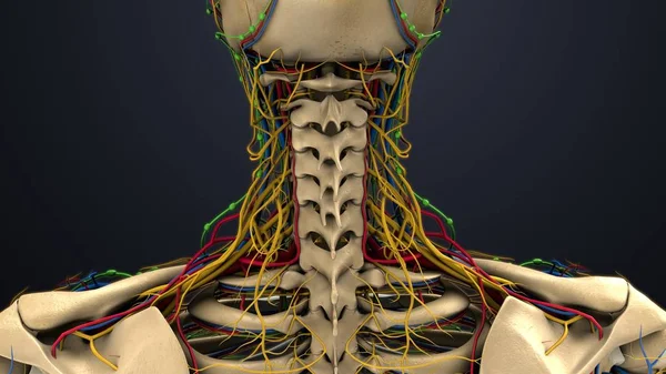
The cervical nerves are the spinal nerves from the cervical vertebrae in the cervical segment of the spinal cord
Cervical Nerves Spinal Nerves Cervical Vertebrae Cervical Segment Spinal Cord — Stock Photo, Image
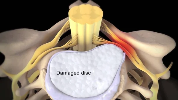
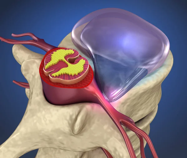
Spinal cord under pressure of bulging disc
Spinal cord under pressure of bulging disc — Stock Photo, Image
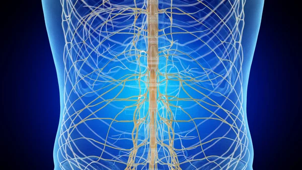
3D illustration male nervous system.
3D illustration male nervous system. — Stock Photo, Image
This system includes all the nervous structures that leave the central nervous system to the rest of our body.
System Includes All Nervous Structures Leave Central Nervous System Rest — Stock Photo, Image
Woman suffering from pain
Woman suffering from pain — Stock Photo, Image
Spinal cord under pressure of bulging disc, 3D illustration
Spinal Cord Pressure Bulging Disc Illustration — Stock Photo, Image
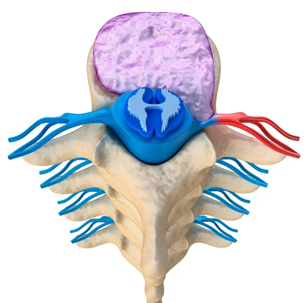
Hernaited disc in human spine in details
Hernaited Disc Human Spine Details — Stock Photo, Image
View of male nervous system
View of male nervous system — Stock Photo, Image
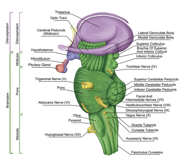
Brainstem, brain stem, lateral view, adjoining and structurally continuous with the spinal cord, parts of the diencephalon, motor and sensory innervation to the face and neck via thecranial nerves
Brainstem, brain stem, lateral view, adjoining and structurally continuous with the spinal cord, parts of the diencephalon, motor and sensory innervation to the face and neck via thecranial nerves — Stock Photo, Image
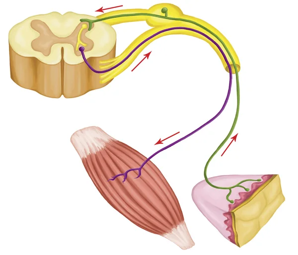
Somatic motor reflex, somatic nervous system, peripheral nervous system, voluntary control of body movements via skeletal muscles, afferent and efferent nerves
Somatic motor reflex, somatic nervous system, peripheral nervous system, voluntary control of body movements via skeletal muscles, afferent and efferent nerves — Stock Photo, Image
Human organ system anatomy 3d illustration
Human Organ System Anatomy Illustration — Stock Photo, Image
Male nervous system, illustration.
Male Nervous System Illustration — Stock Photo, Image
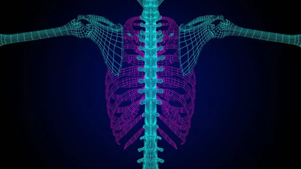
Rib cage anatomy 3D Illustration with wire frame skeleton for medical concept
Rib Cage Anatomy Illustration Wire Frame Skeleton Medical Concept — Stock Photo, Image
Onion epidermus micrograph
Onion epidermus micrograph — Stock Photo, Image
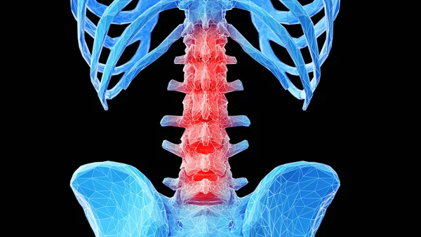
Human Skeleton Vertebral Column Lumbar Vertebrae Anatomy 3D Illustration
Human Skeleton Vertebral Column Lumbar Vertebrae Anatomy Illustration — Stock Photo, Image
The study Plant tissue of under the microscope for classroom education.
Study Plant Tissue Microscope Classroom Education — Stock Photo, Image
Blood vessels of foot, computer illustration
Blood Vessels Foot Computer Illustration — Stock Photo, Image
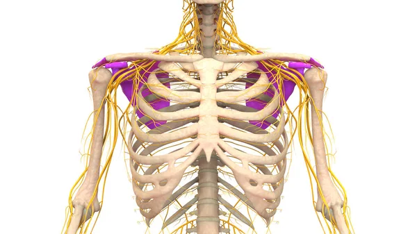
Human Skeleton System Pectoral (Shoulder) Girdle Bone Joints Anatomy. 3D
Human Skeleton System Pectoral Shoulder Girdle Bone Joints Anatomy — Stock Photo, Image
Vertebra on x ray human body, cg medicine 3d illustration
Vertebra Ray Human Body Medicine Illustration — Stock Photo, Image
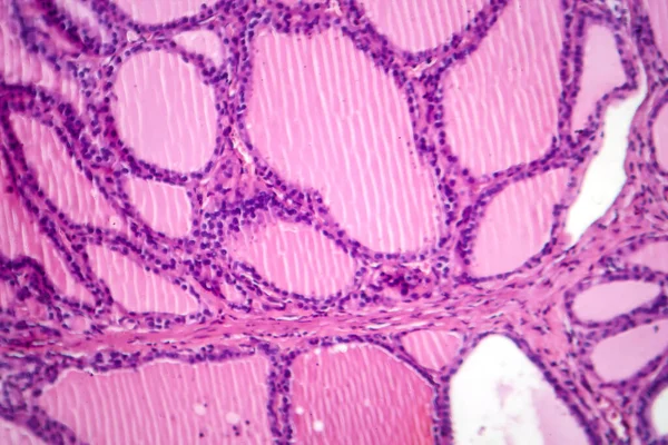
Photomicrograph of a toxic goiter tissue sample under a microscope, revealing hypertrophy of thyroid follicular cells, increased vascularity, and colloid depletion.
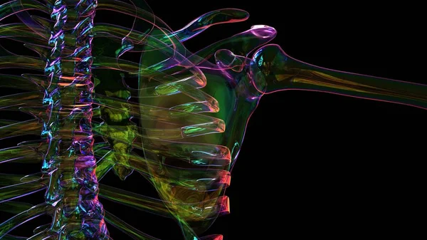
Photomicrograph Toxic Goiter Tissue Sample Microscope Revealing Hypertrophy Thyroid Follicular — Stock Photo, Image
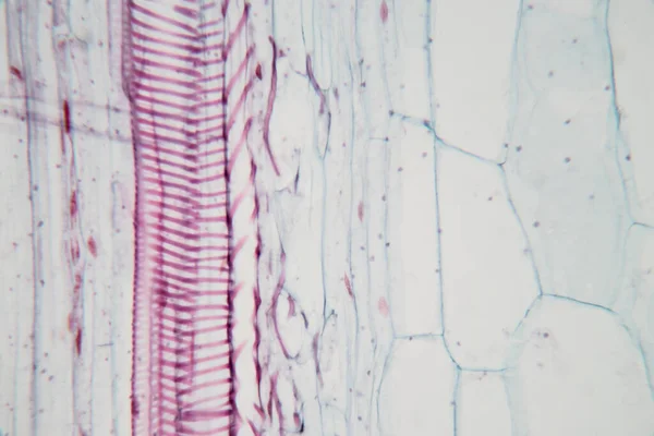
Dandelion taproot with milk tubes 100x along
Dandelion Taproot Milk Tubes 100X — Stock Photo, Image
Grunge abstract texture, background. Copy space
Grunge Abstract Texture Background Copy Space — Stock Photo, Image
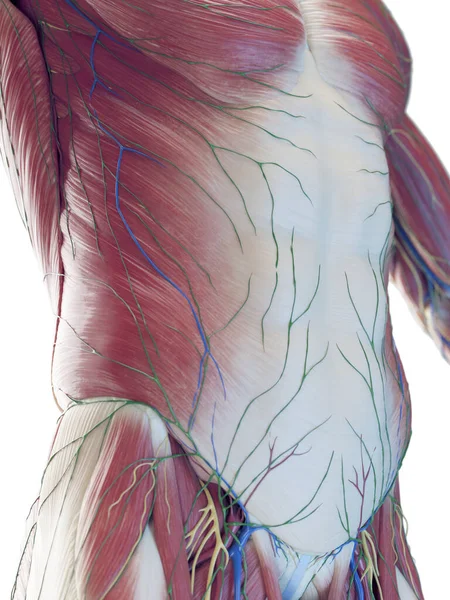
Male abdominal muscles, illustration
Male Abdominal Muscles Illustration — Stock Photo, Image
Bright color wall background
Bright color wall background — Stock Photo, Image
Micrograph plant root tip tissue cell
Micrograph plant root tip tissue cell — Stock Photo, Image
Photomicrograph of fibroadenoma, showcasing benign glandular and fibrous tissue growth within the breast, a common noncancerous tumor.
Photomicrograph Fibroadenoma Showcasing Benign Glandular Fibrous Tissue Growth Breast Common — Stock Photo, Image
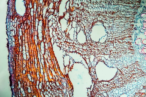
Male nervous system, illustration.
Male Nervous System Illustration — Stock Photo, Image
Cerebellum, Thalamus, Medulla oblongata, Spinal cord and Motor Neuron human under the microscope in Lab.
Cerebellum Thalamus Medulla Oblongata Spinal Cord Motor Neuron Human Microscope — Stock Photo, Image
Male nervous system, illustration.
Male Nervous System Illustration — Stock Photo, Image
Grungy abstract textured background
Grungy Abstract Textured Background — Stock Photo, Image
Abstract background. Monochrome texture. Black and white tones.
Abstract Background Monochrome Texture Black White Tones — Stock Photo, Image
Slime molds, as a group, are polyphyletic under the microscope for education.
Slime Molds Group Polyphyletic Microscope Education — Stock Photo, Image
Background - Stacked Stone Wall.
Background Stacked Stone Wall — Stock Photo, Image
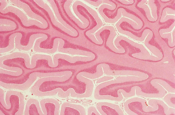
Cross section of a cerebellum. Light micrograph.
Cross Section Cerebellum Light Micrograph — Stock Photo, Image
Painted car tire close-up texture background
Painted Car Tire Close Texture Background — Stock Photo, Image
Human Skeleton Anatomy For medical concept 3D Illustration
Human Skeleton Anatomy Medical Concept Illustration — Stock Photo, Image
Trachea, light micrograph. Hyaline cartilage from trachea showing chondrocytes, collagen fibers and matrix.
Trachea Light Micrograph Hyaline Cartilage Trachea Showing Chondrocytes Collagen Fibers — Stock Photo, Image
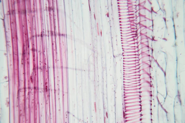
Stem with sieve cells under the microscope
Stem with sieve cells under the microscope — Stock Photo, Image
Carrot root across 100x
Carrot Root 100X — Stock Photo, Image
Characteristics of fungi living in wood as a group, are polyphyletic under the microscope for education.
Characteristics Fungi Living Wood Group Polyphyletic Microscope Education — Stock Photo, Image
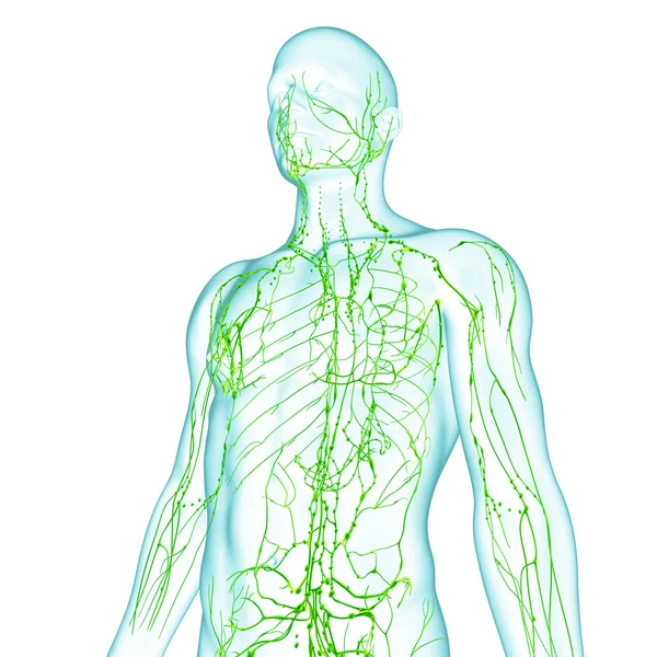
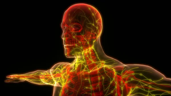
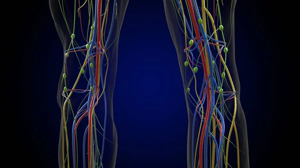
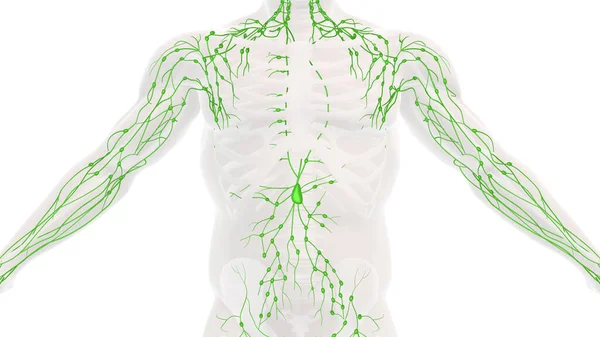
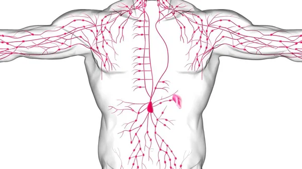
Lymphatic system of male in green
Lymphatic system of male in green — Stock Photo, Image
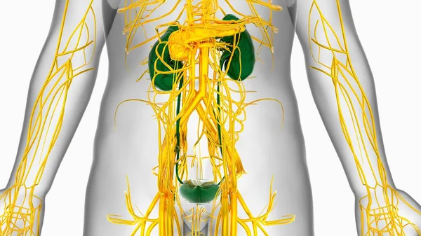
Human Kidneys Anatomy For Medical Concept 3D Illustration
Human Kidneys Anatomy Medical Concept Illustration — Stock Photo, Image
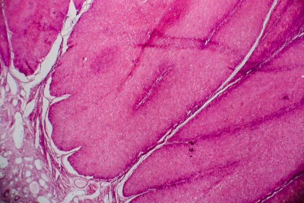
Condyloma acuminatum, also known as genital warts. Light micrograph, photo under microscope
Condyloma Acuminatum Also Known Genital Warts Light Micrograph Photo Microscope — Stock Photo, Image
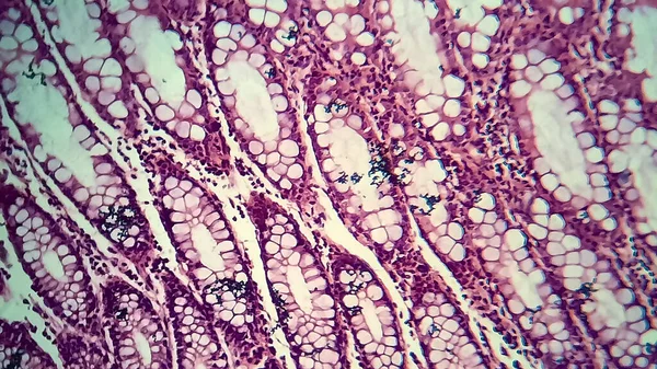
Bacillary dysentery, light micrograph, photo under microscope showing presence of bacteria and accumulation of inflammatory cells in intestinal epithelium
Bacillary Dysentery Light Micrograph Photo Microscope Showing Presence Bacteria Accumulation — Stock Photo, Image
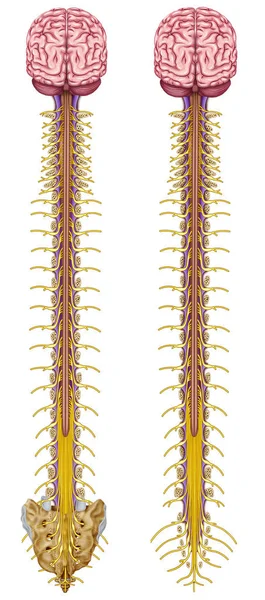
Spinal Cord
Spinal Cord — Stock Photo, Image
Human back bones, illustration.
Human Back Bones Illustration — Stock Photo, Image
Abstract background. Monochrome texture. Black and white textured background.
Abstract Background Monochrome Texture Black White Textured Background — Stock Photo, Image
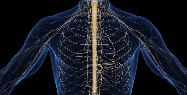
3d rendered medically accurate illustration of the nervous system
Rendered Medically Accurate Illustration Nervous System — Stock Photo, Image
Human organ system anatomy 3d illustration
Human Organ System Anatomy Illustration — Stock Photo, Image
Human Internal System Lymph Nodes Anatomy. 3D
Human Internal System Lymph Nodes Anatomy — Stock Photo, Image
Human lymph nodes anatomy for medical concept 3D illustration
Human Lymph Nodes Anatomy Medical Concept Illustration — Stock Photo, Image
Human Heart With Circulatory System Anatomy For Medical Concept 3D Illustration
Human Heart Circulatory System Anatomy Medical Concept Illustration — Stock Photo, Image
Watercolor pink decorative texture. Abstract background
Watercolor Pink Decorative Texture Abstract Background — Stock Photo, Image
3d rendered medically accurate illustration of a male brain
Rendered Medically Accurate Illustration Male Brain — Stock Photo, Image
Histology of cardiac muscle under microscope view
Histology Cardiac Muscle Microscope View — Stock Photo, Image
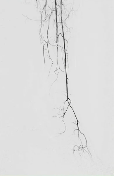
Femoral artery angiogram or angiography at lower extremity area.
Femoral Artery Angiogram Angiography Lower Extremity Area — Stock Photo, Image
Abstract background. Monochrome texture. Black and white textured background
Abstract Background Monochrome Texture Black White Textured Background — Stock Photo, Image
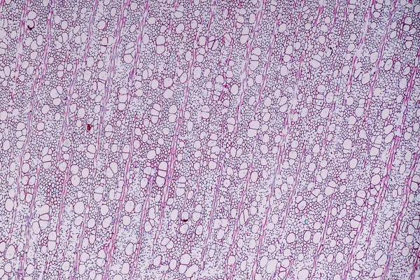
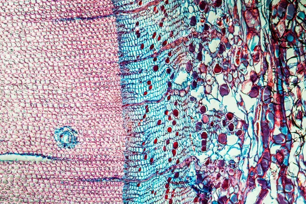
Pine tree trunk across 100x
Pine Tree Trunk 100X — Stock Photo, Image
Bacillary dysentery, light micrograph, photo under microscope showing presence of bacteria and accumulation of inflammatory cells in intestinal epithelium
Bacillary Dysentery Light Micrograph Photo Microscope Showing Presence Bacteria Accumulation — Stock Photo, Image
Human Anatomy For medical concept 3D Illustration Render
Human Anatomy Medical Concept Illustration Render — Stock Photo, Image
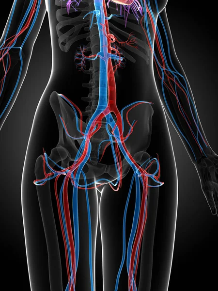
3d rendered illustration of the female vascular system
Rendered Illustration Female Vascular System — Stock Photo, Image
3d rendered medically accurate illustration of the nervous system
Rendered Medically Accurate Illustration Nervous System — Stock Photo, Image
Anatomy of the human lymphatic system
Anatomy Human Lymphatic System — Stock Photo, Image
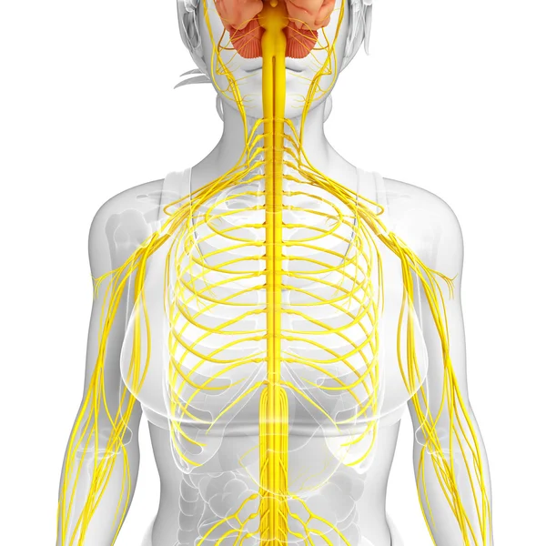
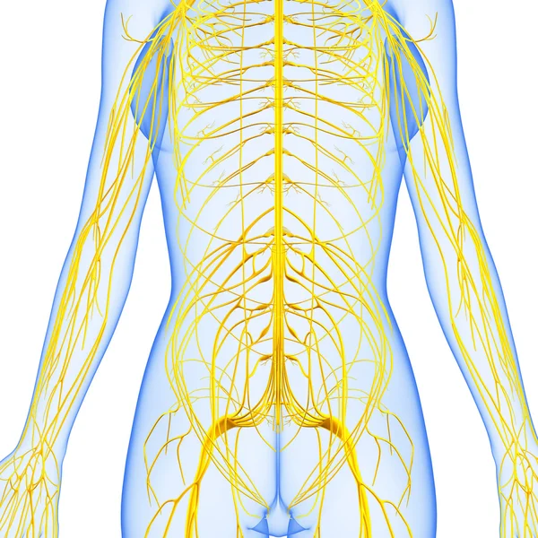
Female nervous system artwork
Female nervous system artwork — Stock Photo, Image
Microscope view, abstract background of blue and purple surface
Microscope View Abstract Background Blue Purple Surface — Stock Photo, Image
Pine trunk across 100x
Pine Trunk 100X — Stock Photo, Image
Abstract surface, microbiology cells
Abstract Surface Microbiology Cells — Stock Photo, Image
Human Muscle Body Anatomy
Human Muscle Body Anatomy — Stock Photo, Image
Condyloma acuminatum, also known as genital warts. Light micrograph, photo under microscope
Condyloma Acuminatum Also Known Genital Warts Light Micrograph Photo Microscope — Stock Photo, Image
The study Plant tissue of under the microscope for classroom education.
Study Plant Tissue Microscope Classroom Education — Stock Photo, Image
Cardiovascular system of neck and head
Cardiovascular system of neck and head — Stock Photo, Image
Abstract background. Monochrome texture. Black and white textured background.
Abstract Background Monochrome Texture Black White Textured Background — Stock Photo, Image
Purulent meningitis, light micrograph, photo under microscope
Purulent Meningitis Light Micrograph Photo Microscope — Stock Photo, Image
Human Lymph Nodes Anatomy For Medical Concept 3D Illustration
Human Lymph Nodes Anatomy Medical Concept Illustration — Stock Photo, Image
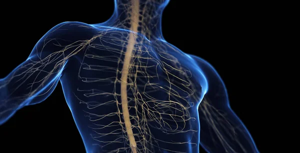
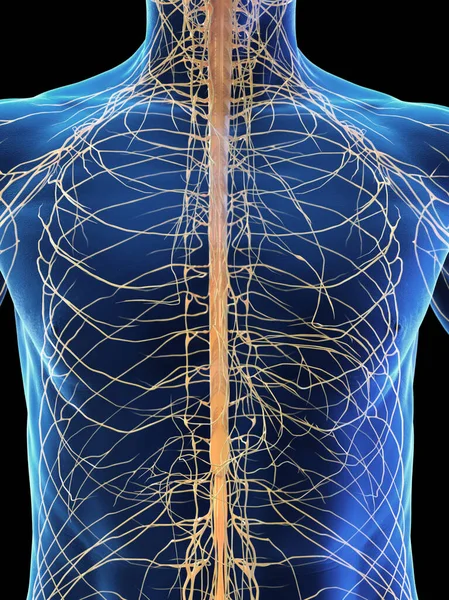
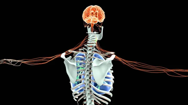
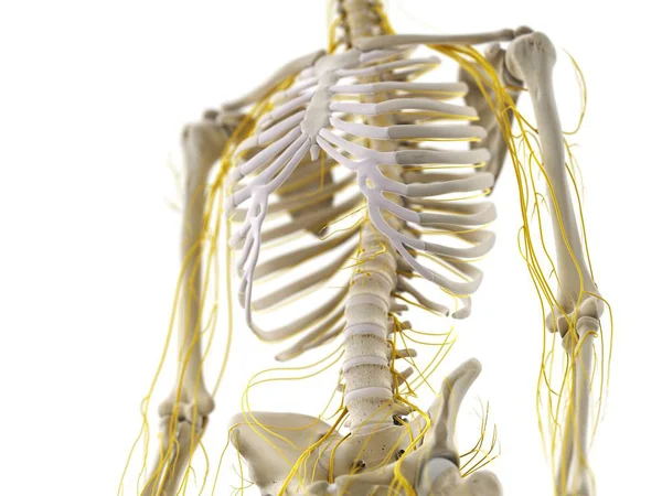
Thorax nerves, computer illustration
Thorax Nerves Computer Illustration — Stock Photo, Image
3d art illustration of Nervous system
3d art illustration of Nervous system — Stock Photo, Image
The study Plant tissue of under the microscope for classroom education.
Study Plant Tissue Microscope Classroom Education — Stock Photo, Image
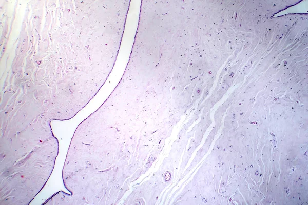
A longitudinal section of human nerve cells under the microscope.
Longitudinal Section Human Nerve Cells Microscope — Stock Photo, Image
Condyloma acuminatum, also known as genital warts. Light micrograph, photo under microscope
Condyloma Acuminatum Also Known Genital Warts Light Micrograph Photo Microscope — Stock Photo, Image
Abstract colorful texture background, copy space wallpaper
Abstract Colorful Texture Background Copy Space Wallpaper — Stock Photo, Image
Abstract background. Monochrome texture. Black and white textured background.
Abstract Background Monochrome Texture Black White Textured Background — Stock Photo, Image
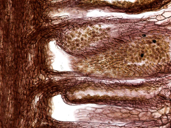
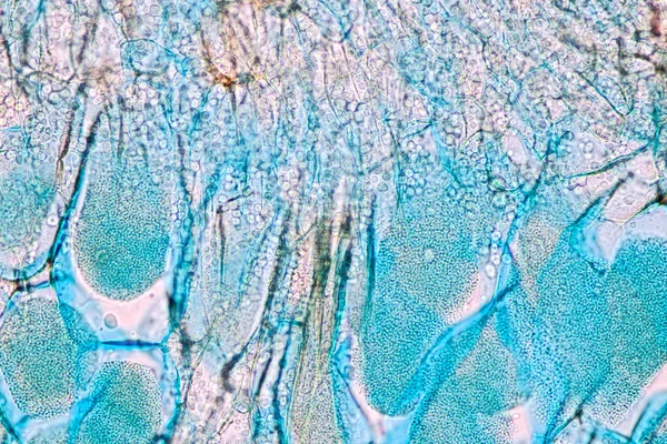
Host cells with spores (mold) are inside wood under the microscope for education.
Host Cells Spores Mold Wood Microscope Education — Stock Photo, Image
Abstract grunge background. Monochrome texture. Black and white textured background
Abstract Grunge Background Monochrome Texture Black White Textured Background — Stock Photo, Image
Grunge abstract texture, background. Copy space
Grunge Abstract Texture Background Copy Space — Stock Photo, Image
Female body of Nervous and digestive system artwork
Female body of Nervous and digestive system artwork — Stock Photo, Image
Creative abstract hand drawn oil painting. Paint stroke texture on canvas. Wallpaper for web and game design. Grunge background. Macro image of artwork. Modern art. Contemporary art.
Creative Abstract Hand Drawn Oil Painting Paint Stroke Texture Canvas — Stock Photo, Image
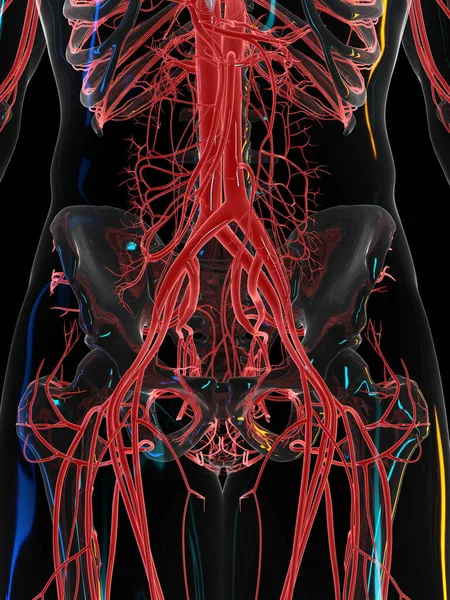
Vascular system on black background, illustration.
Vascular System Black Background Illustration — Stock Photo, Image
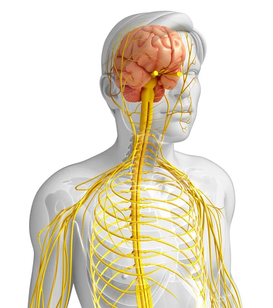
Male nervous system artwork
Male nervous system artwork — Stock Photo, Image
Human Cervical Vertebra
Human Cervical Vertebra — Stock Photo, Image
Abstract background. Monochrome texture. Black and white textured background.
Abstract Background Monochrome Texture Black White Textured Background — Stock Photo, Image
Monochrome texture with black and white background.
Monochrome Texture Black White Background — Stock Photo, Image
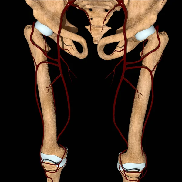
This 3d illustration shows the femoral artery on bones
Illustration Shows Femoral Artery Bones — Stock Photo, Image
3D Illustration Human Brain Anatomy With Nervous System For Medical Concept
Illustration Human Brain Anatomy Nervous System Medical Concept — Stock Photo, Image
Lower spinal cord, illustration.
Lower Spinal Cord Illustration — Stock Photo, Image
Cerebellum view from microscope
Cerebellum View Microscope — Stock Photo, Image

Discover the Best Spinal Nerves Images for Your Next Project

Choosing the right visual for your project can make all the difference in making it successful. If you're looking for high-quality spinal nerves images, then you've come to the right place! Our collection offers a wide variety of images that can be used in various projects, from medical brochures to educational videos.

Our collection includes both JPG and EPS file formats that you can easily download and use for different purposes. Whether you're a healthcare professional or an educator, you'll find our images highly useful in bringing your projects to life.

Different Types of Spinal Nerves Images You Can Find Here

Our collection offers a wide range of spinal nerves images that you can choose from. These include images of the spinal cord and its branches, detailed illustrations of the spinal anatomy, and 3D representations of the spinal nerves. We also have images of spinal disorders and conditions that can help you explain complex medical concepts to your audience.

If you're looking for specific images, such as cervical or lumbar spinal nerves images, you can easily find them by using our search filters. These filters allow you to sort the images based on their resolution, orientation, and other criteria, making it easier for you to find exactly what you need.

Where to Use Spinal Nerves Images

Spinal nerves images can be used in various projects, such as medical websites, educational textbooks, and healthcare brochures. They're also highly useful for presentations and lectures, where you need to explain complex medical concepts in a simple and easy-to-understand manner.

Our collection of spinal nerves images is also perfect for healthcare practices and clinics that want to create informative and engaging infographics or posters. By using these images, you can educate your patients about their medical condition and help them understand their treatment options better.

Tips for Using Spinal Nerves Images Effectively

When using spinal nerves images in your projects, it's essential to choose the right image for the right purpose. If you're using images for educational purposes, make sure they're accurate, clear, and easy to understand. If you're creating marketing materials, choose images that are visually appealing and attention-grabbing.

Another essential tip when using images is to be mindful of copyright laws. Always make sure you have the proper permissions to use images in your projects, and always credit the source.

Start Exploring Our Spinal Nerves Images Collection Today

Our collection of spinal nerves images offers an excellent selection of high-quality visuals that you can use in your projects. Whether you're working on a medical brochure or an educational video, our collection has something for everyone. Get started today and bring your projects to life with our stunning spinal nerves images.