Tailbone Stock Photos

100,000 Tailbone pictures are available under a royalty-free license

  • Best Match
  • Fresh
  • Popular
1-100 of 100,000
Tailbone Pain Relief, Coccydynia. Coccyx pain during pregnancy
Tailbone Pain Relief Coccydynia Coccyx Pain Pregnancy — Stock Photo, Image
Low back anatomy. Pelvis , hip joints, tailbone. Doctor Chiropractor explains causes of back pain to Patient
Low Back Anatomy Pelvis Hip Joints Tailbone Doctor Chiropractor Explains — Stock Photo, Image
Close-up of a young girls spine base editing by a male physiotherapist of visceral massage. The hands of the doctor set and knead the tailbone of the patient
Close-up of a young girls spine base editing by a male physiotherapist of visceral massage. The hands of the doctor set and knead the tailbone of the patient — Stock Photo, Image
Close up of physiotherapist applying pressure on lumbar spine.Young woman laying on spa bed with therapist doing manipulative treatment on back.
Close Physiotherapist Applying Pressure Lumbar Spine Young Woman Laying Spa — Stock Photo, Image
Therapist applying pressure on female tailbone.
Therapist applying pressure on female tailbone. — Stock Photo, Image
Close-up of a young girls spine base editing by a male physiotherapist of visceral massage. The hands of the doctor set and knead the tailbone of the patient
Close-up of a young girls spine base editing by a male physiotherapist of visceral massage. The hands of the doctor set and knead the tailbone of the patient — Stock Photo, Image
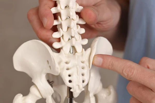
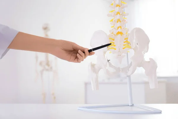
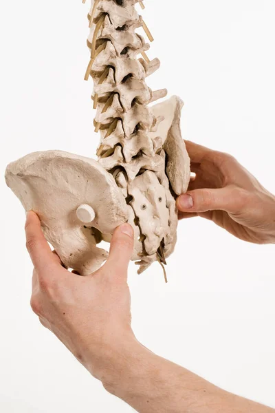
Orthopedist pointing on human spine model in clinic, closeup
Orthopedist Pointing Human Spine Model Clinic Closeup — Stock Photo, Image
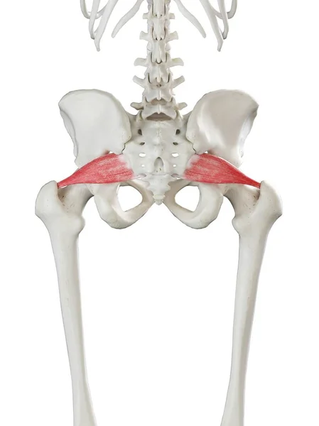
Human skeleton model with detailed Piriformis muscle, digital illustration.
Human Skeleton Model Detailed Piriformis Muscle Digital Illustration — Stock Photo, Image
3D Illustration of Human Body Organs, Spine
Illustration Human Body Organs Spine — Stock Photo, Image
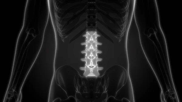
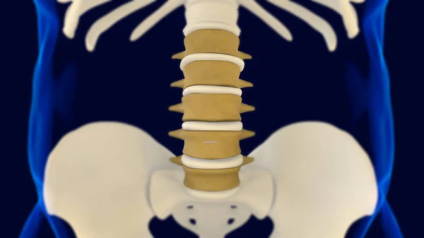
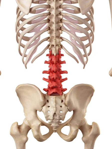
Lumbar Vertebrae Anatomy for medical concept 3D Illustration
Lumbar Vertebrae Anatomy Medical Concept Illustration — Stock Photo, Image
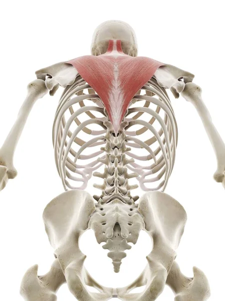
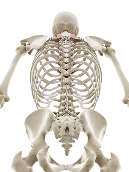
Trapezius muscle, computer illustration
Trapezius Muscle Computer Illustration — Stock Photo, Image
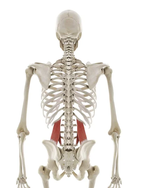
Quadratus lumborum muscle, computer illustration
Quadratus Lumborum Muscle Computer Illustration — Stock Photo, Image
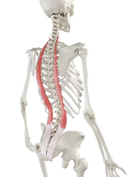
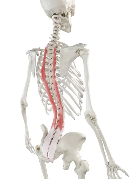
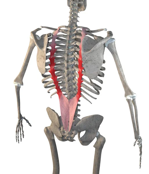
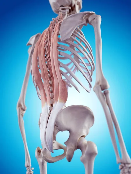
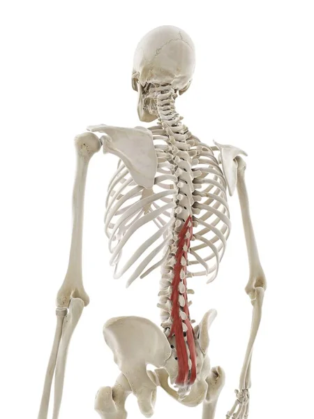
Human skeleton with red colored Iliocostalis muscle, computer illustration.
Human Skeleton Red Colored Iliocostalis Muscle Computer Illustration — Stock Photo, Image
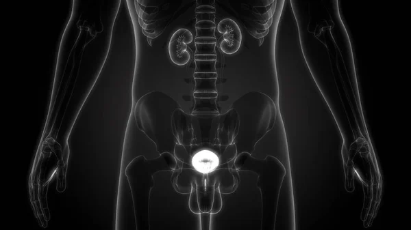
Human Body Organs (Kidneys with Urinary Bladder)
Human Body Organs Kidneys Urinary Bladder — Stock Photo, Image
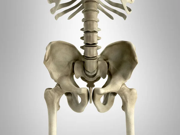
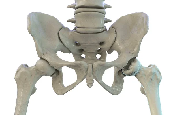
3D rendering medical illustration of the pelvis bone on grey
Rendering Medical Illustration Pelvis Bone Grey — Stock Photo, Image
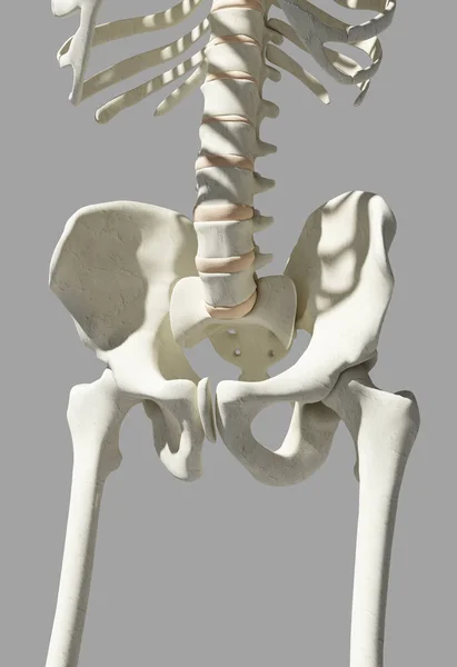
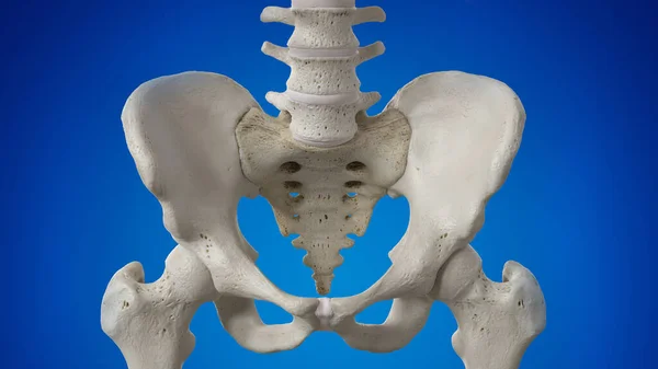
Hip Skeleton on blue background.
Hip Skeleton on blue background. — Stock Photo, Image
Quadratus lumborum muscle, computer illustration
Quadratus Lumborum Muscle Computer Illustration — Stock Photo, Image
Human skeleton with Sacrotuberous ligaments, computer illustration.
Human Skeleton Sacrotuberous Ligaments Computer Illustration — Stock Photo, Image
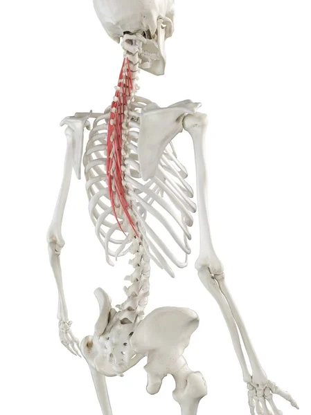
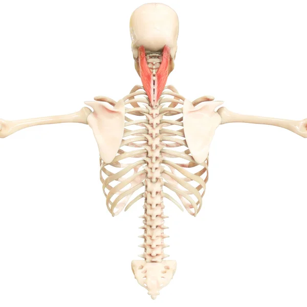
Human skeleton with red colored Semispinalis thoracis muscle, computer illustration.
Human Skeleton Red Colored Semispinalis Thoracis Muscle Computer Illustration — Stock Photo, Image
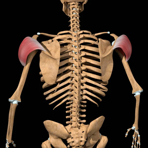
This 3d illustration shows the back view of the posterior head of the deltoid muscles
Illustration Shows Back View Posterior Head Deltoid Muscles — Stock Photo, Image
Scapula Bone anatomy Human skeleton 3D Rendering For Medical Concept
Scapula Bone Anatomy Human Skeleton Rendering Medical Concept — Stock Photo, Image
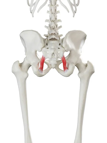
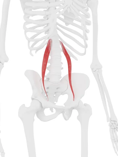
Human skeleton with red colored Psoas minor muscle, digital illustration.
Human Skeleton Red Colored Psoas Minor Muscle Digital Illustration — Stock Photo, Image
Rotatores muscles, computer illustration
Rotatores Muscles Computer Illustration — Stock Photo, Image
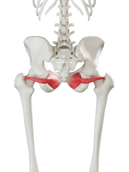
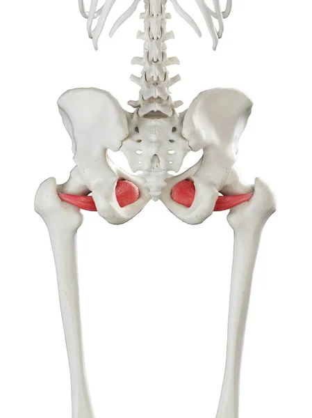
Human skeleton model with detailed Obturator internus muscle, digital illustration.
Human Skeleton Model Detailed Obturator Internus Muscle Digital Illustration — Stock Photo, Image
Human skeleton anatomy Radius Bone 3D Rendering For Medical Concept
Human Skeleton Anatomy Radius Bone Rendering Medical Concept — Stock Photo, Image
Human hip bones structure
Human hip bones structure — Stock Photo, Image
Human skeleton with detailed red Sacrotuberous ligament muscle, digital illustration.
Human Skeleton Detailed Red Sacrotuberous Ligament Muscle Digital Illustration — Stock Photo, Image
Human skeleton anatomy Ulna Bone 3D Rendering For Medical Concept
Human Skeleton Anatomy Ulna Bone Rendering Medical Concept — Stock Photo, Image
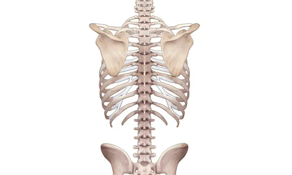
Human Skeleton System Axial Skeleton Anatomy. 3D
Human Skeleton System Axial Skeleton Anatomy — Stock Photo, Image
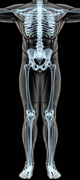
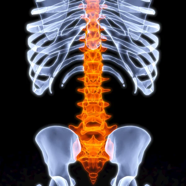
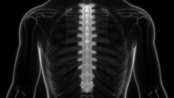
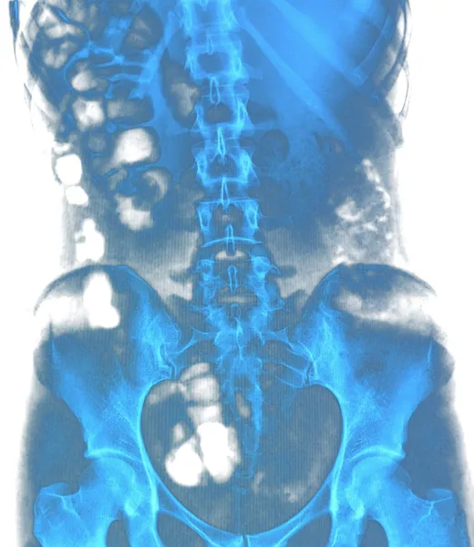
Man's body under X-rays
Man's body under X-rays — Stock Photo, Image
Human skeleton model with detailed Longissimus thoracis muscle, digital illustration.
Human Skeleton Model Detailed Longissimus Thoracis Muscle Digital Illustration — Stock Photo, Image
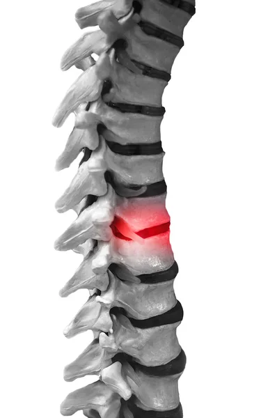
Thoracic and lumbar sections of spine
Thoracic and lumbar sections of spine — Stock Photo, Image
Lumbar spine structure
Lumbar spine structure — Stock Photo, Image
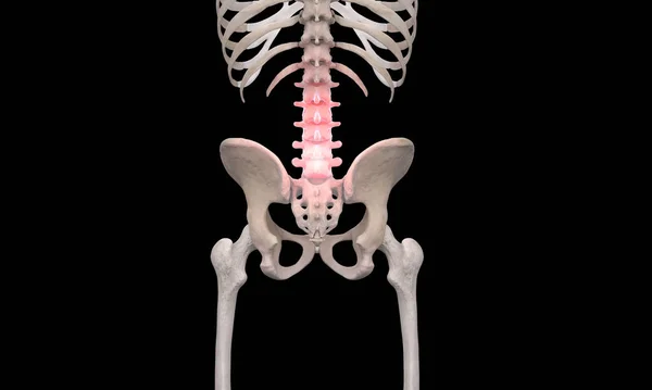
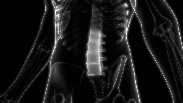
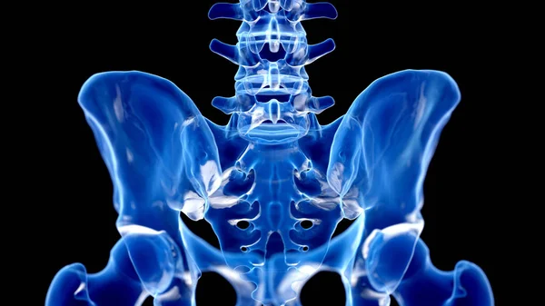
Skeleton pelvic back pain on black background
Skeleton Pelvic Back Pain Black Background — Stock Photo, Image
Skeleton
Skeleton — Stock Photo, Image
Quadratus lumborum muscle, computer illustration
Quadratus Lumborum Muscle Computer Illustration — Stock Photo, Image
Human skeleton model with detailed Multifidus muscle, digital illustration.
Human Skeleton Model Detailed Multifidus Muscle Digital Illustration — Stock Photo, Image
Human Muscular System Torso Muscles Capitus Muscle Anatomy. 3D
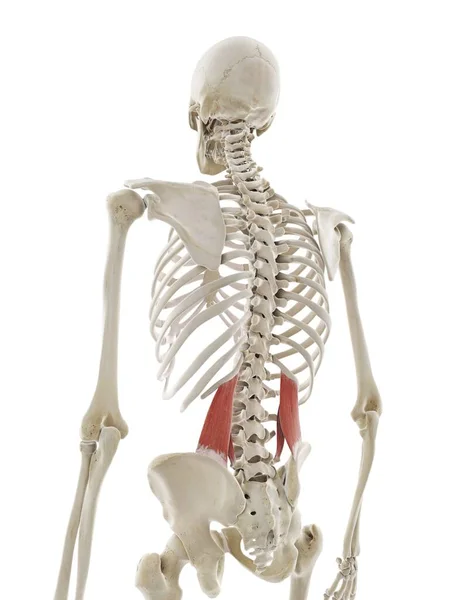
Human Muscular System Torso Muscles Capitus Muscle Anatomy — Stock Photo, Image
Scoliosis of the spine
Scoliosis of the spine — Stock Photo, Image
Human skeleton model with detailed Longissimus thoracis muscle, digital illustration.
Human Skeleton Model Detailed Longissimus Thoracis Muscle Digital Illustration — Stock Photo, Image
Serratus posterior superior muscle, computer illustration
Serratus Posterior Superior Muscle Computer Illustration — Stock Photo, Image
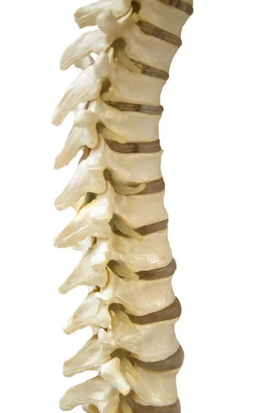
Human Spinal-column model
Human Spinal-column model — Stock Photo, Image
Serratus posterior superior muscle, computer illustration
Serratus Posterior Superior Muscle Computer Illustration — Stock Photo, Image
Human Skeleton System Bone Joints Anatomy. 3D
Human Skeleton System Bone Joints Anatomy — Stock Photo, Image
Male pelvic bones, illustration.
Male Pelvic Bones Illustration — Stock Photo, Image
Posterior pelvic spine region on black background
Posterior Pelvic Spine Region Black Background — Stock Photo, Image
This 3d illustration shows the pectineus muscles on skeleton on a white background
Illustration Shows Pectineus Muscles Skeleton White Background — Stock Photo, Image
Illustration of human anatomy, illustration.
Illustration Human Anatomy Illustration — Stock Photo, Image
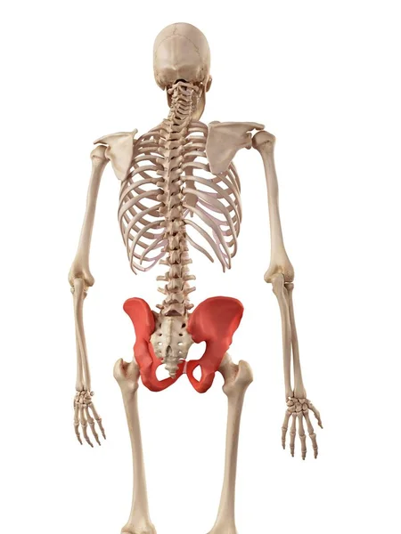
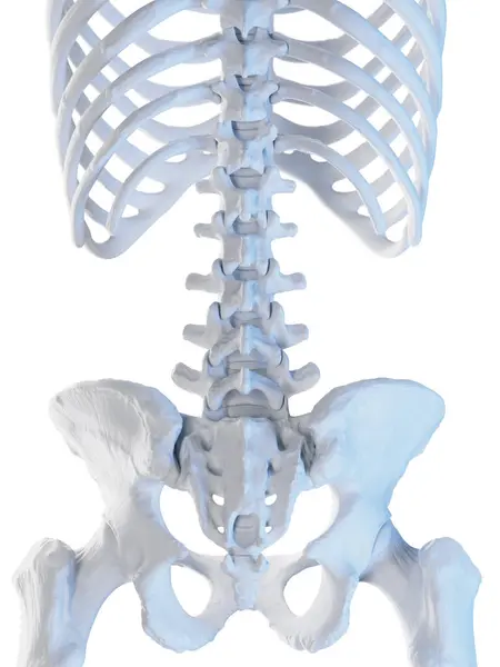
Skeleton pelvic back view on white background
Skeleton Pelvic Back View White Background — Stock Photo, Image
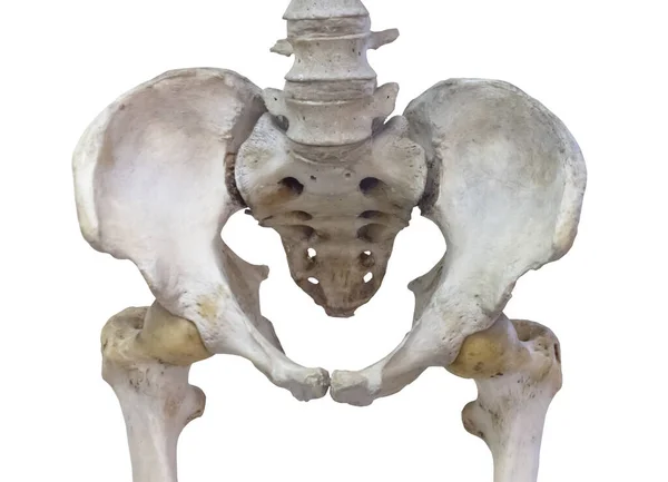
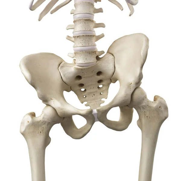
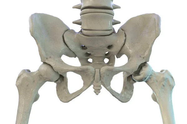
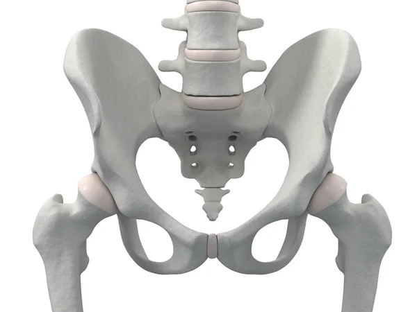
Pelvic bone with femoral neck joint isolated on white background.
Pelvic bone with femoral neck joint isolated on white background. — Stock Photo, Image
3D Illustration of Human Body Organs, Spine
Illustration Human Body Organs Spine — Stock Photo, Image
Human sacrum and pelvic girdle
Human sacrum and pelvic girdle — Stock Photo, Image
3D Illustration, Muscle is a soft tissue, Muscle cells contain proteins , producing a contraction that changes both the length and the shape of the cell. Muscles function to produce force and motion.
Illustration Muscle Soft Tissue Muscle Cells Contain Proteins Producing Contraction — Stock Photo, Image
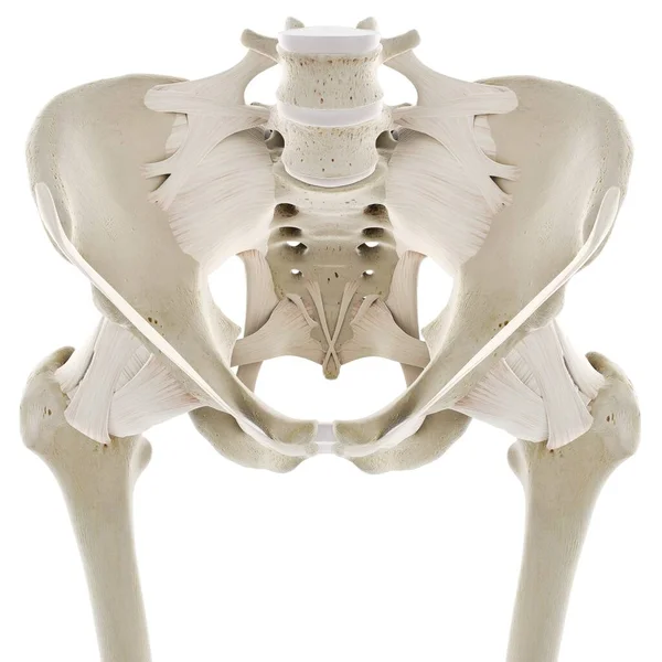
Ligaments of hip, computer illustration
Ligaments Hip Computer Illustration — Stock Photo, Image
Human Skeleton System Bone Joints Anatomy. 3D
Human Skeleton System Bone Joints Anatomy — Stock Photo, Image
This 3d illustration shows the longissimus thoracis muscles on skeleton on a white bacground
Illustration Shows Longissimus Thoracis Muscles Skeleton White Bacground — Stock Photo, Image
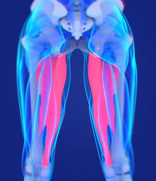
Adductor Muscles anatomy model
Adductor Muscles anatomy model — Stock Photo, Image
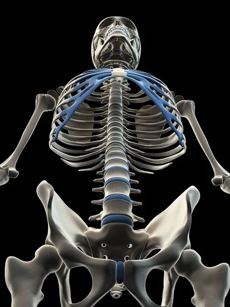
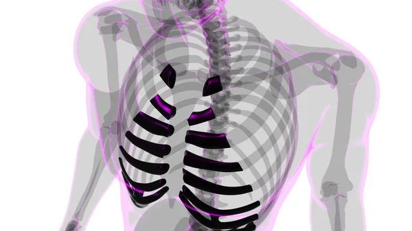
Rib cage and costal cartilage
Rib cage and costal cartilage — Stock Photo, Image
Human skeleton model with detailed Superior gemellus muscle, computer illustration.
Human Skeleton Model Detailed Superior Gemellus Muscle Computer Illustration — Stock Photo, Image
Human Muscular System Torso Muscles Capitus Muscle Anatomy. 3D
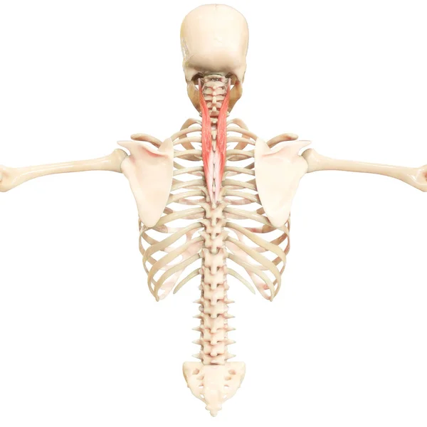
Human Muscular System Torso Muscles Capitus Muscle Anatomy — Stock Photo, Image
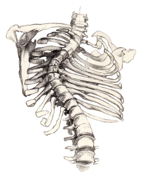
Painful hips
Painful hips — Stock Photo, Image
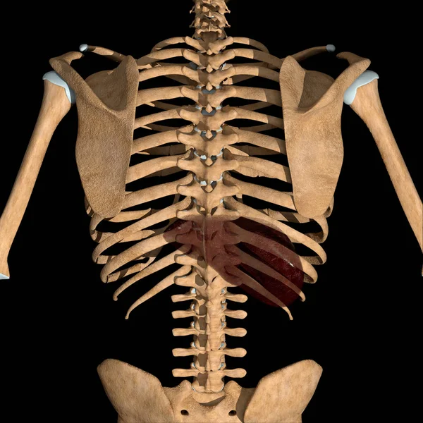
This is a 3d illustration of the back view of the human liver position in skeleton
Illustration Back View Human Liver Position Skeleton — Stock Photo, Image
3d rendered illustration of tilted pelvis in human skeleton.
Rendered Illustration Tilted Pelvis Human Skeleton — Stock Photo, Image
Vertebral Column of Lumbar Vertebrae Human Skeleton System Anatomy 3d rendering
Vertebral Column Lumbar Vertebrae Human Skeleton System Anatomy Rendering — Stock Photo, Image
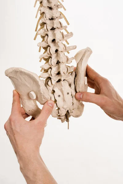
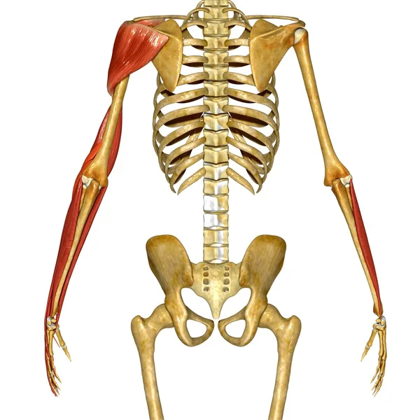
Human skeleton with red colored Biceps muscle, computer illustration.
Human Skeleton Red Colored Biceps Muscle Computer Illustration — Stock Photo, Image
Human Muscle Body with skeleton
Human Muscle Body with skeleton — Stock Photo, Image
Pelvic skeleton x-ray on white background
Pelvic Skeleton Ray White Background — Stock Photo, Image
Vertebral Column of Human Skeleton Anatomy X-ray 3D rendering
Vertebral Column Human Skeleton Anatomy Ray Rendering — Stock Photo, Image
Skeleton
Skeleton — Stock Photo, Image
Hip bone on white background, illustration.
Hip Bone White Background Illustration — Stock Photo, Image
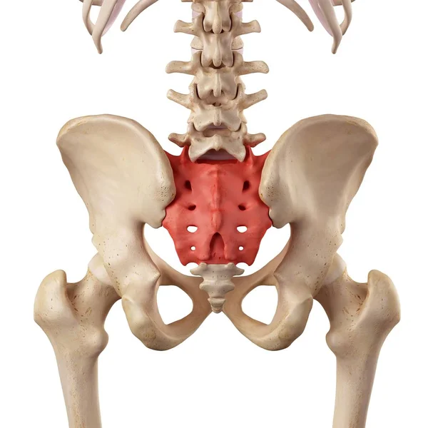
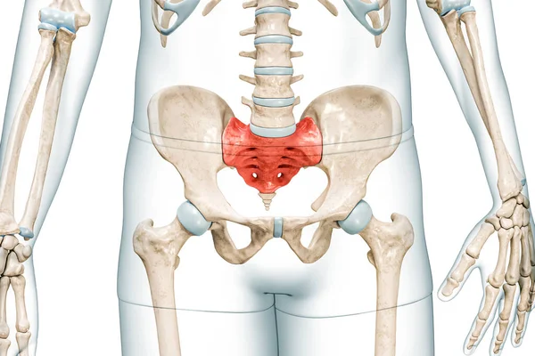
Sacrum bone in red color with body front view 3D rendering illustration isolated on white with copy space. Human skeleton anatomy, medical diagram, osteology, skeletal system, science concepts.
Sacrum Bone Red Color Body Front View Rendering Illustration Isolated — Stock Photo, Image
Musculature system of human back,
Musculature system of human back, — Stock Photo, Image
Human back anatomy
Human back anatomy — Stock Photo, Image
Bones protect and support vital organs and work with muscles to help the body move. 3D rendering
Bones Protect Support Vital Organs Work Muscles Help Body Move — Stock Photo, Image
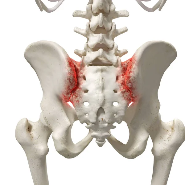
Realistic digital illustration showing arthritis in human sacrum.
Realistic Digital Illustration Showing Arthritis Human Sacrum — Stock Photo, Image
Posterior lumbar spine, illustration.
Posterior Lumbar Spine Illustration — Stock Photo, Image
Sacrum on blue background, illustration.
Sacrum Blue Background Illustration — Stock Photo, Image
Computer illustration of a left femur bone (right) affected by Legg-Calve-Perthes Disease. This childhood hip disorder affects the blood supply to the femoral head.
Computer Illustration Left Femur Bone Right Affected Legg Calve Perthes — Stock Photo, Image
Femur bone affected by Legg-Calve-Perthes Disease, a childhood hip disorder that affects the blood supply to the femoral head, 3D illustration shows affected left femur bone (right side of the image)
Femur Bone Affected Legg Calve Perthes Disease Childhood Hip Disorder — Stock Photo, Image
Hand Muscle, Human Anatomy
Hand Muscle, Human Anatomy — Stock Photo, Image
Human Spinal-column model
Human Spinal-column model — Stock Photo, Image
Spinal column encloses the spinal cord and fluid surrounding spinal cord. Spinal column or backbone model with bones, muscles, tendons, and other tissues on white background
Spinal Column Encloses Spinal Cord Fluid Surrounding Spinal Cord Spinal — Stock Photo, Image
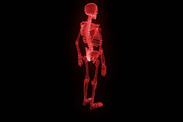
Human Skeleton Wireframe Hologram in Motion. Nice 3D Rendering
Human Skeleton Wireframe Hologram in Motion. Nice 3D Rendering — Stock Photo, Image
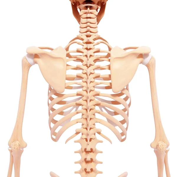
Upper body skeletal structure
Upper body skeletal structure — Stock Photo, Image
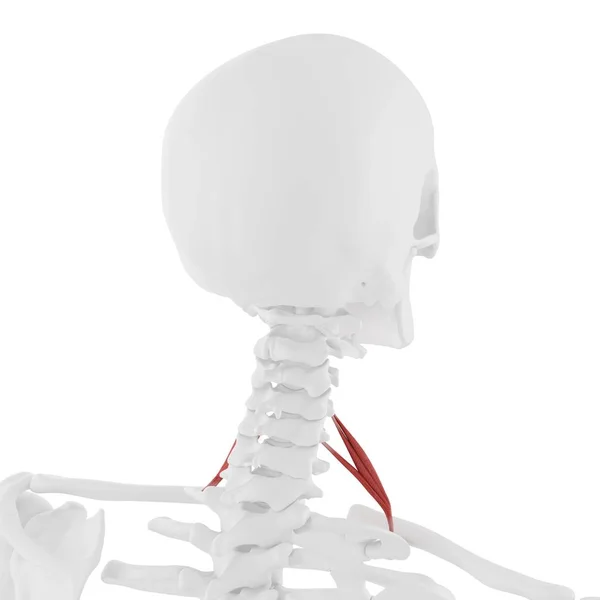
Human skeleton with red colored Scalene anterior muscle, digital illustration.
Human Skeleton Red Colored Scalene Anterior Muscle Digital Illustration — Stock Photo, Image
Multifidus muscle, computer illustration
Multifidus Muscle Computer Illustration — Stock Photo, Image
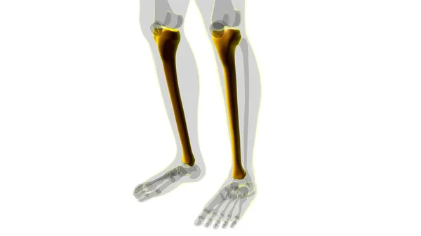
Human skeleton anatomy Tibia Bone 3D Rendering For Medical Concept
Human Skeleton Anatomy Tibia Bone Rendering Medical Concept — Stock Photo, Image
This 3d illustration shows the obturator externus muscles on skeleton
Illustration Shows Obturator Externus Muscles Skeleton — Stock Photo, Image
Male skeletal system, illustration
Male Skeletal System Illustration — Stock Photo, Image
Pelvic girdle and sacrum
Pelvic girdle and sacrum — Stock Photo, Image
Female Body Anatomy. 3D illustration
Female Body Anatomy Illustration — Stock Photo, Image
Human skeleton model with detailed Obturator externus muscle, digital illustration.
Human Skeleton Model Detailed Obturator Externus Muscle Digital Illustration — Stock Photo, Image
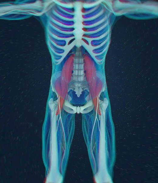
Psoas anatomy model
Psoas anatomy model — Stock Photo, Image
Human skeleton anatomy Costal Cartilage 3D Rendering For Medical Concept
Human Skeleton Anatomy Costal Cartilage Rendering Medical Concept — Stock Photo, Image
Spine xray
Spine xray — Stock Photo, Image
Nerves of head, computer illustration
Nerves Head Computer Illustration — Stock Photo, Image
3d render medical illustration of the human spine
3d render medical illustration of the human spine — Stock Photo, Image
3D Illustration, Muscle is a soft tissue, Muscle cells contain proteins , producing a contraction that changes both the length and the shape of the cell. Muscles function to produce force and motion.
Illustration Muscle Soft Tissue Muscle Cells Contain Proteins Producing Contraction — Stock Photo, Image
Spinal column or backbone model with bones, muscles, tendons, and other tissues on white background. Spinal column encloses the spinal cord and fluid surrounding spinal cord
Spinal Column Backbone Model Bones Muscles Tendons Other Tissues White — Stock Photo, Image

Related image searches

Tailbone Images: A Comprehensive Library for Accurate and High-Quality Visuals

Tailbone images are essential for anatomy and medicine-related projects. These images help illustrate the anatomy and various medical problems associated with the tailbone. At our platform, you can find an extensive collection of high-resolution tailbone images that are perfect for your projects. Our library is filled with a range of images of different angles, positions, and conditions that are easily customizable.

The Types of Tailbone Images Available

We have a comprehensive library of tailbone images that cater to different requirements and needs. Our collection includes X-ray images, MRI scans, and detailed illustrations of the tailbone region. You will find images of various disorders like fractures, dislocations, degenerative changes, infections, and tumors.

Our stock images are available in JPG, AI, and EPS formats, which are widely used across different platforms. You can easily integrate our images into your project and manipulate them according to your needs. Our images are compatible with different software, which improves the feasibility of using these images for any project.

Where can Tailbone Images be Used

Tailbone images are versatile and can be used for different purposes. These images are widely used in medical and health-related projects such as research papers, patient education materials, and presentations. Health professionals like doctors and chiropractors also use these images for their clinics' websites.

Moreover, some designers and art instructors use these images for anatomical studies and illustrations. Additionally, tailbone images can be used for creating informative posters or flyers about medical problems related to the coccyx.

The Importance of Choosing the Right Tailbone Images for Your Project

Choosing the right image for your project is crucial. The right image can help convey your message more effectively, while the wrong one can lead to misinterpretation. It is essential to consider the context of the image, its quality, and its relevance to your project.

Our platform features tailbone images that are both accurate and of high-quality, ensuring a better visual experience for your project. It is also important to ensure that the image is legally obtained and that you're not infringing on any copyrights. Our images are royalty-free, so you can use them confidently without any legal repercussions.

Using Tailbone Images: Tips & Tricks

When using tailbone images, it is important to pay attention to the details that you want to showcase and the visuals' placement. It's essential to make sure the image is placed in the right position so that it is more visible and readable. You can also customize the image with colors or other graphics to produce an engaging final product.

Moreover, you can also overlay graphics over the image to create a more interactive element for your project. You can add annotations or labels highlighting specific features or provide more context to the image with explanatory texts.

The Bottom line

With our comprehensive library of tailbone images, you have access to visuals that help convey your message in a compelling and engaging way. Whether you're using it for educational purposes or a professional presentation, our high-quality images cater to a variety of project requirements. With a range of formats available, integration with third-party tools is seamless.

Choosing the right image to complement the tone and message of your project is crucial to creating a lasting impression. Take your project's visual appeal to the next level and use our tailbone images today.