Rabbit Teeth Grow Continuously Throughout Life Why Ameloblasts Can Seen — Stock Photo, Image

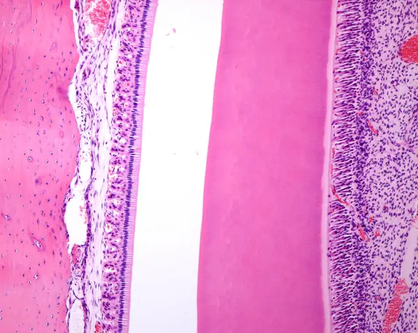
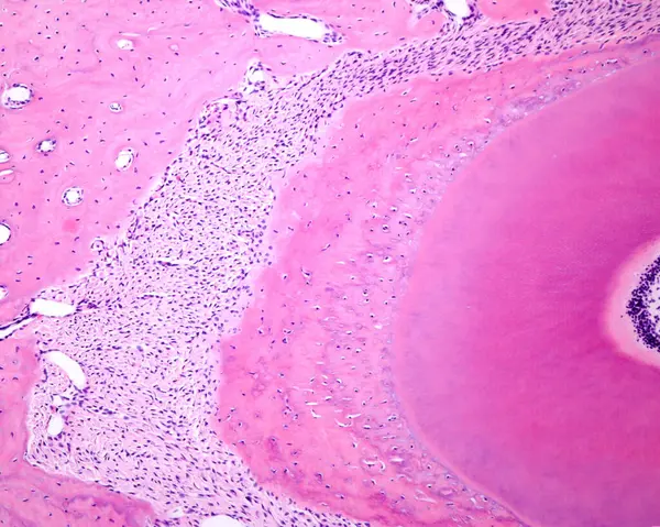
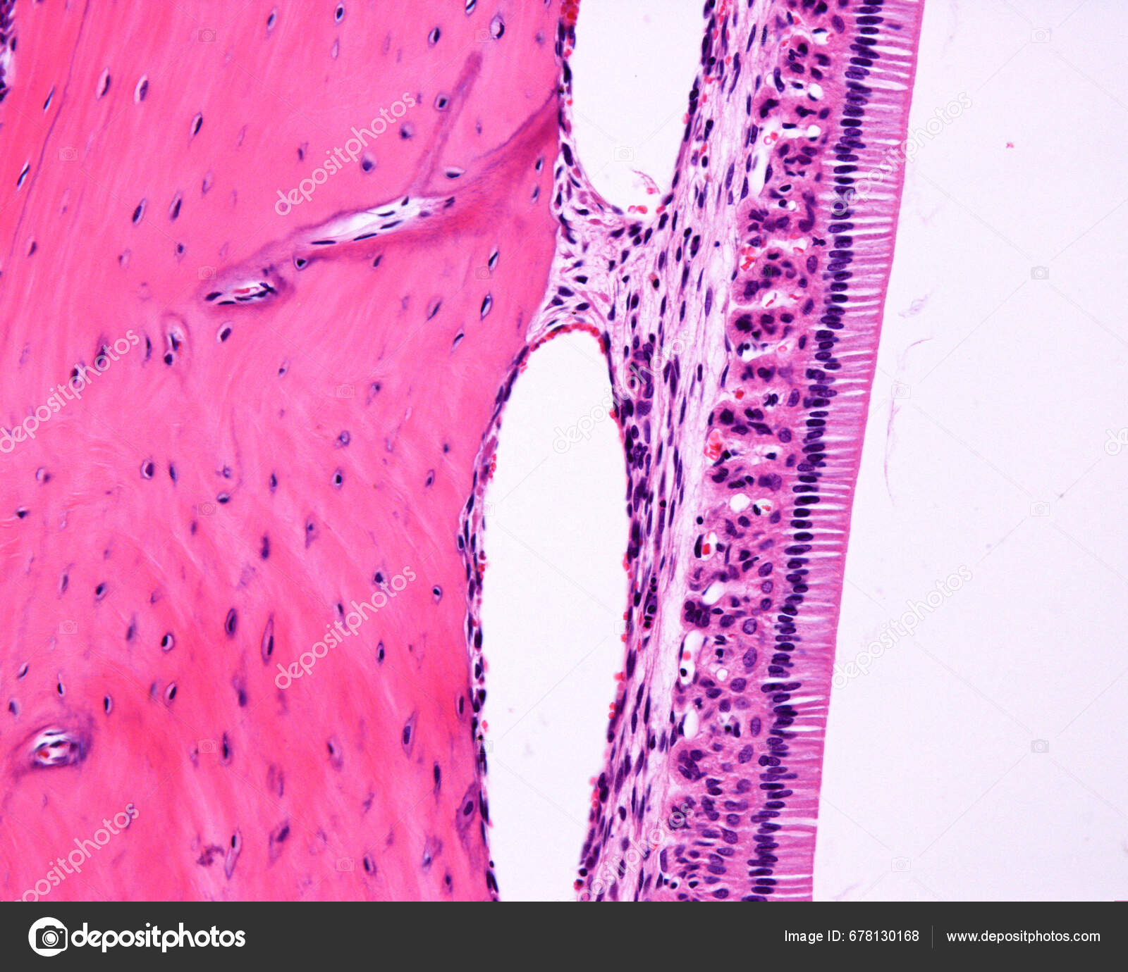
Rabbit teeth grow continuously throughout life. This is why ameloblasts can be seen in an adult tooth. Light microscope micrograph showing amelobasts arranged as a single row of very tall cells. The enamel appears empty on the right because it is tot — Photo

Rabbit teeth grow continuously throughout life. This is why ameloblasts can be seen in an adult tooth. Light microscope micrograph showing amelobasts arranged as a single row of very tall cells. The enamel appears empty on the right because it is tot

 — Photo by jlcalvo@ucm.es

Same Series:

Rabbit molar tooth showing from left to right: alveolar bone, ameloblasts, enamel void region (that appears empty because it is totally dissolved during tissue processing) dentin, predentin, odontoblasts and dental pulp with enlarged blood vessels. R
Rabbit Molar Tooth Showing Left Right Alveolar Bone Ameloblasts Enamel — Stock Photo, Image
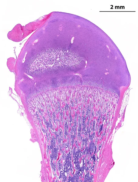
Low power light microscope micrograph showing a developing long bone (femur). On top, the epiphysis is made by hyaline cartilage that shows an initial secondary ossification centre. On bottom, the secondary diaphyseal ossification centre. The round l
Low Power Light Microscope Micrograph Showing Developing Long Bone Femur — Stock Photo, Image
Cross section of an artery, showing an atheroma plaque which reduces drastically the blood vessel lumen, situated at the right.
Atheroma plaque — Stock Photo, Image
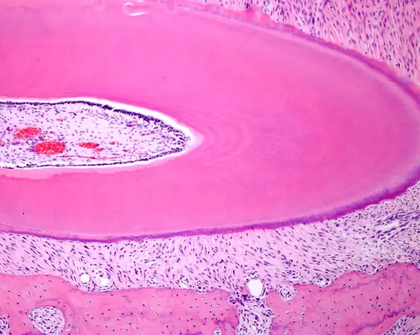
Cross section (slightly oblique) of a tooth root showing from the left border: dental pulp, row of odontoblast cell bodies, predentine, dentin, acellular cementum, periodontal ligament and alveolar bone.
Cross Section Slightly Oblique Tooth Root Showing Left Border Dental — Stock Photo, Image
Skeletal striated muscle fibers in cross section showing the presence of several nuclei (multinucleated cells) located in the cell periphery. Light microscope photomicrograph.
Striated muscle fibers — Stock Photo, Image
Human brain. Acute meningitis for Haemophilus influenzae. The meninges appear very thickened by an extensive infiltration of inflammatory cells. Light microscope micrograph. Hematoxylin & eosin stain
Acute meningitis — Stock Photo, Image
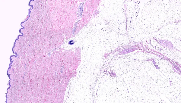
Low power light microscope micrograph of thin skin showing form left to right, the epidermis, a very thick dermis showing sweat glands and a hair follicle in the deep region, and the adipose tissue of the hypodermis. Hematoxylin-eosin.
Low Power Light Microscope Micrograph Thin Skin Showing Form Left — Stock Photo, Image
Cross sectioned tooth root showing from the right border: odontoblast cell bodies, predentine, dentin, cellular cementum, periodontal ligament and alveolar bone.
Cross Sectioned Tooth Root Showing Right Border Odontoblast Cell Bodies — Stock Photo, Image
Simple columnar epithelium of the small intestine. The apical surface shows a well developed brush border. In the center, the "cup" of three goblet cell are seen. Light microscope micrograph. H&E stain.
Small intestine epithelium — Stock Photo, Image
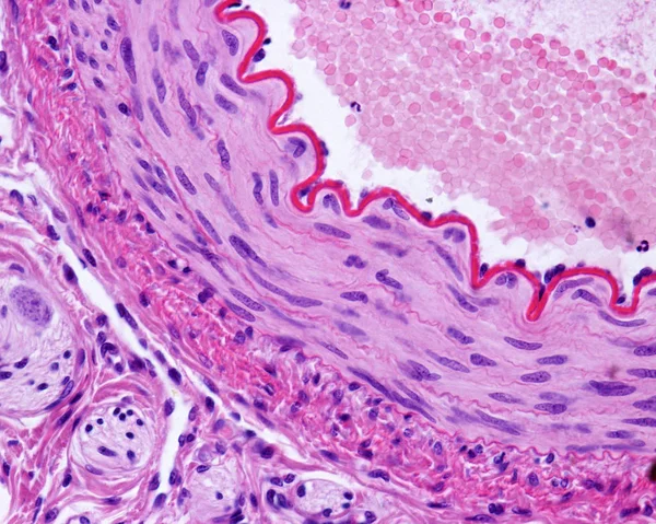
Light micrograph of a cross-sectioned muscular artery, showing a thick and wavy internal elastic lamina, a middle layer with smooth muscle fibers, and an outer connective tissue adventitia. Hematoxylin & eosin stain
Muscular artery — Stock Photo, Image
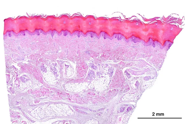
Low power light microscope micrograph of human glabrous skin. From top, the epidermis, a keratinized stratified squamous epithelium showing a prominent horny layer, the dermis and hypodermis. The hypodermis is identified by the presence of adipose ti
Low Power Light Microscope Micrograph Human Glabrous Skin Top Epidermis — Stock Photo, Image
A septum of connective tissue, with numerous adipocytes (fat cells) which contain a large lipid droplet. The sinuous structures correspond to nerves. Light micrograph. Hematoxylin & eosin stain
Adipose tissue — Stock Photo, Image
Low power light microscope micrograph of a cat thyroid. The thyroid parenchyma is composed of many follicles filled with colloid. On the right side, a parathyroid gland is located inside the thyroid. Light microscope micrograph. H&E stain.
Low Power Light Microscope Micrograph Cat Thyroid Thyroid Parenchyma Composed — Stock Photo, Image
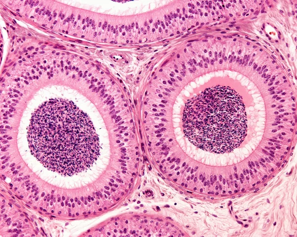
Two sections of the epididymal duct surrounded by concentric layers of fibromuscular tissue. The pseudostratified epithelium consists of tall columnar cells showing stereocilia, and basal cells. The lumen is filled with sperm cells.
Epididymis. Pseudostratified epithelium — Stock Photo, Image
Brown adipocytes stained with hematoxylin & eosin. They contain many small lipid droplets distributed throughout the cytoplasm, thus acquiring a spongy appearance.
Brown adipocytes — Stock Photo, Image
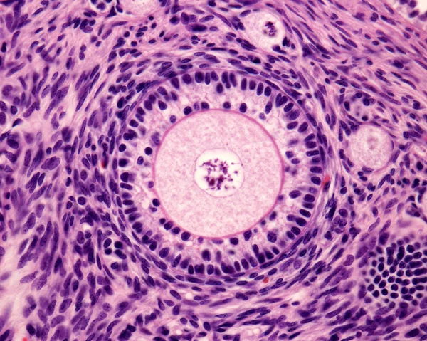
Light microscope micrograph of an ovary. Primary follicle with a big round ovocyte surrounded by the zona pellucida and two layers of granulosa cells.
Primary ovary follicle — Stock Photo, Image

Usage Information

You can use this royalty-free photo "Rabbit teeth grow continuously throughout life. This is why ameloblasts can be seen in an adult tooth. Light microscope micrograph showing amelobasts arranged as a single row of very tall cells. The enamel appears empty on the right because it is tot" for personal and commercial purposes according to the Standard or Extended License. The Standard License covers most use cases, including advertising, UI designs, and product packaging, and allows up to 500,000 print copies. The Extended License permits all use cases under the Standard License with unlimited print rights and allows you to use the downloaded stock images for merchandise, product resale, or free distribution.

You can buy this stock photo and download it in high resolution up to 3840x3072. Upload Date: Sep 28, 2023