Fundus Stock Photos

100,000 Fundus pictures are available under a royalty-free license

  • Best Match
  • Fresh
  • Popular
1-100 of 100,000
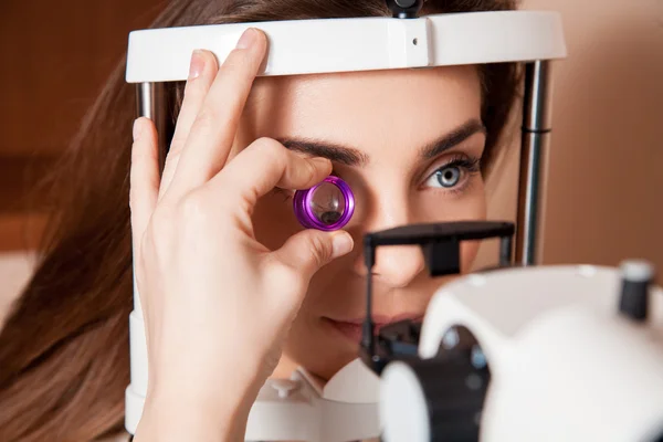
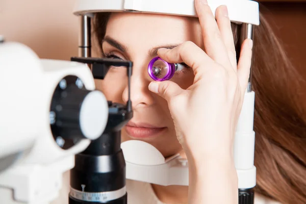
Beautiful adult woman patient during an eye examination at the e
Beautiful adult woman patient during an eye examination at the e — Stock Photo, Image
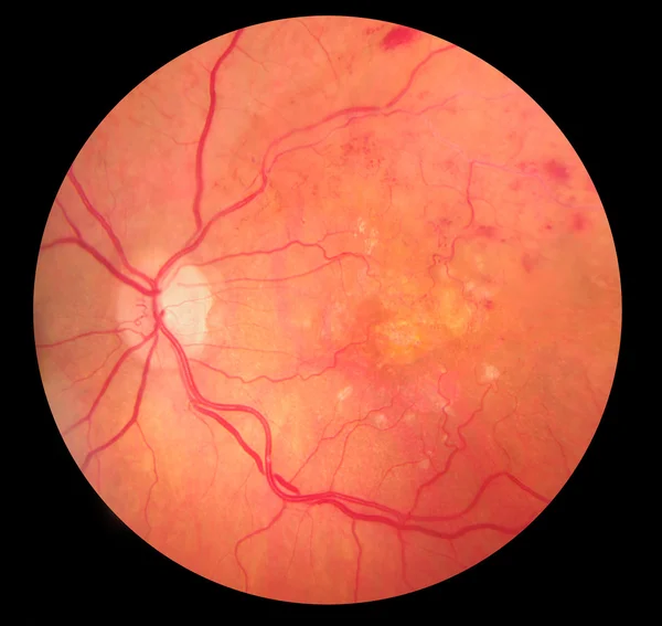
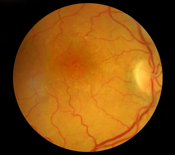
Medical Fundus photo of retinopathy hemmorhage
Medical Fundus photo of retinopathy hemmorhage — Stock Photo, Image
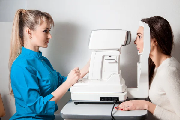
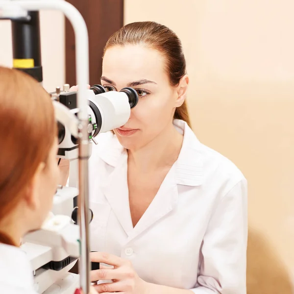
Ophthalmologist woman In Exam Room With Young beautiful brunette
Ophthalmologist woman In Exam Room With Young beautiful brunette — Stock Photo, Image
Retina
Retina — Stock Photo, Image
Retina
Retina — Stock Photo, Image
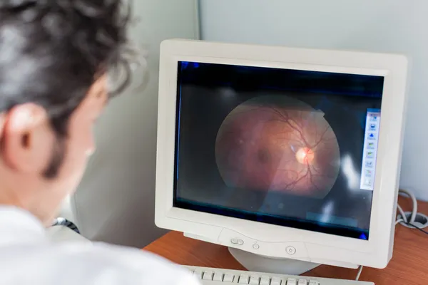
Retinal exam
Retinal exam — Stock Photo, Image
View inside human eye showing retina, optic nerve and macula. Health concept
View inside human eye showing retina, optic nerve and macula. Health concept — Stock Photo, Image
Orange food isolate
Orange food isolate — Stock Photo, Image
Human Retina - Optic Nerve
Human Retina - Optic Nerve — Stock Photo, Image
Ophthalmologist doctor in exam optician laboratory with female patient. Eye care
Ophthalmologist doctor in exam optician laboratory with female patient. Eye care — Stock Photo, Image
Woman check her vision
Woman check her vision — Stock Photo, Image
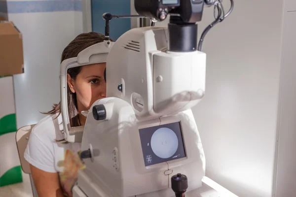
Retinal camera
Retinal camera — Stock Photo, Image
Medical Fundus photo of retinal pathology, hemmorhages, vein occ
Medical Fundus photo of retinal pathology, hemmorhages, vein occ — Stock Photo, Image
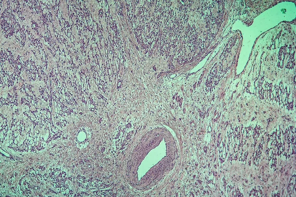
Inflamed tissue under the microscope 100x
Inflamed Tissue Microscope 100X — Stock Photo, Image
Endometrial adenocarcinoma, light micrograph, photo under microscope
Endometrial Adenocarcinoma Light Micrograph Photo Microscope — Stock Photo, Image
Spinal ganglion from microscopy
Spinal Ganglion Microscopy — Stock Photo, Image
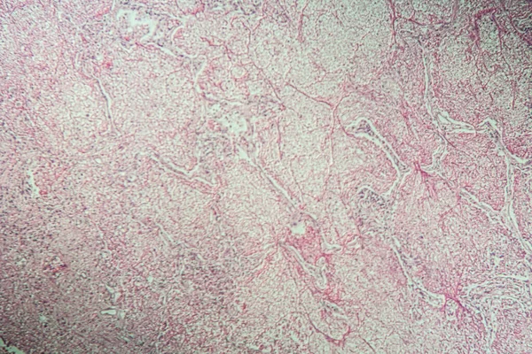
Micrograph of liver tissue
Micrograph of liver tissue — Stock Photo, Image
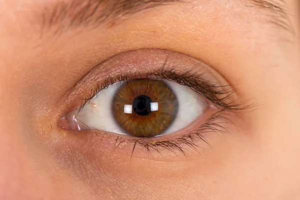
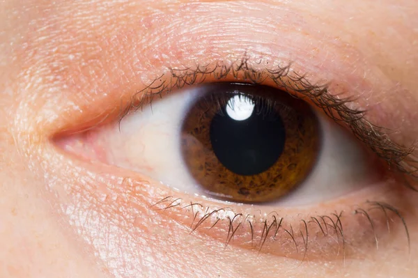
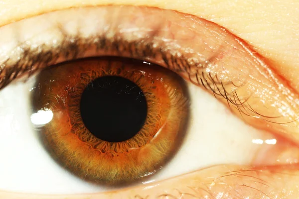
Extreme close up picture of a young woman's brown eye
Extreme Close Picture Young Woman Brown Eye — Stock Photo, Image
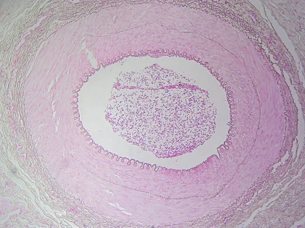
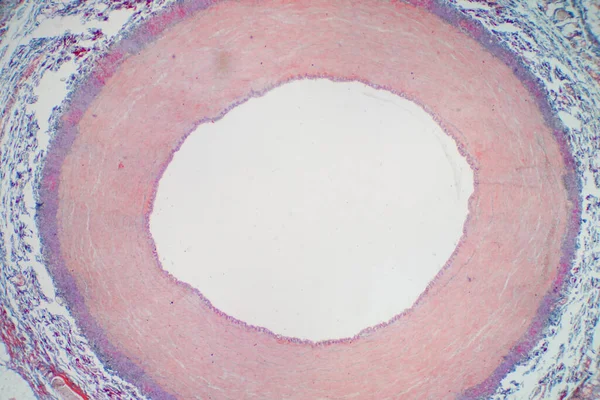
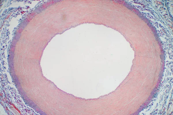
The Anatomy of Blood Vessels: Muscular Artery in High Detail
Anatomy Blood Vessels Muscular Artery High Detail — Stock Photo, Image

Abstract black ink stain. Chinese ink background on watercolor paper.
Abstract Black Ink Stain Chinese Ink Background Watercolor Paper — Stock Photo, Image
Neurofibromatosis, light micrograph, photo under microscope. A group of genetic disorders with formation of tumors in the nervous system
Neurofibromatosis Light Micrograph Photo Microscope Group Genetic Disorders Formation Tumors — Stock Photo, Image
Spinal ganglion view from microscope
Spinal Ganglion View Microscope — Stock Photo, Image
Artery blood vessel under microscope view for education histology. Histological for human physiology.
Artery Blood Vessel Microscope View Education Histology Histological Human Physiology — Stock Photo, Image
Liver cells of a rabit with Coccidia parasites.
Liver cells of a rabit with Coccidia parasites. — Stock Photo, Image
Glossy pink background stock images
Glossy pink background stock images — Stock Photo, Image
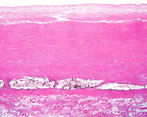
Human aorta. Dissecting aneurysm or aortic dissection. Low magnification micrograph showing a dissecting aneurysm located in the media layer of the aorta wall. Macrophages loaded with iron pigment appear in the detachment zone.
Human Aorta Dissecting Aneurysm Aortic Dissection Low Magnification Micrograph Showing — Stock Photo, Image
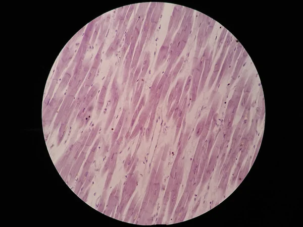
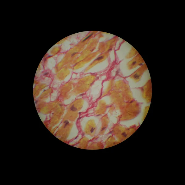
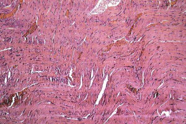
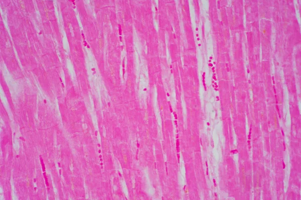
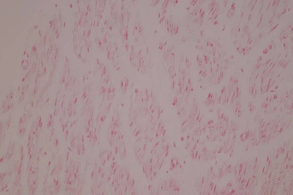
Histology of cardiac muscle under microscope view
Histology Cardiac Muscle Microscope View — Stock Photo, Image
Macro of One brown woman eye
Macro One Brown Woman Eye — Stock Photo, Image
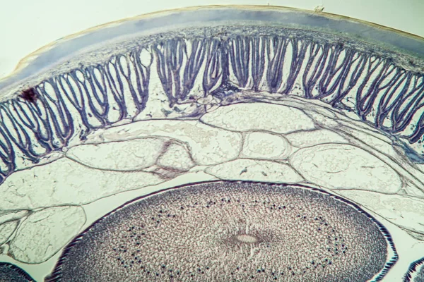
Microscopic photography of the insect tissue
Microscopic Photography Insect Tissue — Stock Photo, Image
Microscopic section of liver tissue
Microscopic section of liver tissue — Stock Photo, Image
Photomicrograph of lung adenocarcinoma, illustrating malignant glandular cells characteristic of the most common type of lung cancer.
Photomicrograph Lung Adenocarcinoma Illustrating Malignant Glandular Cells Characteristic Most Common — Stock Photo, Image
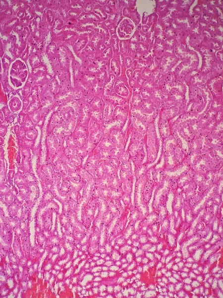
Red eye isolated
Red eye isolated — Stock Photo, Image
Microscopic photo of many things found around
Microscopic Photo Many Things Found — Stock Photo, Image
Abstract ink background. Marble style. Black paint stroke texture on white paper. Wallpaper for web and game design. Grunge mud art. Macro image of pen juice. Dark Smear
Abstract Ink Background Marble Style Black Paint Stroke Texture White — Stock Photo, Image
Watercolor stain circle, brown stain
Watercolor Stain Circle Brown Stain — Stock Photo, Image
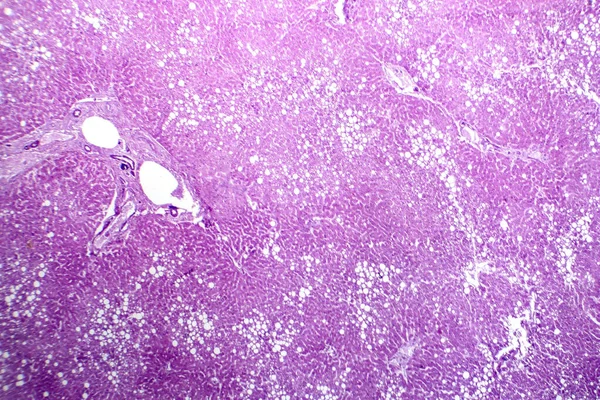
Photomicrograph of hepatic steatosis, revealing fat accumulation in liver cells, known as fatty liver disease.
Photomicrograph Hepatic Steatosis Revealing Fat Accumulation Liver Cells Known Fatty — Stock Photo, Image
Metal texture with scratches and cracks
Metal Texture Scratches Cracks — Stock Photo, Image
Tie Dye Pattern. Blue Artistic Fabric. Heart
Tie Dye Pattern. Blue Artistic Fabric. Heart — Stock Photo, Image
Testicle cross-section under the microscope 100x
Testicle Cross Section Microscope 100X — Stock Photo, Image
Microscopic cells texture, abstract background
Microscopic Cells Texture Abstract Background — Stock Photo, Image
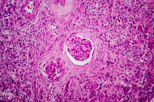
Chronic glomerulonephritis, light micrograph, photo under microscope
Chronic Glomerulonephritis Light Micrograph Photo Microscope — Stock Photo, Image
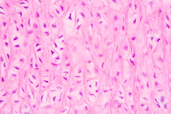
Trachea, light micrograph. Hyaline cartilage from trachea showing chondrocytes, collagen fibers and matrix.
Trachea Light Micrograph Hyaline Cartilage Trachea Showing Chondrocytes Collagen Fibers — Stock Photo, Image
Bone cells of a bone seen from the microscope.
Bone Cells Bone Seen Microscope — Stock Photo, Image
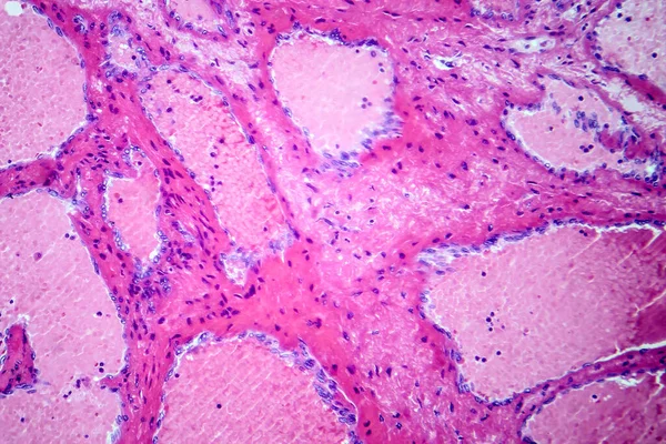
Photomicrograph of hepatic cavernous hemangioma, depicting dilated blood vessels in the liver tissue, characteristic of a benign tumor.
Photomicrograph Hepatic Cavernous Hemangioma Depicting Dilated Blood Vessels Liver Tissue — Stock Photo, Image
Meningioma, a slow-growing tumor that forms from the meninges, light micrograph, photo under microscope
Meningioma Slow Growing Tumor Forms Meninges Light Micrograph Photo Microscope — Stock Photo, Image
Microbiology, microscopic cells surface
Microbiology Microscopic Cells Surface — Stock Photo, Image
Cicatrical myocardial tissue
Cicatrical myocardial tissue — Stock Photo, Image
Motor nerves in spinal tissue.
Motor Nerves Spinal Tissue — Stock Photo, Image
Non-Hodgkin's lymphoma, light micrograph, photo under microscope. High magnification
Non Hodgkin Lymphoma Light Micrograph Photo Microscope High Magnification — Stock Photo, Image
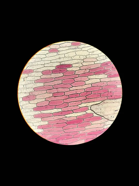
Biological preparation of onion plant cells under a microscope with the phenomenon of plasmolysis
Biological Preparation Onion Plant Cells Microscope Phenomenon Plasmolysis — Stock Photo, Image
Purulent meningitis, light micrograph, photo under microscope
Purulent Meningitis Light Micrograph Photo Microscope — Stock Photo, Image
Horse roundworm in cross section with eggs 100x
Horse Roundworm Cross Section Eggs 100X — Stock Photo, Image
Human eye close-up
Human eye close-up — Stock Photo, Image
Watercolor painted paper background texture
Watercolor painted paper background texture — Stock Photo, Image
View at human sperm under microscope in labaratory
View Human Sperm Microscope Labaratory — Stock Photo, Image
Thyroid follicular carcinoma, light micrograph, photo under microscope
Thyroid Follicular Carcinoma Light Micrograph Photo Microscope — Stock Photo, Image
Photomicrograph of hepatic steatosis, revealing fat accumulation in liver cells, known as fatty liver disease.
Photomicrograph Hepatic Steatosis Revealing Fat Accumulation Liver Cells Known Fatty — Stock Photo, Image
Microscopic detail showing a rats heart detail
Microscopic detail showing a rats heart detail — Stock Photo, Image
Histology of human cardiac muscle under microscope view for education, Human tissue histology.
Histology Human Cardiac Muscle Microscope View Education Human Tissue Histology — Stock Photo, Image
Monochrome texture. Image including effect of black and white tones. Abstract background.
Monochrome Texture Image Including Effect Black White Tones Abstract Background — Stock Photo, Image
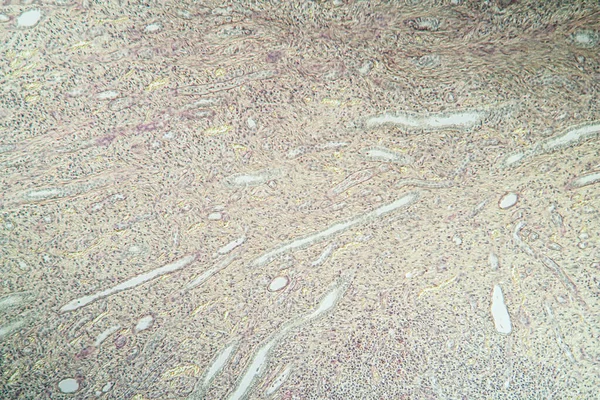
Hydronephrosis kidney inflammation in tissue section 100x
Hydronephrosis Kidney Inflammation Tissue Section 100X — Stock Photo, Image
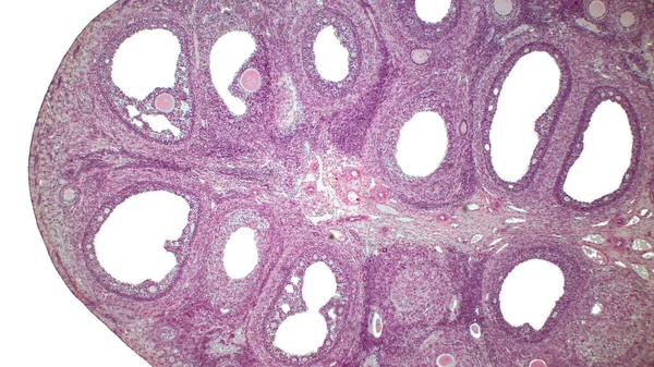
Light micrograph of the Cat's ovarian. Micrograph of ovary showing primordial, primary and secondary follicles isolated on white background. Hematoxylin and eosin stain.
Light Micrograph Cat Ovarian Micrograph Ovary Showing Primordial Primary Secondary — Stock Photo, Image
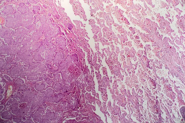
Diseased tissue under the microscope 100x
Diseased Tissue Microscope 100X — Stock Photo, Image
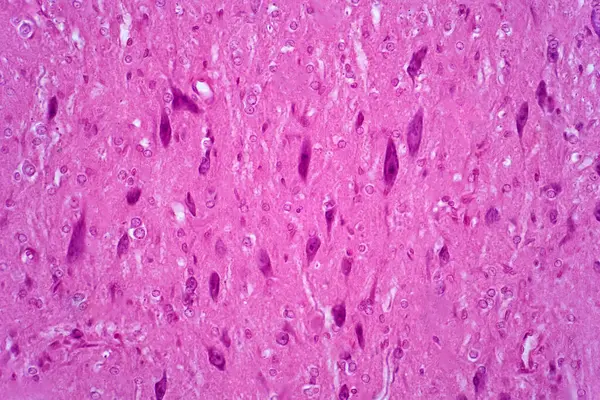
Light microscope of Motor neurons of the spinal cord are part of the central nervous system. Haematoxylin and eosin staining technique for human tissue.
Light Microscope Motor Neurons Spinal Cord Part Central Nervous System — Stock Photo, Image
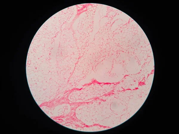
Human fat body tissue under microscope view
Human Fat Body Tissue Microscope View — Stock Photo, Image
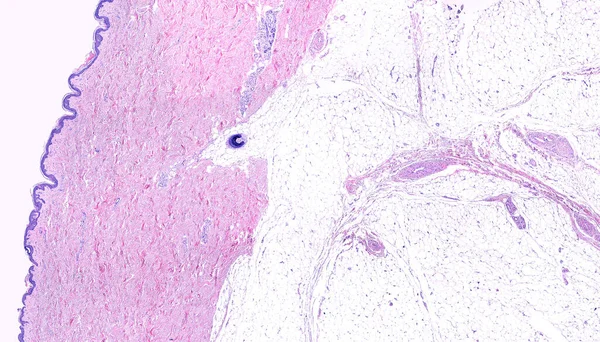
Low power light microscope micrograph of thin skin showing form left to right, the epidermis, a very thick dermis showing sweat glands and a hair follicle in the deep region, and the adipose tissue of the hypodermis. Hematoxylin-eosin.
Low Power Light Microscope Micrograph Thin Skin Showing Form Left — Stock Photo, Image
Large grunge textures and backgrounds
Large grunge textures and backgrounds — Stock Photo, Image
Iris nevi at eye test
Iris nevi at eye test — Stock Photo, Image
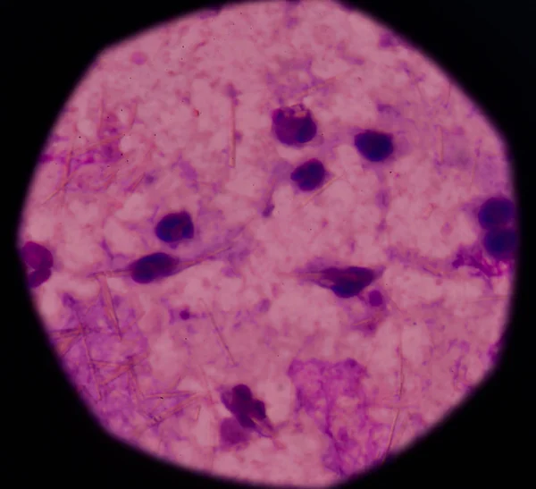
White blood cells with Uric acid crystal needle shape
White blood cells with Uric acid crystal needle shape — Stock Photo, Image
Bowen's Disease Tumor under the microscope 100x
Bowen Disease Tumor Microscope 100X — Stock Photo, Image
Human fat body tissue under microscope view
Human Fat Body Tissue Microscope View — Stock Photo, Image
Insightful look eyes boy
Insightful look  eyes boy — Stock Photo, Image
Microbiology, microscopic cells surface
Microbiology Microscopic Cells Surface — Stock Photo, Image
Abstract black ink stain. Chinese ink background on watercolor paper.
Abstract Black Ink Stain Chinese Ink Background Watercolor Paper — Stock Photo, Image
Artery blood vessel under microscope view for education histology. Histological for human physiology.
Artery Blood Vessel Microscope View Education Histology Histological Human Physiology — Stock Photo, Image
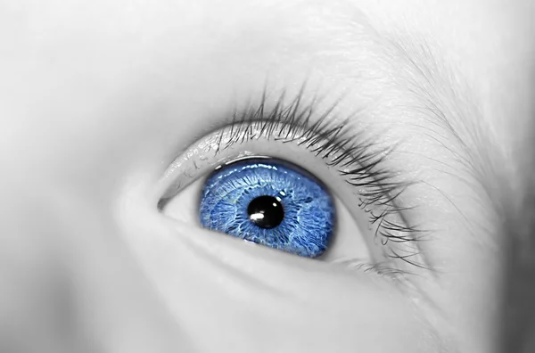
Eye of a female face with blue eyes
Eye Female Face Blue Eyes — Stock Photo, Image
Iris nodule at eye test
Iris nodule at eye test — Stock Photo, Image
Histopathology of Japanese encephalitis, light micrograph, photo under microscope
Histopathology Japanese Encephalitis Light Micrograph Photo Microscope — Stock Photo, Image
Muscle cells
Muscle cells — Stock Photo, Image
Close up human cells with microscope.
Close up human cells with microscope. — Stock Photo, Image
Micrograph of small intestinum tenue tissue
Micrograph of small intestinum tenue tissue — Stock Photo, Image
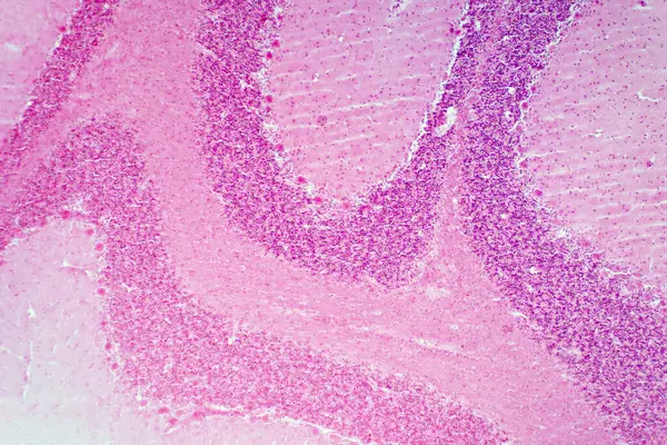
Cerebellum tissue, light micrograph. Haematoxylin and eosin stain.
Cerebellum Tissue Light Micrograph Haematoxylin Eosin Stain — Stock Photo, Image
Cliated epithelium of human under the microscope in Lab.
Cliated Epithelium Human Microscope Lab — Stock Photo, Image
Anatomy and Histological Bone, Elastic cartilage human and Joint of human foetus under the microscope for education.
Anatomy Histological Bone Elastic Cartilage Human Joint Human Foetus Microscope — Stock Photo, Image
Girl's blue eye close up
Girl Blue Eye Close — Stock Photo, Image
A young woman's eye - close up
A young woman's eye - close up — Stock Photo, Image
Hippie Water Effects. Artistic Design for Cloth
Hippie Water Effects. Artistic Design for Cloth — Stock Photo, Image
Thyroid follicular carcinoma, light micrograph, photo under microscope
Thyroid Follicular Carcinoma Light Micrograph Photo Microscope — Stock Photo, Image
Abstract ink background. Marble style. Black paint stroke texture on white paper. Wallpaper for web and game design. Grunge mud art. Macro image of pen juice. Dark Smear
Abstract Ink Background Marble Style Black Paint Stroke Texture White — Stock Photo, Image
Anatomy and Histological Ovary and Testis human cells under microscope.
Anatomy Histological Ovary Testis Human Cells Microscope — Stock Photo, Image
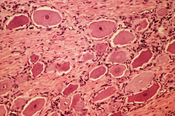
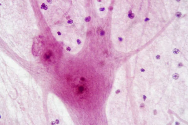
Motor neurons, light micrograph. Motor neurons of the spinal cord are part of the central nervous system. Haematoxylin and eosin stain.
Motor Neurons Light Micrograph Motor Neurons Spinal Cord Part Central — Stock Photo, Image
Eyeball with iris pupil
Eyeball Iris Pupil — Stock Photo, Image
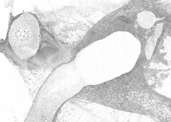
Longitudinal section of a porcine embryo head under the microscope.
Longitudinal Section Porcine Embryo Head Microscope — Stock Photo, Image
Abstract colorful stains on background
Abstract Colorful Stains Background — Stock Photo, Image
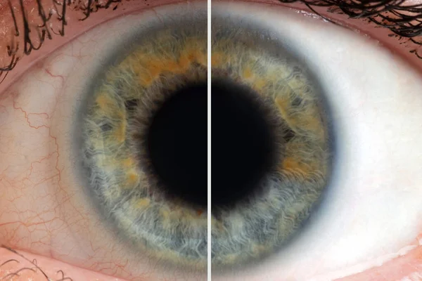
Human eye with blood vessels before and after
Human eye with blood vessels before and after — Stock Photo, Image
Education Spinal cord and Motor Neuron under the microscope in Lab.
Education Spinal Cord Motor Neuron Microscope Lab — Stock Photo, Image
Histology of human kidney under microscope view
Histology Human Kidney Microscope View — Stock Photo, Image
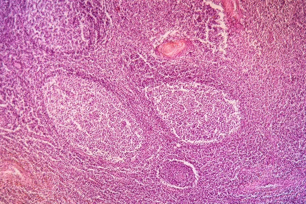
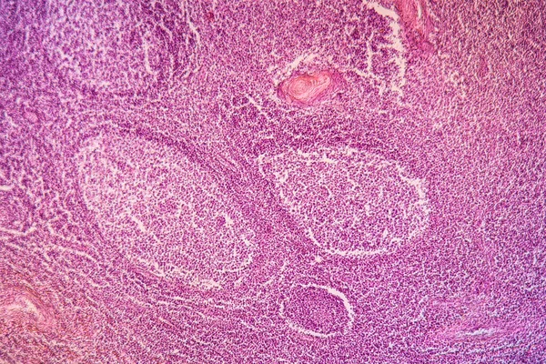
In Depth Look at Lymph Nodes: Magnified Histology Image in High Detail
Depth Look Lymph Nodes Magnified Histology Image High Detail — Stock Photo, Image
Poly nephritis, tissue under the microscope 100x
Poly Nephritis Tissue Microscope 100X — Stock Photo, Image
Motor Neuron and nerve cells under the microscope in Lab.
Motor Neuron Nerve Cells Microscope Lab — Stock Photo, Image

Explore the Wide Range of Fundus Images for Medical Projects

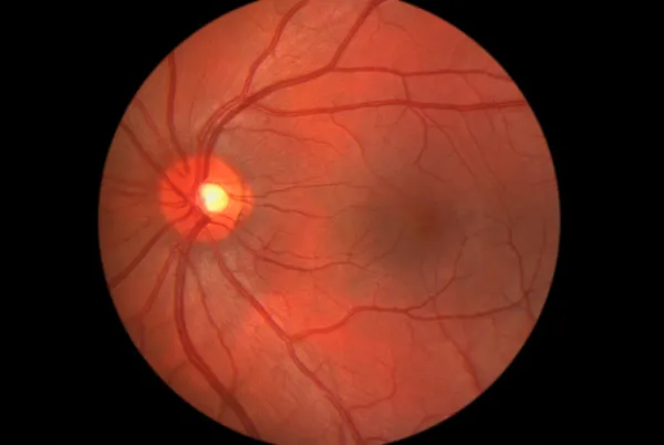
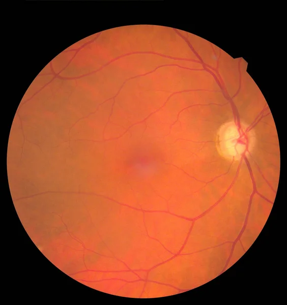
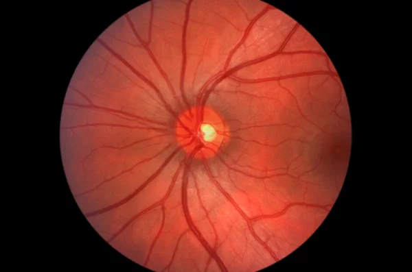
If you are looking for high-quality fundus images to use in your medical projects, you have come to the right place. Our stock image collection features a wide range of fundus images, which display the inner surface of the eye, including the retina and optic disc. These images are available in various file formats, including JPG, AI, and EPS, making it easy to work with them across different platforms.

Types of Fundus Images Available

Our collection of fundus images includes a diverse range of options, including color and grayscale images. These images are taken using different techniques, including direct and indirect ophthalmoscopes, slit-lamp biomicroscopy, and fundus photography. The images capture different parts of the retina, such as the macula, the optic disc, and the peripheral retina, enabling you to choose the right image for your project.

Where to Use Fundus Images

Fundus images are widely used in the medical field, especially in ophthalmology, where they are helpful in diagnosing and managing various eye diseases. These images are also used in academic and research projects, including medical publications, presentations, and training materials.

When choosing a fundus image for your project, it is important to consider the context and audience. For instance, a colored fundus image may be more suitable for a patient education brochure, while a grayscale image may be better suited for a medical research paper.

Tips for Using Fundus Images Effectively

When using fundus images, it is crucial to ensure they are of high quality so that they accurately represent the eye's anatomy. Additionally, you should ensure that the image's orientation and labeling are consistent with the intended use.

Incorporating fundus images into your medical project can enhance its visual appeal and make it more engaging for the audience. The images can be used to draw attention to specific areas of the eye and highlight abnormalities, improving the viewer's understanding of the medical condition being discussed.

Conclusion

Our collection of fundus images provides a diverse range of high-quality images that can be used for different medical projects. Whether you are creating patient education materials or academic research papers, you will find the right image to suit your needs. With these tips, you can use fundus images effectively and create compelling medical projects that enhance viewer understanding and engagement.